
Citation: Anmol M, et al. A Comparative Study to Access Cephalometric and Arch Width Characteristics of Class II Division 1 and Division 2 Malocclusions among a Indian and Turkish Population. J Dental Sci 2017, 2(3): 000133.
*Corresponding author: Anmol M, Associate Professor, Department of Public Health Dentistry, Surendera Dental College and Research Institute, Sri Ganganagar, India, Tel: 9899963523; Email: dranmolmathur@gmail.com
Objective: To investigate the cephalometric and dental arch width in Indian population and to compare the same with Turkish population.
Material and Methods: The dental arch dimensions of study cast of selected sample among Indian population was collected and the data was entered in the spreadsheet for the statistical analysis using SPSS software version 20.Inter examiner variability was tested among the trained dentist. The reliability between the examiners was tested two days prior to the study.
Results: The difference of all the parameters was found to be significant when compared between Indian and Turkish population.
Conclusion: The study yielded a database about dental arch width with different definitions by which different studies can be compared.
Keywords: Malocclusion; Arch width; Cephalometrics
In the clinical dental field, dental arch size and shape are of particular interest to the field of forensic, orthodontist and prosthodontics. In the anthropologic field, studies on dental arches have been conducted directly or indirectly. Direct method involved measurements [1]. Various landmarks have been argued by different investigators who used the dimension of the arches across the permanent canines, premolars and 1st molar at the cusp tips, central fossae or contact points or the greatest distance between buccal surface [1-10]. As one of the most frequently encountered orthodontic problems, Class II malocclusions have been examined in many cephalometric studies [11] which used dental study casts to compare arch dimensions of untreated Class IIdivision 1 and division 2 groups and concluded that in ClassII division 2 subjects the maxillary and mandibular inter-canine distances were greater than the control reference population, whereas inter-molar distances were normal. On the other hand, in the Class II division 1 group the inter-canine and inter-molar distances were found to be smaller than average. Unlike the indicated studies, [11- 13] that division 2 subjects showed a reduced intercanine width.
A study comparing various study casts and cephalometric measurements of adults with normal occlusions and adults with Class II division1 malocclusions revealed that the Class II division 1 group had a tendency to a posterior crossbite [14]. In a more recent investigation, the craniofacial morphology in Class II division 1 children with and without a deep bite was evaluated, and the results showed that an anterior mandibular growth rotation occurred especially in subjects with a lack of incisor support [15]. When Class II division 2malocclusions were considered, some studies have found no maxilla-mandibular dento-alveolar discrepancy [16,17]. However, Pancherz, et al. [18] stated that mandibular retrusion was a common characteristic not only of Class II division1 subjects but also of division 2 subjects. Examination of these investigations revealed that no definite dental and skeletal differences appear to exist [18-20]. The absence of any clear-cut differences may be due to several factors such as insufficient sample size, lack of homogeinity in the age groups and variation in the dento-skeletal selection criteria. The aim of this study was to determine which dental and skeletal factors are different between Class II division 1 and division2 subjects among Indian population and their comparison with the Turkish population.
Materials and MethodsDental study cast were obtained as part of multidisciplinary survey in a cross sectional, randomly selected sample from Udaipur city. Teeth found to be carious missing, restored at the measurement landmark, hypoplastic, worn or malformed or orthodontically moved were excluded from the present investigation. The ethical clearance was granted by the ethical committee of Darshan Dental College. The study subjects were obtained from patients visiting in dental clinics from September to March and were voluntarily invited to participate in the study. The procedures followed were in accordance with the ethical standards of the responsible committee on human experimentation (institutional or regional) and with the Helsinki Declaration of 1975, as revised in 2000. There were certain criteria for inclusion. It included no history of previous orthodontic treatment, presence of the permanent dentition (including second permanent molars), Bilateral half unit Class II or greater canine and molar relationships for both groups, Proclination of the maxillary anterior teeth with an overjet of more than7 mm without an open bite for Class II division1 subjects and retroclination of the maxillary anterior teeth (at least of the two central incisors) and a deep bite (complete vertical coverage by a maxillary central incisor of the crown of the corresponding mandibular incisor) for Class II division 2 cases. Those who could not give their consent were included in the exclusive criteria.
Dental cast measurementsA universal digital caliper was used to measure the transverse widths of the upper and lower dental casts to the nearest 0.01 mm. The distance between the mesiobuccalcusp tips of the molars, buccal cusp tips of the first and second premolars and cusp tips of the canines were measured in order to determine the inter-molar, interpremolar and inter-canine measurements (Figure 1). Damage cast which made data questionable, also were omitted. Only study cast with permanent dentition were included in the study. Dental arch were recorded manually to the nearest 0.01mm after initial calibration had been provided by another orthodontist. The error for the method was calculated for all parameter through double determination method [21]. The method error for manual measurements of arch dimensions was within 0.1mm.
Cephalometric measurementsThe radiographs were scanned into a digital format at 300 dpi, and displayed on a high resolution monitor. All the scanned bitmap images of the radiographs were then digitized and processed by one investigator.
Statistical methodPaired t-test was employed to compare intra observer measurements. The two tailed p- values was greater than 0.05 and was considered non-significant. For the analysis of the studied sample, an SPSS computer program was used. Unpaired Student t-test was performed to test if there were any significant differences among the sexes, occlusal group and ethnic groups. In order to evaluate measurement error, 20 dental casts and lateral cephalometric radiographs were selected at random and the experimental procedure was repeated by the same investigator. All measurements of the study models and cephalograms had intra-class correlation coefficients greater than 0.92 and 0.95, respectively (Table 1).
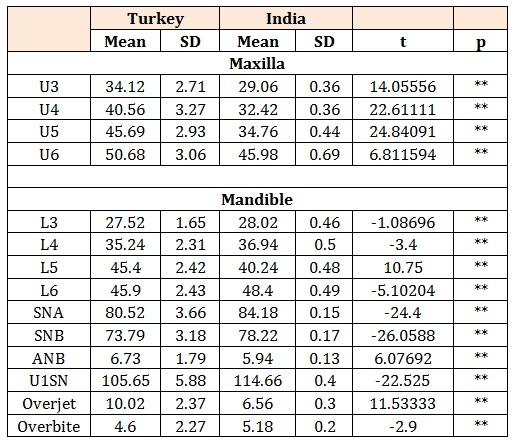
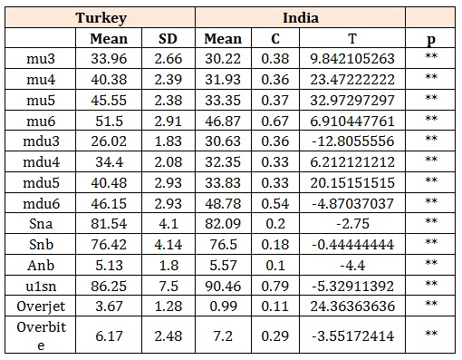
ResultsTable 2 illustrates a comparative study of mean and standard deviation values of linear and angular measurements for present sample of Indian population among classII division 1and division 2malocclusion and their mean difference. In the maxillary arch, linear measurements, that is, inter-canine and inter-molar width is greater in division II malocclusion (30.22 mm and 46.87mm respectively) as compared to division I (29.06mm and45.98 mm respectively). With respect to angular measurements; SNA, SNB, ANB angles are found to be greater in division I (84.18, 78.22 and 5.94 degrees respectively) as compared to division II (82.09, 76.5, 5.57 degrees respectively). Table 3 denotes the significant differences between class II division 1 malocclusion among Turkish and Indians. Maxillary linear measurements are greater in Turkish population when compared to Indians. All values are found to be significant. Unlike maxillary, mandibular linear measurements are greater for Indian population with an exception of second inter-premolar width. Above table reveals that over jet is greater for Turkish population (10.02 mm) as compared to Indians (6.56 mm). Overbite on other hand is greater for Indian population (5.18 mm) as compared to Turkish population (4.6 mm). Table 4 illustrates that maxillary linear parameters are found to be significantly greater for Turkish population as compared to Indians. SNA and SNB angle for Turkish population is (81.54 and 76.42 degrees respectively) while that for Indian population is (82.09 and 76.5 degrees respectively)revealing the fact that they are significantly higher for Indian population.
DiscussionA Group of people of Udaipur visiting dental clinics with dento-skeletal characteristics of class II patients were randomly chosen for measurements to minimize the alteration of dental arch dimensions, due to attrition, restoration or caries. Efforts were made to ensure randomization and adequate sample size, to ensure validity and adequate clinical significance of prediction equation. This investigation studied the dento-skeletal characteristics of Class II patients using lateral cephalometric radiographs and dental casts. Ninety six untreated Class II division 1 and 2patients were compared. The sample consisted of subjects in the permanent dentition to ensure minimal changes in arch widths due to growth [22]. Class II division 2 malocclusions are seen less frequently in the population; the sample size was therefore kept as large as possible, as previous studies have highlighted an insufficient sample size to be a limiting problem when evaluating Class IIdivision 2 malocclusions [13,16,17]. Comparisons of data on dental arch dimensions from different studies are hampered by the fact that it is not easy to tabulate all data on different landmarks. Moreover different authors chose different sample groups for different measurements. It has also been shown that individual dental arch dimensions change with age [3-9,23-25]. This study uses definition for dental width that allows different studies to be compared. The aim of our study was to examine the arch dimension and angles in Indian population and comparison between Turkish and Indian subjects [13,16,17].
Comparison of dental arch width at canine between class II division 1 and division 2 subjects show a significant difference. Mean difference between them was 1.16mm and it was higher in division 2(30.22mm) in comparison to division1 (29.06mm). In the mandibular arch mean difference was 2.61mm. It was again higher for division 2 (30.63mm) in comparison with division 1(28.02mm). Maxillary inter-molar width and the distobuccal cusp of 1st molar were found to be wider in division 2(46.87mm) as compared to division 1(45.98mm). In the maxillary arch the inter-molar width shows mean difference of 0.89mm. A constant value for upper and lower inter-molar width has also been found after 14 years. Bishara, et al. [22] demonstrated a mean increase of 0.2mm in upper inter-molar width, but they were observing individuals between 25 and 45 years which could be of reflection of predisposition for this age group. The patient selection criteria were based only on visual evaluation of dental casts. In assigning Class II division1cases, the mean over jet (6.56 mm) in a Class II division 1sample was taken as a guide, [23] and proclination of the maxillary anterior teeth with an over jet of more than7 mm was adopted as the criterion to ensure a distinction from the division 2 group. Additionally, open bite subjects were excluded since this may be a result of deleterious oral habits such as lip, tongue or thumb sucking and tongue thrusting, which can influence dental and skeletal morphology. For the definition of Class II division 2 cases, retroclination of the maxillary anterior teeth (at least of the two central incisors) and a deep bite were needed. The cephalometric dental findings for the upper and lower incisors supported the selection criteria of the groups. Mean difference in the over jet between division1 and 2 was found to be 5.57mm while with respect to overbite it was 2.02mm. The data on SNA angle indicated that the maxilla was normally positioned in both sample groups. It was84.18 degrees in division 1 while 82.09 degrees in division 2. Their mean difference was 2.09 degrees. Likewise, Pancherz, et al. [18] found a small SNA angle (maxillary retrusion) in Class II groups, whereas Rothstein [25] noted a protrusive maxilla. The differences in the methods of registering maxillary position may explain the various finding [19].
SNB angle in the division 1 and 2 groups was 78.22 and76.5 degrees, respectively. This finding, [16,17] stated that in Class II division 2 cases, the mandible is not posteriorly displaced. On the contrary, in a study by Pancherz, et al. [18] SNB angle in both the division1 and 2 groups was found to be smaller than the reference data. A reason for the dissimilar results for mandibular position may be explained by the age difference between the samples. Pancherz, et al. [18], who found that the division 2 group presented a smaller SNB angle than the division 1 group, concluded that this trend resulted from the constriction of the retroclined anterior maxillary dentition on the mandibular structures. The present data also shows that the Class II division 2 groups have a more concave profile with a prominent chin. The skeletal Class II division 1 pattern in the Indian sample is more pronounced than in the Turkish sample, reflected by the significantly larger ANB angle i.e. 6.73 and 5.94 degrees respectively. Subsequently the ethnic comparison of differences in patients with Class II Division 1 malocclusion has to be interpreted with some caution. For the similar comparison between Indian and Turkish classII division 2 malocclusion the angle was found to be (5.57 and5.13) respectively.
ConclusionThe study yielded a data base about dental arch width with different definition by which different studies can be compared. The craniofacial pattern and morphology of Indian class II division 1 and division 2 differed significantly from Turkish population norm and form.
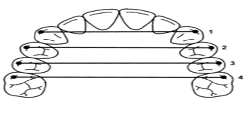
Figure 1: Measurements carried out on the dental casts. 1 Inter-canine distance, 2 Inter-first premolar distances, 3 Inter-second premolar distances, 4 Inter-molar distance
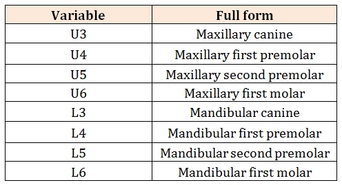
Table 1: Illustrating the variables used in the results section.
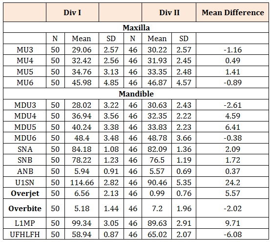
Table 2: Comparison between classII division 1 and division 2 malocclusion among Indians.
** Highly significant
Table 3: A comparative study between classII division1 malocclusion among Turkish and Indian population.
** Highly significant
Table 4: A comparative study between class II division 2 malocclusion between Turkish and Indian population.


