
Citation: Sailesh KM, et al. Pleomorphic Adenoma of Buccal Mucosa: An Uncommon Presentation among Cheek Swellings. J Dental Sci 2017, 2(3): 000134.
*Corresponding author: Sailesh KM, Assistant Professor, HOD Department of Dentistry, AIIMS, Bihar, India, Email: dr.saileshmukul@yahoo.co.in
Cheek mass is a common presenting problem in Oral & Maxillofacial OPD. It may be due to many underlying causes like inflammation, cyst or neoplasm. Common cheek swellings sometimes come out as an uncommon lesion on histopathological examination where the provisional diagnosis is challenged. Therefore the final diagnosis of common small cheek swelling is not complete till its therapeutic removal and histopathological examination. We represent a case of young healthy female who reported with swelling of right side buccal mucosa which was finally diagnosed as pleomorphic adenoma based on histopathologic examination.
Keywords: Pleomorphic Adenoma; Minor salivary gland tumor; Cheek mass
Among all head and neck neoplasm, Salivary gland neoplasm represents3-5% [1] and minor salivary glands tumor account for about 22% of all salivary gland neoplasm [2]. Majority of minor salivary gland tumors reported in literature are malignant with only 18% being benign [3]. Pleomorphic adenoma (PA) is a benign, mixed tumor which most commonly involves the parotid gland. Among tumors of the minor salivary glands palate is the most common intra-oral site (42.8-68.8%) followed by upper lip (10.1%) and cheek (5.5%). Other rare sites include the throat (2.5%), retro molar region (0.7%), alveolar mucosa and floor of the mouth [4]. Vaidya, et al. [5] conducted a study of 104 minor salivary gland tumor and concluded that there was no gender predilection and the median age of presentation was 45 years [5]. Minor salivary gland tumors are rare in children. They are responsible for only 5-10% of all salivary tumors under 20 years of age [6-8]. The presentation of Pleomorphic adenoma is as a painless, well-delineated, slow growing smooth, firm, mobile swelling that does not cause ulceration of overlying mucosa [9]. This paper describes the diagnosis and management of an asymptomatic slowly growing pleomorphic adenoma of buccal mucosa in a young patient. It is suggested that a practitioner has to be aware of such a rare condition.
Case ReportA 19 year old female patient reported to the department with chief complains of unaesthetically looking painless swelling of right side cheek since one year. The swelling started as a painless mass which gradually increased to the presenting size of 2cm X 1.5 cm. the size remained constant in last 4 months. The swelling was present over the right cheek region at infero anterior border of the mandible. The skin over the anteroinferior aspect of the swelling had blind spot (punctum) (Figure 1). On palpation swelling was soft, non-compressible, non-tender, and non-pulsatile and attached to overlying skin. Swelling was not appreciable intra orally. No signs of inflammation were present. Ultrasound (USG) of the swelling revealed well defined cystic lesion with echogenic content and multiple small anechoic areas, no vascularity was present and provisional diagnosis of sebaceous cyst was made. Surgical excision of lesion was planned under GA .Through intra oral approach an incision was made over the mucosa, the underlying muscle was dissected and the mass was approached. The mass was dissected free from overlying skin. The mass was encapsulated with small multilobulated surface and pale yellowish appearance (Figures 2 & 3). Histopathology report showed areas of chondromyxoid stroma, proliferating ductal epithelial cells, myoepithelial cells, squamous differentiation with keratinization. Histological features were consistent with Pleomorphic Adenoma.
DiscussionWHO pathological classification of salivary gland tumors has proved to be most useful in daily practice and has been adopted worldwide [10]. Pleomorphic adenomas are benign heterogeneous tumors of salivary gland origin. In head and neck region they arise mainly in the major salivary glands mostly parotid (85%), submandibular gland (8%) and intra oral (7%) salivary glands. They are commonly seen in 4 -6 decades of life [8] .The etiology of pleomorphic adenoma is not known. It is of epithelial origin and cytogenetical analyses of pleomorphic adenomas have shown the presence of clonal chromosomal aberrations on the basis of which
Classification in Three Cytogenetic Subgroups have been MadeHistopathological features of pleomorphic adenoma are a variable pattern of epithelium in a loosely fibrous stroma which may be myxoid, chondroid or mucoid. The epithelium is usually arranged in sheets or strands, and ductal structures, often bilayered, are typical. In the minor glands, lesions are often more solid or cellular than those seen in the major glands, and the myoepithelial cells are often polygonal with a pale eosinophilic cytoplasm giving plasmacytoid an epithelioid phenotype [12] as present in our case. The main histologic subgroups have been identified: myxoid (80% stroma), cellular (myoepithelial predominant) and mixed (classic) type [13]. Rarely, a malignant tumor may arise within this tumor, a phenomena known as carcinoma ex Pleomorphic Adenoma [14]. PA should be considered in the differential diagnosis of mass of the cheek in youngsters [15]. Differential diagnosis of intraoral, solid, asymptomatic swelling include dermoid cyst, neurofibroma, rhabdomyosarcoma, lipoma, foreign body reaction, buccal space abscess, mucocele, fibroma, adenoid cystic carcinoma and mucoepidermoid carcinoma [6,16]. The ideal treatment for PA is wide local excision with good safety margins and regular follow up for 3-4 years [17]. Since these tumors are radio resistant, the use of radiation therapy is contraindicated.
To conclude we can say that Pleomorphic adenoma of buccal mucosa is an uncommon provisional diagnosis. It must be considered in differential diagnosis for cheek mass in youngsters as its complete resection with surrounding dispensable normal tissue is paramount. Recurrence after long period of excision and malignant transformation is a concern and therefore long period of follow up is necessary.
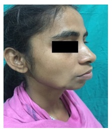
Figure 1: The swelling presented as a painless mass of size of 2 cm X 1.5 c. The skin over the anteroinferior aspect of the swelling had blind spot (punctum).
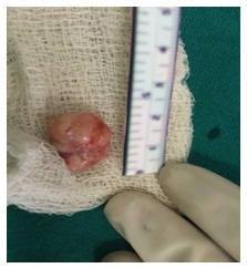
Figure 2: The mass was encapsulated with small multilobulated surface and pale yellowish appearance.
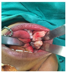
Figure 3: The mass was dissected free from overlying skin.


