
Citation: Chakravarthi S. Evaluation of Virtual Microscope as an Interactive Educational Tool in Microbiology and Pathology in Medical Curriculum. J Microbiol Biotechnol, 2017, 2(2): 000119.
*Corresponding author: Dr. Srikumar Chakravarthi, Deputy Dean, Perdana University, Serdang, 43400 Selangor, Malaysia, Tel: +603 8941 8646; Email: srikumar@perdanauniversity.edu.my
Teaching microbiology and pathology in medical education is predominantly through didactic lectures. Imparting knowledge through other forms of instruction is often lacking in medical education. Other innovative forms of imparting microbiology and pathology education, such as learning through virtual microscope (VM), is necessary in the advancing trend of the medical curriculum. With increasing number of disease processes, and morphological abnormalities like inflammation, necrosis, abscess, metastasis, especially in the study of cancers of diffierent microscopic variants, and infections with diagnostic difficulties, some medical universities are now using more state-of-the-art technology driven software. The ultimate goal of the study was to provide options for students and teachers to use virtual microscope learning modules corresponding to key topics in microbiology and pathology. Through the microbiology and pathology sessions in years 1 and 2 in the medical curriculum, we developed a series of virtual microscopy sessions. A total of nine pre-clinical modules consisting of 224 respondents were done. The students were invited to take part in an evaluation exercise consisting of basic survey questions. Question areas included the most memorable experience, the course's influence on the student-student and teacher-student relationship, usefulness during pre-clinical years of medical school, and skills which would help them in clinical years. The anonymous data were analyzed qualitatively. A significant number of students responded positively for three important themes: (1) the virtual microscope sessions positively influenced more enthusiasm in learning microbiology and pathology (84%), (2) both VM and a clinicopathological discussion in the form of case study were necessary to achieve those skills (76%), and (3) the VM sessions led to a sense of personal development as a student (71%). An interactive discussion with the students revealed that they were interested and quite enthusiastic to gain knowledge by this module, which depicted the microscopic picture with some salient text notes, and they felt that this would also be useful for them in tackling the exams, and in future, during their clinical exposure.
Keywords: Virtual Microscope; Microbiology; Pathology; Pre-Clinical Curriculum; Computer Based Interaction
Compared to didactic lectures, teaching the skill of microbiology and pathology through other forms of instruction is often lacking in medical education. Other innovative forms of imparting microbiology and pathology education, such as learning through virtual microscopy, is necessary in the advancing trend of the medical curriculum [1]. For ages, traditional teaching in medical education, especially in Microbiology and Pathology, has always relied on tissue slides of various organisms and diseases which are studied under the microscope. With increasing number of disease processes, and various tissue changes presented by many disease variants, especially in the study of cancers and infections, some medical universities are now using more state-ofthe-art technology driven software to the knowledge hungry student of the 21st century, both for medical students and also for dentistry, veterinary science students [2,3] and students of experimental pathology in other biomedical professions.
Keeping this in mind, Perdana University, Malaysia, uses Mscope, an interactive virtual microscope extensively for microbiology, histology & pathology teaching. This provides online access to an expanding digital archive of high resolution scanned images of many common and important diseases, infections and cancers of human tissue sections, prepared over several years using many different staining methods. These images have been annotated by trained faculty to highlight key learning points; students can access this using a personal user account. Online evaluations with formative feedback, case atlases, course materials and media library enable students to test their understanding of microbiology and pathology, compare normal and abnormal tissue, and the link between structure and function.
This interactive computer-based technology offers the full range of traditional microscope functionality to the medical students to access from outside university premises, such as from their homes, thereby serving as an enhanced curriculum delivery for distance learning and self-directed learning. The use of virtual microscopes has transformed traditional teaching methods by removing the reliance on physical space, equipment, and specimens to a model that is solely dependent upon computerinternet access. This rich database is enhanced with clinical presentations, laboratory data, comprehensive morphology interpretations, and diagnoses [4].
MScope’s hematology section has a huge collection of diseases and cancers of the blood, and impart an understanding of blood cell morphology and identification for hematology training, representing a variety of diseases, common, rare and unique [5-11]. Glass microscope slides are fragile, fade over time and are extremely difficult to duplicate because they have been created from actual patient samples. Hence, by digitizing the slide sets, this resource is preserved and can be accessed online by more students.
ObjectivesTo organize better computer based virtual microscopy educative sessions for students in learning microbiology, histology and pathology. To encourage the students to utilize the virtual microscopy facility. To enhance small group interactive sessions. The ultimate goal of the study was to provide options for students and teachers to use virtual microscope learning modules corresponding to key topics in basic medical sciences.
DesignThrough the microbiology, histology and pathology sessions in years 1 and 2 in the medical curriculum, we developed a series of virtual microscopy sessions. This provides online access to an expanding digital archive of high resolution scanned images of many common and important diseases, infections and cancers of human tissue sections, prepared over several years using many different staining methods (Figure 1-3). These images have been annotated by trained faculty to highlight key learning points; students can access this using a personal user account. The system includes several features to annotate slides with markers that act as visual signposts placed on the slide itself and with descriptions. The lecturer can add information to every slide in the following categories: source tissue/organ type, organism type, stain, preparation, section type, scan level, and diagnosis. All of these data are searchable so that faculty and students can quickly find all slides of a given tissue type or stain for example. The system also has a feature where the lecturer can attach links to other slides and students can easily see a different example of the same tissue or structure. Our VM also permits students and faculty to add random text tags to any slide. This approach allows the entire educational community at our school to add additional searchable terms, create ad hoc or informal collections of slides, or to organize slides in unanticipated ways.
Online evaluations with formative feedback, case atlases, course materials and media library enable students to test their understanding of microbiology, histology, pathology, compare normal and abnormal tissue, and the link between structure and function. The sessions involved facilitating by lecturers. To evaluate the course's effect on the students’ skills, we performed a qualitative evaluation of the students.
MethodsA total of nine pre-clinical modules consisting of 224 respondents were done.
These students were exposed to both conventional microscopy and virtual microscopy sessions for the following modules: general pathology/microbiology, cardiovascular, respiratory, gastrointestinal, renal, endocrine, reproductive and musculoskeletal systems. The students were invited to take part in an evaluation exercise consisting of basic survey questions (questionnaire as in Table 1). Question areas included the most memorable experience, the course's influence on the student-student and teacher-student relationship, and usefulness during pre-clinical years of graduate medical school, and skills which would help them in clinical years. The anonymous data were analyzed qualitatively.
ResultsA significant number of students responded positively for three important themes: (1) the virtual microscope sessions positively influenced more enthusiasm in learning microbiology and pathology (84%), (2) both VM and a clinicopathological discussion in the form of case study were necessary to achieve those skills (76%), and (3) the VM sessions led to a sense of personal development as a student (71%). Other parameters, such as the course's influence on the student-student and teacher-student relationship, usefulness during preclinical years of medical school, and skills which would help them in clinical years, were all rated above 80%. In addition, students responded that the training in observation and description skills they learned were unique.
The use of the VM system reduced the amount of time the lecturer spent teaching the module, but did not reduce the number of laboratory sessions or the number of required faculty. Due to the efficiencies and workflow of the VM system, laboratory sessions were reduced from three hours to two hours without reducing the number of slides taught in each session. The laboratory duration reduced the overall facilitating time by 26%, saving 40 hours per year in faculty time and freeing up an additional 40 hours of laboratory space for other courses.
Though we did not formally assess the workflow changes that enabled the reduction in laboratory session duration, reports from faculty credited the continuous access to all specimens and the students not having to share slide boxes and wait before they could use a given slide. Student performance on the end of module exam was compared from one year prior to, and one year after the transition from microscopes to the VM. Their performances were better in the USMLE part 1, which contains picture based vignettes in microbiology and pathology components.
DiscussionThis interactive computer-based technology offers the full range of traditional microscope functionality to the medical students to access from outside university premises, such as from their homes, thereby serving as an enhanced curriculum delivery for distance learning and self-directed learning [7]. The use of virtual microscopes has transformed traditional teaching methods by removing the reliance on physical space, equipment, and specimens to a model that is solely dependent upon computer-internet access [5]. This rich database is enhanced with clinical presentations, laboratory data, comprehensive morphology interpretations, and diagnoses [6].
An interactive discussion with the students revealed that they were interested and quite enthusiastic to gain knowledge by this module, which depicted the microscopic features with some salient text notes, and they felt that this would also be useful for them in tackling the exams, and in future, during their clinical exposure.
Overwhelmingly, students wanted virtual microscopy sessions available for study and review during the modules, before exams and also remote access from home. Students identified many advantageous features, which included winning the first place in a national level quiz.
The Virtual Microscope MScope allows medical students to access microscope slides through an online database for distance learning and enhanced curriculum delivery [8]. The Virtual Microscope MScope database offers a high quality resolution of 100X oil immersion, which is necessary for the proper examination of blood cells. The use of virtual microscopes can transform traditional teaching methods by removing the reliance on physical space, equipment, and specimens to a model that is solely dependent upon computer-internet access. This rich database is enhanced with patient clinical presentations, laboratory data, comprehensive slide interpretations, and diagnoses.
There are numerous benefits of MScope. Firstly, it is less expensive as the cost is less than that of replicating, creating, and storing glass slide sets. Secondly, it is of high quality. The digitized slides have a high resolution of 100X oil immersion. Glass slides are fragile, can be easily broken or damaged, and can fade [9]. Thirdly, it is convenient. The slide sets can be accessed through an online database, anywhere, anytime. Hence, there is no dependence on availability of space, equipment, or specimens [10,11]. And fourthly, there is more access, as there is increased availability of slide sets to students, even while outside campus
ConclusionOur virtual microscope system has been an effective solution to the challenges facing traditional laboratories and the novel needs of our revised curriculum. The use of a web-based system empowered learners to have greater control over their content and work together in collaborative groups. The VM system saved faculty time and enhanced student performance during their exams.
After having used virtual microscopy in our University for 3 years since its inception, it is evident that VM technology is effective for the new generation microbiology and pathology education in undergraduate medical teaching, and may be suitably implemented in other student centered pedagogic models, including integrated and problem-based learning curricula and in traditional education system.
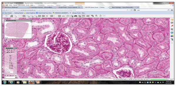
Figure 1: A snapshot during a virtual microscopy session in the renal system showing an in-depth access to an H&E stained tissue section of glomerulus and tubules. The students have the access to zoom in to higher resolutions.
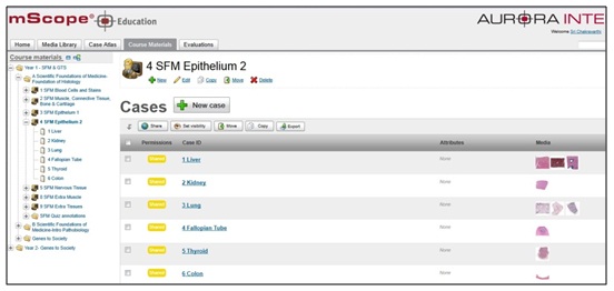
Figure 2: A view of the MScope portal that displays the cases and lesions across each organ system, which is accessible as the students click on each link.
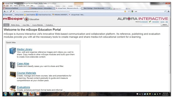
Figure 3: The log in page of the MScope educational portal that shows the interactive menu, which gives access to the enormous knowledge, including case discussion, atlas, course materials and evaluation.
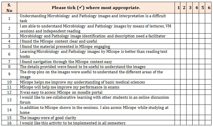
Table 1: Questionnaire provided at the end of each module.
Scoring guide
1= strongly disagree; 2= disagree; 3= somewhat disagree; 4= somewhat agree; 5= agree; 6=strongly agree


