Movement of Intravascular Air Bubble during Esophagectomy
Letter to Editor
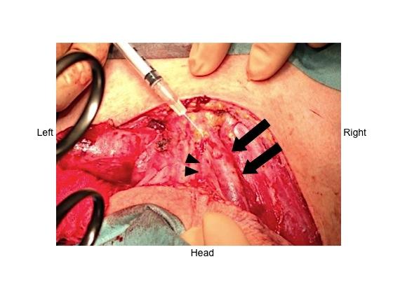
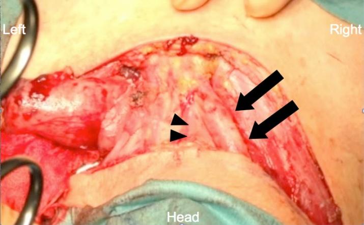
Esophageal cancers may be resected by thoracoscopicor laparoscopic esophagectomy, along with lymph node dissection. Care must be taken, however, during one-lung ventilation, patient position change (from prone to supine), and management of infusion, as each step carries a high risk of air embolism [1]. This report presents a video of a visible air bubble in a 70-year-old man who underwent thoracoscopic and laparoscopic esophagectomy, including three-field lymphadenectomy. Thoracoscopic esophagectomy required 4hours and laparoscopic gastric tube reconstruction 4hours. During subsequent lymphadenectomy, visible bubbles were detected in the right external jugular vein and the communicating vein (Figure1, Video1). The air bubble moved with the jugular pulse but stayed in the same vessels, and an attempt to suction it with a 22-G needle and 2.5-ml syringe was unsuccessful. The patient’s vital signs were stable and the patient was extubated the next day. His postoperative course was uneventful. Air visible ontransesophageal echocardiography during open heart operations frequently moves in parallel with blood flow. However, mobile air in this patient remained in the same vessels, indicating that the timing of posture change and the volume of infusion were critical. Intravenous air can flow unexpectedly into the pulmonary arteries [2], with more than 0.5 ml/kg of air causing critical status [1].
Letter to Editor
Physicians should be aware of this important complication and take steps to prevent it.
Video 1
References
-
Mirski MA, Lele AV, Fitzsimmons L, Toung TJK (2007) Diagnosis and treatment of vascular air embolism. Anesthesiology 106(1): 164−77.
-
Schmitt HJ, Hemmerling TM. (2002) Venous air emboli occur during release of positive end- expiratory pressure and repositioning after sitting position surgery. Anesth Analg 94(2): 400−403.
- Editorial on Multimodal Analgesia
- Surgical Incision Site Local Anaesthetic Infiltration and Superior Hypogastric Plexus Block in Total Abdominal Hysterectomy Under General Anaesthesia- A Placebo-Controlled, Randomized Clinical Trial
- Supraglottic Airway Insertion in Semi Fowler Position Due to Severe Thoracic Hyperkyphosis: A Case Report
- Anaesthetic Management of Cardiac Myxoma Patient with Systemic Involvement: A Case Report
- Current Problems in Pulmonary Respiratory Distress Syndrome (Literature Review)
- Evolution of Perioperative Hemodynamic Monitoring from the Hand on Pulse to Hypotension Prediction Index