Formulation of Methotrexate Loaded Polymeric Zn-Nanoparticle: An Efficient Anti-Cancer Nano-Drug
The objective of this research was to formulate, characterize, interpret and study in-vitro drug release of methotrexate (MTX) polymeric nano-drug. Methotrexate is frequently used antineoplastic drug for chemotherapy. There are certain hindrances like increased toxicity problems and brief plasma life that have caused limitations to its usage. Zn/PEG nano-composite was prepared by sol gel formulation method. Zinc was preferred as it has importance in defense of host against cancer development by DNA repair of damaged cells and for maintaining DNA integrity of healthy host cell. PEG is an ideal and important drugcarrier for enhancing therapeutic value of antineoplastic drugs. MTX was loaded in Zn/PEG nano-composite to overcome toxic side effects and to improve plasma half-life of the loaded drug. The volume mean diameter (D) determined by particle size analyzer was less than 100nm. X-ray diffraction, FTIR and UV spectroscopy investigations manifested the successful loading of MTX in Zn/PEG nano-composite. The drug loading content was as high as 15 %. Percentage drug release profile showed that up to 18% MTX was released in 3 days from drug loaded nanoparticle.
Introduction
Methotrexate (MTX) is a promising and widely used anticancer drug belonging to the pharmacological class of anti-metabolites. It is a folic acid antagonist and has effective therapeutic value against several types of cancerous cells. It is a drug of choice and key molecule in treatment of colon cancer and various other malignancies like Hodgkin’s and Non-Hodgkin’s lymphoma ,leukemia ,head and neck cancer, breast cancer and lung cancer [1]. The MTX chemotherapeutic action is by the inhibition of dihydrofolate reductase, the enzyme that is responsible for biosynthesis of purines and pyrimidines. This inhibition of enzyme results in disruption of DNA, impairment of tumor growth or apoptosis [2]. The tumor cells are more sensitive to the activity of anti- folates as they need ten times more thymidine triphosphate then healthy cells [3, 4]. The chemical structure of MTX is shown in Figure 1. Despite the MTX effective antineoplastic activity, its usage is hampered due to multi drug resistance in certain cancer cells due to cellular efflux of the molecule [5], poor-solubility and toxic effects of drug to normal tissues. One way to solve these problems is by conjugation and encapsulation of drug with polymeric nanoparticles. These polymeric nanoparticles provide excellent solubilization to hydrophobic drugs, long circulation in blood, specificity, target delivery and reduce side effects to healthy cells. There are large numbers of cancer nanoparticle-based therapeutics like Abraxane (Paclitaxel-albumin Nanoparticle), Genexol- PM (Methoxy-PEG-poly(D, L-lactide) taxol) and Oncaspar (PEG–L-asparaginas) available on the market. All these nanoparticles are being effectively used.
![Figure 1: Despite the MTX effective antineoplastic activity, its usage is hampered due to multi drug resistance in certain cancer cells due to cellular efflux of the molecule [5], poor-solubility and toxic effects of drug to normal tissues. One way to solve these problems is by conjugation and encapsulation of drug with polymeric nanoparticles. These polymeric nanoparticles provide excellent solubilization to hydrophobic drugs, long circulation in blood, specificity, target delivery and reduce side effects to healthy cells. There are large numbers of cancer nanoparticle-based therapeutics like Abraxane (Paclitaxel-albumin Nanoparticle), Genexol- PM (Methoxy-PEG-poly(D, L-lactide) taxol) and Oncaspar (PEG–L-asparaginas) available on the market. All these nanoparticles are being effectively used.](/fulltextimages/5476/fig_1.png)
Polyethylene glycol is an ideal and remarkable part as a drug carrier and many rationales are proposed because of greater water solubility, regulated permeability via biological barriers, controlling of drug release, prolonged existence in blood, killing tumors, modified antitumor effect and lessened protein immunogenicity [6, 7, 8, 9, 10]. A widespread of biological molecules like enzymes, lipids, proteins and drugs of low molecular weight are conjugated with PEG to benefit from these advantages. Polyethylene glycol conjugates of camptothecin C, doxorubicin [11] and paclitaxel are developed to enhance water solubility and plasma half-life of the all three drugs [12]. The retention time of nanoparticles is prolonged due to its conjugation with PEG by preventing it to be taken by mononuclear phagocyte system.
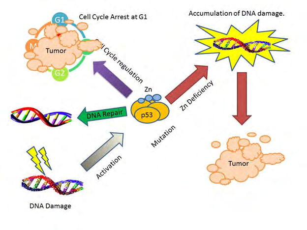
Human body requires large number of trace elements and there deficiency can lead to abnormal body condition one of which is cancer. Zinc is one of the most abundant and essential cofactor of proteins that inhibits the induction and development of cancer. It is important for transcriptional activation and antioxidant resistance mechanism. The p53 tumor suppressor transcription factor have single zinc ion at its DNA binding interface. Deficiency of zinc causes p53 mutation, leading to DNA damage and progression of tumor. Zinc will maintain the p53 integrity, cell cycle arrest G1, DNA repair and finally tumor apoptosis [13, 14]. In this work, we have formulated polymeric nano anticancer drug loaded with Zn nanoparticles with enhanced drug released.
Experimental
“Polyethylene Glycol 2000 (Merck)”, “Sodium Hydroxide pellets (Merck)”, “Zinc Nitrate Hexahydrate (DaeJung)”, “Standard Methotrexate (USP)”, “Dimethyl Formide (AC tabscan)”, “Hydrochloric Acid (Merck)”, “Methotrexate Tablet”, “Potassium Dihydrogen Phosphate (Sigma Aldrich).”
Instrument Used
FTIR (IR affinity 1) was used for structural characterization of synthesized nanoparticles in a range starting from 400cm-
1 to 4000cm-1. X-ray diffraction (Panalytical powder X-ray diffractometer) was used to determine crystal structure and crystallite size by using Cukα radiations having wavelength 1.54Å, Transmission Electron Microscopy (JEM-ARM200F UHR) to study morphology and calculation of particle size. UV-visible Spectrophotometer (UV 1800 Shimadzu) for quantification of drug in nanoparticles. Dissolution Assembly (TDT-08L Electro Lab) for study in-vitro drug release in phosphate buffer. The nanoparticle size analyzer (BT 90) by the use of dynamic light scattering calculated the nanoparticle diameter using Stokes Einstein Equation.
Preparation of Zn/PEG Nano-Composite
The nano-composite was prepared by employing sol gel method [15]. Took 10 ml of 0.02M polyethylene glycol 2000 (pH =7.5) and added equal volume of 0.04M NaOH (pH 10.5). The solution was sonicated for 30 min using ultra- sonicator. Clear solution was obtained having (pH=12). Then added 0.02M Zn(NO3)2.6H2O at a rate of 0.2ml/5min during magnetic stirring on hot plate. The reaction completed in 4 hrs. Colloidal solution was formed which formed precipitates on standing. The centrifugation was done at 13000 rpm for 2 to 3 min and washed several times with water. The precipitates formed were dried in an oven at 45°C.White colored powder formed was stored in vial and characterized.
Loading of Methotrexate in Zn/PEG Nano- particle
Weighed approximately 10mg of Zn/PEG nano- composite obtained previously and dispersed in 10 ml of water. The solution was sonicated for 1 hr. in ultrasonic bath. Colloidal solution was formed. Simultaneously dissolved approximately 10 mg of methotrexate in 60% of dimethyl formide (DMF) in water and added at a rate of 0.2ml/5min in the above solution. Yellow color colloidal solution was formed which was centrifuged at 13000 rpm and dried at 70°C.The yellow colored dried nanoparticles were then stored in tightly closed vial for characterization.
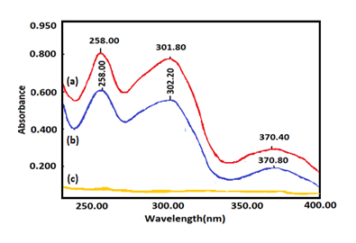
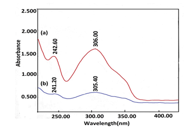
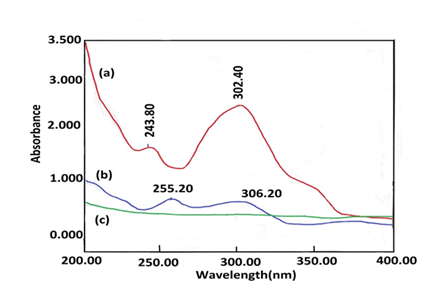
Identification of Drug Loading by UV Analysis
To identify the loading of methotrexate in nanoparticles and verify the interference in quantification of methotrexate, the nanoparticles were dissolved in three different media i.e. acid, basic and neutral media. Drug loaded nanoparticle, pure methotrexate, PEG 2000, Zn(NO3)2.6H20 and Zn/PEG nano- composite absorbance was measured in the region 230nm to 400nm. Approximately 10 mg of methotrexate standard was dissolved in 0.1M NaOH to produce 100ml, filtered and diluted 10 ml to 100 ml with 0.1M NaOH. Similarly 5.0 mg of methotrexate standard was dissolved in 50ml of 0.1M HCl. Pipetted 3ml and diluted to 10 ml with the same solvent. Then approximately 5 mg of drug loaded nanoparticle was dissolved in 50ml of 0.1M NaOH and 0.1M HCl, separately. Dissolved 5 mg of drug loaded nanoparticle in sufficient quantity of purified water and made the volume up to 50 ml.
FTIR of Methotrexate Loaded Polymeric Nanoparticle
FTIR of methotrexate polymeric nanoparticles along with methotrexate pure, PEG, PEG/Zn nano-composite were subjected to the characterization by FTIR (IR affinity 1).
Drug Content Determination
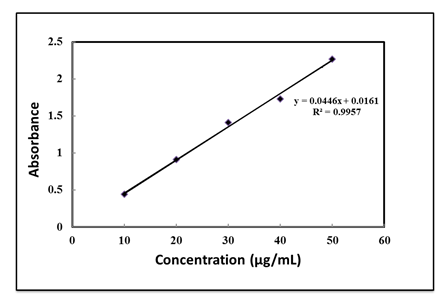
To determine the methotrexate in nanoparticles, 5.0mg of MTX loaded nanoparticle was dissolved in 50ml 0.1 M HCl .Took 1 ml of this solution and made up the volume to 10ml with 0.1M HCl.5.3 mg of methotrexate standard was dissolved in 50 ml of 0.1 M HCl. Different concentrations of MTX standard solutions were prepared by dilution with 0.1 M HCl (10, 20, 30, 40, 50 µg/ml) [16]. Absorbance was measured by UV-visible Spectrophotometer (UV 1800 Shimadzu) .The drug content was determined by plot of calibration curve. Linear regression analysis was done.
XRD Analysis
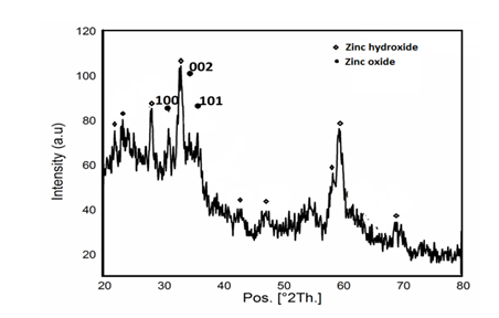
XRD measurements were obtained by X-ray diffraction (Panalytical powder X-ray diffractrometer) using 83 PAN analytical MPD XPERT PRO to determine crystal structure and crystallite size by using Cukα radiations having wavelength 1.54Å.10 mg of nanoparticles was scanned from 10º to 70º at 25ºC.
Dynamic light scattering: The PEG/Zn composite and drug loaded nanoparticles were dispersed in water, sonicated and irradiated by stream of laser light by BT 90 nanoparticle size analyzer .The diameter of nanoparticle was calculated with the help of Stokes Einstein equation.
Transmission electron microscopy: In this technique an ultrathin layer of PEG/Zn nano-composite was magnified and detected by Transmission Electron Microscopy (JEM- ARM200F UHR) in order to determine the morphology and particle size of PEG/Zn nano-composite.
Dissolution Studies
Dissolution Assembly (TDT-08L Electro Lab) was used for study of in-vitro drug release in phosphate buffer. Approximately 50 mg of nanoparticles containing methotrexate were suspended in 500ml of 0.2 M potassium dihydrogen phosphate with pH 7.125 and stirred on a dissolution apparatus at 75 rpm at 37°C. At predetermined time intervals, 15 ml aliquots were withdrawn and dissolved methotrexate was determined at 302 nm. A physical mixture containing 250 mg of methotrexate standard dissolved in 100 ml of 0.1M potassium dihydrogen phosphate (Sigma Aldrich). Took aliquot of 2 ml of standard solution and make volume up to 10ml with same solvent, as a reference solution for dissolution test.
Result and Discussion
UV Analysis
UV spectra of all compounds used in synthesis were obtained. The Figures 2 & 3 depicts that the spectrum of nanoparticle obtained showed absorbance at the same maxima and minima as that of the standard [17, 18]. However in Figure 4 the maxima of nanoparticle shifted towards higher wavelength. Hence the solvent in which the absorbing specie is dissolved also influence the spectrum with increasing solvent polarity.
FTIR Spectra
Figure 5 represented the FTIR spectra of polyethylene glycol, PEG/Zn nano-composite, pure MTX and MTX in PEG/ Zn nano-composite.
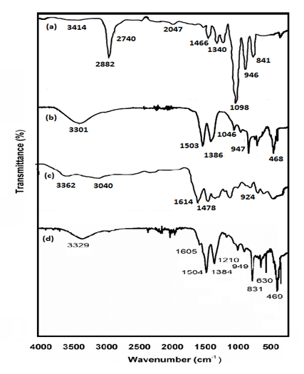
Figure 5 (d) showed the successful loading of MTX in PEG/Zn nano-composite. The peaks in the range of 3000 cm-1 to 3500 cm-1 represented O-H stretching. In pure MTX the peak at 1614cm1 corresponded to C=O stretching from COOH group and C=O stretching from amidic group. However in methotrexate loaded nanoparticle the peak shifted to a considerably lower frequency 1605 cm-1 due to chelation of zinc with drug. The peak at 1386 cm-1 and 1384 cm-1 showed interaction between Zn and PEG by the bending vibrations of CH2 [18]. The peak at 1210 cm-1 corresponded to C-N stretching vibration of R-NH2 [19]. The sharp peak at 630 cm-1 was also intensified in MTX loaded nanoparticle showing Zn-O stretching vibrations [20]. The peak at 468 cm-1 and 469 cm-1 represented stretching vibrations of Zn-O [11].
Drug Content Determination
The calibration curve of MTX standard was obtained by charting different standard methotrexate concentration (10 to 50µg/ml) at x axis versus values of absorbance at y axis 306 nm, see Figure 6. The correlation coefficient was 0.9957. The percentage assay of drug was 15.45%.
XRD Analysis
In Figure 7 the peaks present at 2θ= 31.76° (100), 34.48° (002) and 36.31° (101) represented the formation of zinc oxide nanoparticle with hexagonal crystal structure [11]. The major peaks at 21.96°,28.07°, 32.95°,58.2° and 59.47° might be due to Zn(OH)2 that could be result of unreactive excess of OH¯ of NaOH and Zn+2 of Zn(NO)3. The Zn(OH)2 peak may also be due to interaction of Zn+2 with OH-1 of water. The mean crystallite size was 1.047 nm, as found from Scherer’s equation [22]. The XRD pattern represented that the MTX loaded in zinc/PEG nanoparticles had amorphous nature.
Dynamic Light Scattering
The volume mean diameter of Zn/PEG nano-composite was 137nm and that of methotrexate nanoparticle was 73.7 nm.
TEM Analysis
Average size particle calculated length was 53.88nm and width 12.19 nm. Zn was present in the form of nanotubes and PEG as flakes as, illustrated in Figure 8.
In-vitro Drug Release Studies
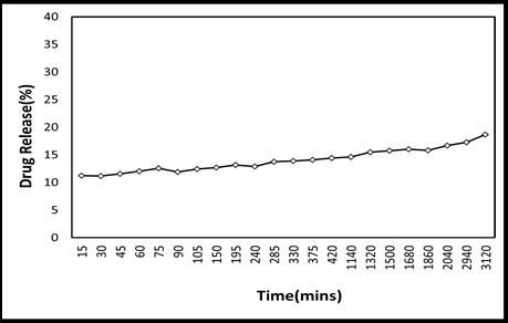
The percentage of methotrexate release in phosphate buffer from nanoparticles after 3 days was approximately 18%, see Figure 9(a). It is also reported that release of drug at pH 7.4 is less as compared to pH 5 and pH 6 [11]. Generally, the release profile depends on number of parameters most importantly pH, particle size and dehydration of polymers [23, 24]. The sustained release of drug may also be due to the strong chelation between drug and zinc in nanoparticle. The release of methotrexate from tablet after 3 days was 42%, as shown in Figure 9(b). The pure drug release after 5 minutes was 71% and 100% after 30 min as illustrated in Figure 9(c) [25].
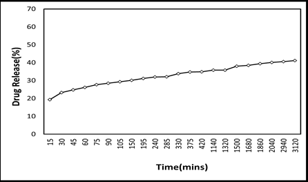
Figure 9 (a): Percentage Release of Methotrexate from Nanoparticle.
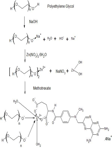
![Figure 10: Methotrexate acts as a ligand and zinc as a complex generator. Firstly, PEG was chelated with Zn+2 to form PEG/ Zn nano-composite. Secondly, the obtained PEG/Zn nano- composite was then reacted with MTX. The MTX molecules act as a ligand and reacted with COO- anions to form MTX loaded nanoparticle [26]. The MTX have functional groups like di-amino, pteridine ring, alkanoyl and two carboxylic group that favors it to be used as an effective ligand and carrier for complex agents [20]. As it is well reported that metals ions have low affinity for nitrogen atom the more likely coordination site will be that of COO-. This fact and coordination mode of zinc metal was further supported by FTIR characterization. It is evident from the FTIR spectrum of pure drug and nanoparticle that the peaks in the range of (1700-1600cm-1) C=O stretching from COOH group disappeared in the drug loaded nanoparticle. An absorption band above 1700cm-1 was not observed, which is due to COOH group. Furthermore, the interaction between zinc and oxygen was also verified by the XRD pattern as depicted in Figure 7 [27-30].](/fulltextimages/5476/fig_10.png)
Figure 9 (b): Percentage Release of Methotrexate from Tablet.
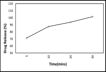
Figure 9 (c): Percentage Release of Pure Methotrexate.
This revealed prolonged released of drug from the
nanoparticle as compared to conventional dosage form and pure drug. This prolong release may reduce the toxic effects of drug on the body and can effectively cure the cancerous cells. The drug loaded polymeric nanoparticle having controlled and steady release are therapeutically effective and significant [20].
Proposed Mechanism through which Zn/PEG Nano-composite interacted with MTX
The proposed mechanism through which methotrexate interacted with PEG/Zn nano-composite is displayed in Figure 10. Methotrexate acts as a ligand and zinc as a complex generator. Firstly, PEG was chelated with Zn+2 to form PEG/ Zn nano-composite. Secondly, the obtained PEG/Zn nano- composite was then reacted with MTX. The MTX molecules act as a ligand and reacted with COO- anions to form MTX loaded nanoparticle [26]. The MTX have functional groups like di-amino, pteridine ring, alkanoyl and two carboxylic group that favors it to be used as an effective ligand and carrier for complex agents [20]. As it is well reported that metals ions have low affinity for nitrogen atom the more likely coordination site will be that of COO-. This fact and coordination mode of zinc metal was further supported by FTIR characterization. It is evident from the FTIR spectrum of pure drug and nanoparticle that the peaks in the range of (1700-1600cm-1) C=O stretching from COOH group disappeared in the drug loaded nanoparticle. An absorption band above 1700cm-1 was not observed, which is due to COOH group. Furthermore, the interaction between zinc and oxygen was also verified by the XRD pattern as depicted in Figure 7 [27, 28, 29, 30].
Conclusion
In the present study, I have first successfully formulated the PEG/Zn nano-composite followed by loading of MTX in nanoparticles via sol gel method. The spectroscopic studies showed the successful loading of methotrexate in the PEG/ Zn nano-composite The UV Spectrophotometer analysis showed that the drug contents were 15.45µg/ml. The FTIR spectra of methotrexate loaded polymeric nanoparticle indicated that all relevant functional group moieties were present. The drug was safely loaded into PEG polymer and Zn ion had chelation with MTX. The average crystallite size of drug loaded nanoparticle was 1.047 nm. The volume mean diameter (D) methotrexate nanoparticle was 73.7 nm. The dissolution studies concluded that after in-vitro release study of methotrexate loaded nanoparticle a prolong and steady state release pattern was achieved as compared to the methotrexate conventional dosage form and this nano dosage form can be successfully used in anticancer treatment.
Acknowledgement
The corresponding author (Muhammad Akhyar Farrukh) would like to thank Higher Education Commission (HEC) Pakistan for providing funds through Project No. 20-3142/ NRPU/R&D/HEC/ & Project No. 20-2660/NRPU/R&D/HEC/ and The World Academy of Sciences (TWAS), Italy. TWAS Research Grant No. 11-028 RG/MSN/AS_C to establish Nano- Chemistry Lab.
References
-
Santra S, Kaittanis C, Santiesteban OJ, Perez JM (2011) Cell-Specific, activable and theranostic prodrug for dual target cancer imaging and therapy. Journal of the American Chemical Society 133(41): 16680-1688.
-
Nagaj J, Kolkowska P, Bykowska A, Komarnicka UK, Kyziol A, et al. (2015) Interaction of Methotrexate, an anticancer drug, with copper ions: coordination pattern, DNA cleaving properties and cytotoxic studies. Medicinal Chemistry Research 24: 115-123.
-
Perran NE, Cabezas-Herrera J, Garcia-Canovas F, Durrant MC, Thorneley RNF, et al. (2005) The antifolate activity of tea catechins. Cancer Research 65(6): 2059-2064.
-
Lorico A, Toffoli G, Boiocchi M, Erba E, Broggini M, et al. (1988) Accumulation of DNA strand breaks in cells exposed to methotrexate or N10-propargyl-5,8- dideazafolic acid. Cancer Research 48(8): 2036-2041.
-
Chen Y, Sha X, Zhang W, Zhong W, Fan Z, et al. (2013) Pluronic mixed micelles overcoming methotrexate multidrug resistance: in vitro and in vivo evaluation. International Journel Nanomedicine 8: 1463-1476.
-
Yousefi G, Foroutan SM, Zarghi A, Shafaati A (2010) Synthesis and characterization of methotrexate polyethylene glycol esters as a drug delivery system. Chemical and Pharmaceutical Bulletin 58(2): 147-153.
-
Yang X, Zhang Q, Wang Y, Chen H, Zhang H, et al. (2008) Self-aggregated nanoparticles from methoxy poly (ethylene glycol)-modified chitosan: synthesis; characterization; aggregation and methotrexate release in vitro. Colloids and Surfaces B: Biointerfaces 61(2): 125-131.
-
Akhtar MJ (2012) Zinc oxide nanoparticles selectively induce apoptosis in human cancer cells through reactive oxygen species. Int J Nanomedicine 7: 845-857.
-
Hanley C, Layne J, Punnoose A, Reddy KM, Coombs I, et al. (2008) Preferential killing of cancer cells and activated human T cells using ZnO nanoparticles. Nanotechnology 19(29).
-
Deng X, Luan Q, Chen W, Wang Y, Wu M, et al. (2009) Nanosized zinc oxide particles induce neural stem cell apoptosis. Nanotechnology 20(11).
-
Hariharan R, Senthilkumar S, Suganthi A, Rajaraja M (2012) Synthesis and characterization of doxorubicin modified ZnO/PEG nanomaterials and its photodynamic action. Journal of Photochemistry and Photobiology B: Biology 116: 56-65.
-
Riebeseel K, Biedermann E, Loser R, Breiter N, Hanselmann R, et al. (2002) Polyethylene glycol conjugates of methotrexate varying in their molecular weight from MW 750 to MW 40000: synthesis, characterization, and structure-activity relationships _in_ _vitro_ and _in vivo._ Bioconjugate chemistry 13(4): 773-785.
-
Ho E (2004) Zinc deficiency, DNA damage and cancer risk. The Journal of nutritional biochemistry 15(10): 572-578.
-
Prasad AS, Beck FWJ, Snell DC, Kucuk O (2002) Zinc in cancer prevention. Cancer and metastasis Reviews 21(3- 4): 291-295.
-
Brinker CJ (2013) Sol-gel science: the physics and chemistry of sol-gel processing. Academic press.
-
Millene CMS, Mara DCV, Fatima PAD, Armando SC, Silvia LF, et al. (2013) Development and Validation of Spectrophotometric method for determination of Methotrexate Incorporated into PLGA Implant. International Journal of Drug Development and Research 5(1): 1-7.
-
(MHRA) TB (2014) In British Pharmacopoeia. TSO.
-
Convention US (2009) USP 32 NF 27: United States Pharmacopeia [and] National Formulary, Volume 2. United States Pharmacopeial Convention.
-
Shameli K, Ahmad MB, Jazayeri SD, Sedaghat S, Shabanzadeh P, et al. (2012) Synthesis and characterization of polyethylene glycol mediated silver nanoparticles by the green method. Int J Mol Sci 13(6): 6639-6650.
-
Dai CF, Li SP, Li XD (2015) Synthesis of nanostructured methotrexate/hydroxyapatite: Morphology control, growth mechanism, and bioassay explore. Colloids and Surfaces B: Biointerfaces 136: 262-271.
-
Nejati KZ, Zolfaghar R, Rafat P (2011) Synthesis of ZnO nanoparticles and investigation of the ionic template effect on their size and shape. International Nano Letters 1(2): 75-81.
-
Monshi AM, Foroughi MR, Monshi MR (2012) Modified Scherrer equation to estimate more accurately nano- crystallite size using XRD. World Journal of Nano Science and Engineering 2(3): 154.
-
Zhang J, Misra RDK (2007) Magnetic drug-targeting carrier encapsulated with thermosensitive smart polymer: core-shell nanoparticle carrier and drug release response. Acta Biomaterialia 3(6): 838-850.
-
Yuana Q, Venkatasubramaniana R, Hein S, Misra RDK (2008) A stimulus-responsive magnetic nanoparticle drug carrier: magnetite encapsulated by chitosan- grafted-copolymer. Acta Biomaterialia 4(4): 1024-1037.
-
Li Y, Kwon SG (1999) Micelle-like structures of poly (ethylene oxide)-block-poly (2-hydroxyethyl aspartamide)-methotrexate conjugates. Colloids and surfaces B: Biointerfaces, 16(1-4): 217-226.
-
Noditi GA (2014) Methotrexate as coordination complex ligand: study of interaction with Zn (II). Digest Journal of Nanomaterials & Biostructures (DJNB) 9(1): 251-260.
-
Ahmad MB (2011) Green synthesis and characterization of silver/chitosan/polyethylene glycol nanocomposites without any reducing agent. International journal of molecular sciences 12(8): 4872-4884.
-
Susan B (1989) The Merck Index. NJ: RSC publishing.
-
Dutta J (2012) Synthesis and characterization of γ-irradiated PVA/PEG/CaCl2 hydrogel for wound dressing. American Journal of Chemistry 2(2): 6-11.
-
Fulias AC, Popoiu C, Vlase G, Vlase T, Oneti D, et al. (2014) Thermoanalytical and spectroscopic study on methotrexate–active substance and tablet. Digest Journal of Nanomaterials and Biostructures 9(1): 93-98.
- Pattern of Gonadal Hormones in Oral Testosterone-Supplimented Male Wistar Rats with Diabetes-Induced Hypogonadism
- Re-Evaluation of the Genotoxicity of Currently Used Food Dyes in Mouse Multiple Organs Via Continuous Administration by Drinking Using the Comet Assay
- Pharmacogenetics of Type 2 Diabetes Mellitus: Linking Genetic Variability to Drug Efficacy and its Cardiovascular Outcomes
- Exploratory Proteomic Profiling of SARS-CoV-2 Infected THP-1 Macrophages Reveals Alterations in Inflammatory Response and Cellular Metabolism
- Study of Genotoxicity of Hepatocarcinogens in Multiple Organs in Mice by Feeding and Drinking Using the Comet Assay
- Spirulina Polypeptides Inhibit the Growth of Human Lung Tumor (H460) Cells