Formulation and Characterization of Solid Lipid Nanoparticles for Topical Delivery of Antibacterial Drug
The aim of this study was to prepare and evaluate solid lipid nanoparticles for the skin delivery of Ciprofloxacin. Ciprofloxacin loaded solid lipid nanoparticles (SLNs) have been successfully developed by using a microemulsion technique. Three different formulations were prepared It was found that variation in the amount of ingredients had profound effects on the Ciprofloxacin loading capacity, mean particle size, size distribution of charge, morphology, and drug-lipid compatibility. At optimized process conditions, Ciprofloxacin loaded SLNs showed spherical particles with a mean particle size of 250 ɳm and 60% Ciprofloxacin incorporation efficacy was achieved. The SLN sustained the drug release for 6 h in vitro. The results suggest formulation could be used as a potential topical formulation for fungal infection.
Introduction
Ciprofloxacin is a therapeutically important antibacterial agent that has been widely used in clinics for a variety of serious fungal infections [1]. The drug may be given orally or intravenously. The oral bioavailability depends upon several factor including aqueous solubility, drug permeability, dissolution rate, first pass metabolism and susceptibility to efflux mechanisms. An interesting alternative approach for ITZ encapsulation may be the use of solid lipid nanoparticles (SLNs) owing to their application for several drugs for topical therapy of fungal skin diseases [2, 3, 4]. The SLNs offered a great potential for the administration of active molecules and simultaneously improved their therapeutic efficacy with great feasibility of incorporation of lipophilic and hydrophilic drugs, enhanced their physical stability, low cost compared to liposomes and ease of scale-up and manufacturing [5, 6, 7, 8]. Moreover, their lipid core made from physiological lipids having high biocompatibility and biodegradability with their potential in epidermal targeting, follicular delivery and controlled release of active moiety with increased skin hydration due to greater occlusivity have been very well established [9, 10].
Material and Method
Ciprofloxacin was obtained from Alpha pharmaceuticals Hyderabad; Stearic acid was purchased from Central Drug House (P) Ltd. New Delhi; Di methyl formamide were purchased from Merck Specialties Private Limited Worli Mumbai; all other required chemicals were procured from supplier. To prepare SLNs dispersion, CPFR, Stearic acid, of different compositions Table 1 were dissolved in 10 ml mixture of Di methyl formamide. Organic solvents were completely removed using a rotary evaporator (Laborota 4000, Heidolph, Germany) to form a drug-embedded lipid layer. This layer was melted by heating at 65°C above the melting point of the lipid. Aqueous phase was prepared by dissolving poly vinyl alcohol (PVA) (2% w/v) in double distilled water (10 ml) and heated to same temperature of oil phase. The hot aqueous phase was added to the oil phase and homogenization was performed (at 2500 rpm and 70°C) using a mechanical stirrer for 30 minutes. The coarse oil in water microemulsion so obtained was sonicated using bath soincator for 25 minutes. ITZ loaded SLN was finally obtained by allowing the hot nanoemulsion to cool at room temperature [11].
| Formulation Code | Ciprofloxacin | Stearic acid |
|---|---|---|
| B1 | 0.5%W/W | 5%W/W |
| B2 | 0.75%W/W | 5%W/W |
| B3 | 1%W/W | 5%W/W |
Table 1: Optimization of formulation.
Differential Study Calorimetry
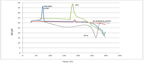
DSC was used to underline the high drug encapsulation potency of the SLN. The higher the enthalpy of the transitions, the more crystalline the SLN and consequently, the more difficult it will be for any drug to be encapsulated. The DSC result is presented in Figure 1.
Particle Size and Zeta Potential Measurements
The particle size of the SLN was determined by phase angle light scattering (PALS) using Zetasizer Nano-Series (Nano-ZS, Malvern Instruments). Samples were diluted with double- distilled filtered water before measurement. The zeta potentials of the formulated SLN were also determined using the same instrument. For the zeta potential measurements, each sample was diluted with bid stilled water and the electrophoretic mobility determined at 25℃. The obtained electrophoretic mobility values were used to calculate the zeta potentials using the software.
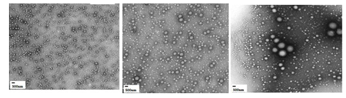
Transmission Electron Microscope
Morphological examination of the SLNs was performed using a transmission electron microscope (TEM). A drop of SLNs suspension was placed on a copper grid with a Form a film. Copper grids coated with Formvar/carbon were glow- discharged for 15 sec to impart hydrophilicity to the surface and facilitate spreading of the SLNs samples onto the grid. Diluted SLN samples were placed on the grid. After 2 min, excess sample was blotted off with filter paper. After 30 sec, excess solution was removed and the grid was dried at room temperature. Samples were viewed in a TEM (Figure 2).
Drug Release
In vitro drug release studies were performed in vertical Franz diffusion cell. Phosphate buffer PH 6.8 (24 ml) was placed in the receiver compartment. A 0.5 gm of SLN of Ciprofloxacin was placed in the donor compartment. A dialysis membrane was used to separate the donor and receiver compartments. The diffusion cells were maintained at (37±0.5ºC) with stirring at 600rpm throughout the experiment. At fixed time intervals, 5 ml of the sample was withdrawn from receiver compartment though side tube and analyzed by UV-Visible spectrophotometer at 275ɳm. Data obtained from in vitro release studies were fitted to various kinetic equations to find out the mechanism of Ciprofloxacin release from SLN.
Entrapment Efficiency
EE was determined by measuring the concentration of free drug (unentrapped) in aqueous medium. The diluted aqueous medium containing the ITZ SLNs was subjected to ultra-filtration to separate the free drug from encapsulated drug. About 1ml of the diluted formulation was placed in the outer chamber and sample recovery chamber was placed on top of the sample and centrifuged at 4000 rpm for 15 min.
The formulation was diluted to 1000 times using the same buffer used in the preparation of the SLN. The SLN along with encapsulated drug remained in the outer chamber while the aqueous phase moved into the sample recovery chamber through filter membrane.
. 100 Wt of drug used inthe formulation Wt of drug aqueous phase EE Wt of drug used in formulation − ×
Result and Discussion
Particle size and zeta potential for topical administration, irritation and wash out may occur on administration of large-sized particles, since smaller particles are better tolerated [11, 12]. Very small particles as nanoparticles possess adhesive properties, which could prolong the release of the drug in the skin, prevent wash out, and increase bioavailability. The zeta potentials of the drug- loaded SLN presented in Table 2 indicate very stable particles. 1.0% ITZ showed the highest absolute zeta potential, it possessed higher crystallinity, lower drug load and higher particle size than other drug-containing SLN. These factors coupled with the tendency to agglomerate eliminate it from being the best SLN in this study.
| Batch of SLN | Zeta Potential mV | Particle size in ɳm |
|---|---|---|
| B1 | -18.7 | 250 |
| B2 | -28.4 | 300 |
| B3 | -30.2 | 323 |
Table 2: Particle size and zeta potential of formulation.
Encapsulation efficiencies above 60% were recorded for the SLN batches the high drug loading was also contributed by the behavior of ITZ shows thermo tropic liquid crystalline behavior and may participate in the microstructure of the system. The encapsulation efficiencies obtained for the drug- loaded SLN are presented in Table 3. This was due to complex formation between lipid and the drug, and the participation of drug in the structures formed at the particle surface.
| Formulation Code | % EE |
|---|---|
| B1 | 64.2 ± 1.5 |
| B2 | 58.0 ± 0.4 |
| B3 | 59.8 ± 1.2 |
Table 3: This was due to complex formation between lipid and the drug, and the participation of drug in the structures formed at
DSC was used to underline the high drug encapsulation potency of the SLN [13]. The higher the enthalpy of the transitions, the more crystalline the SLN and consequently, the more difficult it will be for any drug to be encapsulated.
The DSC result is presented in Figure 1. This showed a slight endothermic signal at about 50℃, confirming their crystallinity [14]. Drug release shows the release profile of ITZ in phosphate buffer (pH 6.8) using dialysis membrane as the release barrier. There was burst release of ITZ from SLNs formulated within the first 125 min. This was due to the more crystalline nature of this SLN, which resulted in more drugs in the periphery and bulk aqueous medium than in the core of the lipid nanoparticles [15, 16]. The profiles of the lipid containing SLN revealed sustained release of the active with regards to topical delivery. Higher quantity of ITZ was released in the batch loaded with higher amount of ITZ.
Conclusion
The high encapsulation efficiency achieved with this novel SLNs formulation indicates this SLN could be an effective drug delivery system for topical active drugs. ITZ- containing SLNs evaluated in this work showed that the performance of their antibacterial application could be improved by formulation as SLN. The studies revealed that the prepared SLNs could be used as a potential carrier for the topical delivery of Ciprofloxacin.
References
-
Müller RH, Mäder K, Gohla S (2000) Solid lipid nanoparticles (SLN) for controlled drug delivery – a review of the state of the art. European Journal of Pharmaceutics and Biopharmaceutics 50(2000): 161- 177.
-
Neubert RHH (2011) Potentials of new nanocarriers for dermal and transdermal drug delivery. Eur Eur J Pharm Biopharm 77(1): 1-2.
-
Gupta M, Tiwari S, Vyas SP (2011) Influence of various lipid core on characteristics of SLNs designed for topical delivery of fluconazole against cutaneous candidiasis. Pharm Dev Technol 18(3): 550-559.
-
Gupta M, Vyas SP (2012) Development, characterization and in vivo assessment of effective lipidic nanoparticles for dermal delivery of fluconazole against cutaneous Candidiasis. Chemistry and Physics of Lipids 165(4): 454-461.
-
Shah AK, Date AA, Joshi MD, Patraval VB (2007) Solid lipid nanoparticles (SLN) of tretinoin potential in topical delivery. Int J Pharm 345(1-2): 163-171.
-
Christopher SJ, Campbell L, Rojas RC, Delgado-Charro MB, Guy RH (2012) Objective assessment of nanoparticle disposition in mammalian skin after topical exposure Christopher. Journal of Controlled Release 162(1): 201- 207.
-
Zhang X, Liu J, Qiao H, Liu H, Jingman Ni, et al. (2010) Formulation optimization of dihydroartemisinin nanostructured lipid carrier using response surface methodology. Powder Technology 197(1-2): 120-128.
-
Wissing S, Muller R (2002) The influence of the crystallinity of lipid nanoparticles on their occlusive properties. International Journal of Pharmaceutics 242(1-2): 377-379.
-
Shah PP, Desai PR, Channer D, Singh M (2012) Enhanced skin permeation using polyarginine modified nanostructured lipid carriers. Journal of Controlled Release 161(3): 735-745.
-
Jensen LB, Petersson K, Nielsen HM (2011) In vitro penetration properties of solid lipid nanoparticles in intact and barrier-impaired skin. European Journal of Pharmaceutics and Biopharmaceutics 79(1): 68-75.
-
Zhanga W, Gaob J, Zhua Q, Zhanga M, Dingb X, et al. (2010) Penetration and distribution of PLGA nanoparticles in the human skin treated with microneedles. International Journal of Pharmaceutics 402 (1-2): 205-212.
-
Patlolla RR, Desai PR, Belay K, Singh MS (2010) Translocation of cell penetrating peptide engrafted nanoparticles across skin layers. Biomaterials 31(221): 5598-5607.
-
Mehnert W, Mäder K (2001) Solid lipid nanoparticles: production, characterization and applications. Adv Drug Deliv Rev 47(2-3): 165-196.
-
Pardeike J, Hommoss A, Müller RH (2009) Lipid nanoparticles (SLN, LNC) in cosmetic and pharmaceutical dermal products. Int J Pharm 366(1-2): 170-184.
-
Müller RH (2007) Lipid nanoparticles: recent advances. Adv Drug Del Rev 59(6): 375-376.
-
Müller RH, Radtke M, Wissing SA (2002) Nanostructured lipid matrices for improved microencapsulation of drug. Int J Pharm 242(1-2): 121-128.
- Effects of 5-HTP and Melatonin on the Sleep Cycle of Medical Students
- Adsorption of Bisphenol A on NH4OH- Modified Rice Husk and Sugar Cane Bagasse Biochar
- Comparative Assessment of the Reinforcement Efficiency of Palm Fruit Fibre and Coconut Fibre in High Density Polyethylene (HDPE) Matrix Composite
- Importance of Bio Compounds Naturally Present in Food with Functionality in Animal Metabolism
- Sub-Acute Study on the Cardiotoxic Effects of Monosodium Glutamate Ingestion in Albino Rat
- Weight Management and Its Natural Solutions: A Review