Phytochemistry, Analgesic and Anti-Inflammatory Activities and Acute Toxicity Studies of Phragmanthera Usuiensis (Oliv.) M.G. Gilbert
Plants have played a significant role in healthcare throughout history by providing medicinal substances. Phragmanthera usuiensis leaves have been used by traditional practitioners for the treatment of oxidative stress-related diseases in the study area, primarily for treating arthritis. The objective of the study was to investigate the phytochemical composition, analgesic effect, anti-inflammatory effect, and acute toxicity of the methanolic extract of P. usuiensis leaves. The phytochemical composition of the methanolic extract of P. usuiensis leaves was studied using qualitative and quantitative methods. The analgesic effect was investigated through the hotplate method, and the anti-inflammatory activity through the carrageenan-induced paw edema model. Investigation of acute toxicity was carried out as per the OECD guidelines on acute oral toxicity – Acute Toxic Classic (ATC) method, with the acute toxicity being administered orally by gavage. Treatment of the animals was initiated at the starting dose level of 300 mg/kg body weight and carried out with six animals (three animals per step). Treatment of animals at the next dose of 2000 mg/kg body weight was delayed until the survival of the previously dosed animals was ensured. The methanolic extracts of P. usuiensis possess several phytochemicals that exhibited promising free analgesic and anti-inflammatory effects. The phytochemicals found in the methanol extract include alkaloids, tannins, glycosides, saponins, steroids, flavonoids, terpenoids, phenols, coumarins, and free sugars. All groups of test animals did not exhibit any serious toxic or lethal effects even at the administration of the limit dose, 2000 mg/kg body weight. The study has successfully validated the use of Phragmanthera usuiensis leaf as having activity that potentially can be used to treat oxidative stress-related disorders like arthritis. Moreover, the study confirmed that these plants exhibit analgesic and anti-inflammatory actives. Additionally, it established the safety of the extracts for use as medicinal agents in the management of oxidative stress.
Introduction
Plants have played a significant role in healthcare throughout history by providing medicinal substances. Plant- derived products have been used to treat various ailments, including aches, swellings, stomach disorders, sores, sexually transmitted infections, nervous system disorders, and mental disorders, among others. However, despite the vast knowledge of traditional medicine, there remains a need for more exhaustive research and documentation of the medicinal potential of various plants. Many studies on plants tend to focus on specific diseases, limiting the discovery of active compounds due to time constraints and economic limitations.
Medicinal plants offer robust antioxidant effects, thanks to the presence of phytochemicals such as flavonoids, terpenoids, alkaloids, and others [1]. These antioxidants protect human cells from oxidative damage caused by free radicals like reactive Oxygen Species (ROS), superoxide anion, hydroxyl radicals, and hydrogen peroxide, which contribute to various diseases, including arthritis, dementia, cancer, hypertension, and diabetes [2, 3]. By scavenging these reactive species, herbal remedies with antioxidants and phytochemicals contribute to safeguarding human health against diseases associated with oxidative stress.
Phragmanthera usuiensis (Oliv.) M.G.Gilbert is a parasitic plant that belongs to the family Loranthaceae. It is native to various regions in Africa, including Kenya. Traditional uses of this plant in Kenyan folk medicine include its application in treating inflammation and oxidative stress-related disorders, such as arthritis. P. usuiensis (Oliv.) M.G.Gilbert contains various compounds that may have potential medicinal properties.
In this study, the plant was collected from Nyamira North Sub-County of Nyamira County, Kenya. It is widely used by the Abagusii folklore in the treatment of pain and inflammation, as seen with arthritis [4]. The leaves of P. usuiensis are collected, dried, crushed, and then boiled in water. One glass of this decoction is consumed daily for 10 days. Fresh decoction has to be prepared every 3 days [5]. Despite its popularity and widespread use, its analgesic, anti-inflammatory, toxicity, and phytochemical properties have not been intensively studied. This study, therefore, aims to investigate the analgesic and anti- inflammatory effects, as well as document the toxicological aspects and phytochemical constituents of P. usuiensis (Oliv.) M.G.Gilbert leaf extracts.
Objectives of the Research
The objective of the study was to provide scientific evidence to validate the continuous usage of Phragmanthera usuiensis (Oliv.) M.G. Gilbert by traditional medicine practitioners in Nyamira County and to investigate the analgesic and anti-inflammatory activity, as well as to evaluate the toxicity and phytochemical composition of its leaf extracts.
Justification of the Research
The World Health Organization (WHO) estimates that 80% or more of the African population depends on plant- and animal-based medicines to meet their healthcare needs [6]. In Kenya, as of 2008, the conventional healthcare system provided services for only 30% of Kenyans. This implies that the remaining 70%, approximately 27 million people, who were outside the national system, had to rely on traditional forms of healthcare [7].
The Abagusii people, residing in the Kisii highlands, have a rich cultural heritage. Traditional healthcare practitioners, particularly herbalists, play a significant role in delivering healthcare services [8, 9]. However, there is limited documentation regarding the use of plants in pain and arthritis management within the Kisii community, as well as the potential risks associated with such usage. Consequently, the current study aims to provide a better understanding of the medicinal plants employed by the community.
Materials and Methods
Plant Material
P. usuiensis was collected in October 2021 from Nyamira North Sub- County in Nyamira County, Kenya, and identification was carried out with the help of a taxonomist from the National Museums of Kenya. The plant specimens were collected, dried, and identified using specified herbarium techniques. Subsequently, they were preserved, and the plant’s voucher specimen was deposited at the East African National Herbarium for future reference. The authentication of the collected plant materials was conducted by comparing the voucher specimen with one from the East African Herbarium at the Nairobi National Museum. The voucher specimen created was subsequently deposited at the museum, and the plant materials were transported to the Mount Kenya University Herbarium. There, they were thoroughly washed with running tap water, sliced into small portions, and air-dried under shade for fourteen days. Before extraction, the samples were ground into a coarse powder.
Reparation of Plant Extract
The extraction was carried out through maceration using methanol and distilled water for the aqueous extract. Approximately 100 g of each part of the ground plant material samples was separately soaked with enough solvent in a 1-liter beaker and then covered with foil for 48 hours. During this period, constant shaking was maintained using a magnetic stirrer (REMI, 2MLH). Afterward, the individual extracts were filtered and reduced in vacuo at 40 °C, eventually being oven-dried at 35 °C.
Phytochemical Screening
Qualitative techniques were employed to detect the phytochemical components present in the leaf extracts of Phragmanthera usuiensis (Oliv.) M.G. Gilbert. Tests for tannins, flavonoids, anthraquinones, alkaloids, terpenoids, saponins, and cardiac glycosides were conducted following procedures outlined by Harborne JB, et al. [10] and Trease and Evans [11] with slight modifications.
Analgesic Activity by Hotplate Method
The analgesic activity of the methanol extract was conducted by hot plate method as described by Yimer T, et al. [12] with minor modifications. The experimental animals that had been deprived of food and water for 12 hours were divided into five groups with each group having five mice (n=5). The groups were assigned as Groups I, II, III, IV and V table 3. Group I was the negative control and was administered with normal saline, Group II was the positive control and administered the reference analgesic drug (diclofenac) and groups III and IV were the test groups and were administered the study plant extract at two different doses of 200 mg/kg bwt and 400 mg/kg bwt, respectively. All the extracts, reference drug and normal saline were administered orally using canula. All animals were placed on the hotplate maintained at 50 ± 2 0 C and the reaction time taken by each individual mouse to respond to the thermal stimuli recorded after,0 minutes 30 minutes, 60 minutes, and 90 minutes. The reaction time cut off was 15 seconds and the responses to stimuli to be noted included paw licking or jumping. The increases in the reaction time for the plant extract as well as the reference analgesic drugs as compared to the negative control was taken as a measure of analgesic activity.
Anti-Inflammatory Activity by Carrageenan Induced Paw Edema Method
The method described by Yimer T, et al. [12] with minor modifications was adopted to investigate the anti- inflammatory activity of the methanol extract. Four different groups of Swiss albino mice were designated as Groups A, B, C, and D, with each group consisting of five mice (n=5). The mice in the negative control group (Group B) were given the vehicle (normal saline). Those in the positive control group (Group B) were given the reference anti-inflammatory drug (diclofenac at 10mg/kg bwt), and the experimental groups (Groups C and D) were administered the plant extract at dosages of 200 mg/kg bwt and 400 mg/kg bwt, respectively.” Top of Form Carrageenan, at a concentration of 1% and prepared in normal saline, was administered by injection into the sub- plantar region of the left hind mouse paw one hour after the oral administration of the plant extract, the reference drug, and normal saline. Paw size was measured at time intervals of 60 minutes, 120 minutes, and 180 minutes after the induction of edema.Top of Form
Acute Toxicity Test
Animal Material: Mature female Swiss albino mice weighing 20-25g (body weight) were employed for acute toxicity studies of crude extracts. The mice used were nulliparous and nonpregnant. These animals were acquired from the University of Nairobi’s Public Health, Pharmacology, and Toxicology Department. The mice were randomly selected and marked on their tails for identification. They were housed separately in polycarbonate cages measuring 35cm (length) x 25cm (width) x 18cm (height) for five days before dosing to allow them to adjust and acclimate to laboratory conditions. The cages were lined with wood shavings, which served as bedding for the animals. The housing temperature was maintained at 25±3°C with a relative humidity of 54±4%. The animals were provided with rat pellets, and water was provided ad libitum.”Top of Form Experimental Design: The procedure for the main test of the Organization for Economic Co-operation and Development (OECD) guidelines on acute oral toxicity, specifically the Acute Toxic Classic (ATC) method, was followed. Prior to dosing, individual animals were fasted for 4 hours, and their weights were recorded before administering the extracts. Animals in the first eight groups were used to test the extracts, while the last group served as the control group. Before dosing, individual animals were fasted for four hours and weighed before oral administration of the extract. Treatment of the animals commenced at the starting dose level of 300 mg/kg body weight, carried out with six animals (three animals per step). Treatment of animals at the next dose of 2000 mg/kg body weight was delayed until the survival of the previously dosed animals was ensured. Physiological saline was administered to the control group. The plant extracts were administered orally by gavage. For restraint, each animal was picked up by the base of the tail, pressed down on a table to immobilize it, and held by the skin at the back of the neck using the thumb and the index finger, with the base of the tail placed between the ring and pinky fingers. Observations of various physical parameters of well-being, such as fur condition, state of the eyes, mucous membranes, salivation, dullness, sleep, tremors, body weight, and mortality, were noted and recorded after 30 minutes, 4 hours, 24 hours, 48 hours, 7 days, and 14 days. After the experiment, all animals that survived were humanely euthanized, and target organs such as the gastrointestinal tract, liver, heart, and kidneys were harvested from the test animals and preserved in 10% formalin for histopathological studies.
Pathological Examination
Tissue Processing and Embedding: Each individual organ was initially fixed in 10% buffered formalin. Subsequently, a dehydration process was performed using graded alcohol baths with concentrations of 50%, 70%, 80%, 90%, and 100%. Following dehydration, the tissues were subjected to dealcoholization using xylene, and infiltration was carried out with molten wax at 62°C for 30 minutes. To complete the embedding process, the tissue cases were placed into cassettes, and molten paraffin wax was added. The wax was allowed to harden, forming tissue blocks, which were then placed in cold water to ensure complete solidification. For this purpose, a continuous supply of paraffin wax was maintained in a molten state at 58°C within a dedicated paraffin wax oven Top of FormTop of Form[13].
Sectioning and Floating Out Sections
Tissues were sectioned using a microtome. The microtome knife was carefully inserted into the microtome and securely clamped at an acute angle to ensure the block would clear the sharpening bevel. Subsequently, the tissue block was placed in the microtome and firmly secured, with its face against the microtome knife. Sections were then cut with deliberate and regular movements to produce a flat ribbon, which was supported by the left hand. This ribbon was gently placed onto the surface of a warm water bath (at 45°C) by gently floating it down in a parallel direction to the water surface. A clean glass slide was then carefully slid at a right angle into the water bath, approaching from the side where the section was located. The slide was moved forward to allow the section to attach to it and was then lifted vertically and slowly out of the water, with the sections still attached. The slide was placed in a rack to allow any excess water to drain, and it was then labeled using a diamond pencil. Subsequently, the slide was left to dry at 58°C for a minimum of 2 hours [13].
Staining and Mounting Stained Sections
Before staining, the sections were initially hydrated to remove the paraffin wax. The sections were, therefore, first treated with xylene and then hydrated by gradually immersing them in alcohol with concentrations of 100%, 90%, 80%, 70%, and 50%, followed by a final step in water. Next, the sections were stained with Harris’ hematoxylin for 4 minutes and subsequently rinsed in running tap water to remove excess stain. They were then counterstained with Putt’s eosin for 2 minutes before being placed in running water to differentiate the eosin. Dehydration was carried out using alcohol baths with concentrations of 50%, 70%, 80%, 90%, and 100% to remove the eosin. Finally, the slides were immersed in a xylene bath to clear the residual alcohol. To mount the stained sections, DPX mounting medium was applied to the center portion of a clean and dry coverslip. The slides were removed from the xylene bath, excess xylene was wiped from the back of the slides, and they were then quickly inverted and gently placed on the DPX mountant applied to the coverslip. The slides were subsequently reverted and placed face up on blotting paper. Following this, the slides were labeled and left to dry on a warm plate at 45°C for 1 hour. Histopathological analyses were conducted with the assistance of a pathologist, who examined the slides under a light microscope and captured images of the tissue structures [13].
Ethical Consideration
Disposal of Experimental Animals
The mice were used and disposed as per the clearance of the University of Nairobi, ethical committee.
Research Approvals
Approval to conduct research was obtained from University of Nairobi graduate school. Biosafety, Animal Welfare and Ethics Committee of the University of Nairobi gave the Ethical clearance (Reference number, FVM BAUEC/2018/168). In the field, written consent was sought from all the herbalists who were willing to participate in the study after the aim of the study was elaborately explained to them. Benefits and potential risks in relation to the study were also communicated to the informants by the researcher prior to the commencement of the study.
Data Management and Statistical Analysis
The experiments were conducted in sets of three, and the results were displayed as mean ± standard deviation. Statistical significance was assessed using a one-way ANOVA test with GraphPad Prism, version 7.0. A p-value of <0.05 was considered statistically significant. The results represent the average of three independent experiments, with each sample tested in triplicate. They were expressed as Mean ± SEM and presented in tables, graphs, and histological images. Data from the histopathological study were presented through photomicrographic images captured using a light microscope.
Results and DiscussionTop of Form
Phytochemical Composition of Phragmanthera Usuiensis
The results of the qualitative phytochemical screening of the methanol crude leaf extract of Phragmanthera usuiensis are presented in Table 1. The detected phytochemicals included phenols, alkaloids, cardiac glycosides, saponins, flavonoids, steroids, terpenoids, coumarins, amino acids and proteins, carbohydrates, and tannins. Fatty acids, gums, mucilage, and anthraquinones were not found in the crude methanol extract. Saponins, carbohydrates, cardiac glycosides, amino acids, and proteins were present in higher concentrations. Steroids were detected in moderate amounts, while tannins, flavonoids (detected by all test methods), phenols, anthraquinones, and coumarins were only present in trace amounts.
| Phytochemicals | Aqueous extract | Methanol extract |
|---|---|---|
| Alkaloids | - | ++ |
| Tannins | +++ | +++ |
| Glycosides | ++ | +++ |
| Saponins | +++ | +++ |
| Flavonoids | +++ | ++ |
| Terpenoids | - | + |
| polyphenols | ++ | +++ |
Table 1: Phytochemical profile of aqueous and methanol leaf extract of _Phragmanthera usiensis._ The aqueous extract was found
Key: +++ = strongly present; ++ = moderately Present: + = mildly present; - = absent Table 1: Phytochemical profile of aqueous and methanol leaf extract of Phragmanthera usiensis. The aqueous extract was found to have alkaloids, tannins, glycosides, saponins, flavonoids, terpenoids and polyphenols. Alkaloids are known for their diverse properties, which encompass analgesic, antispasmodic, antipyretic, among many others [14]. Tannins, on the other hand, act as antioxidants, neutralizing harmful free radicals, and have anti-inflammatory properties that help reduce inflammation. Additionally, tannins contribute to wound healing by promoting tissue repair. Glycosides have notable therapeutic effects, such as cardiotonic, diuretic), and in some cases, demonstrating potential anticancer properties. Saponins have been seen to have cholesterol- lowering activity, antimicrobial and immune-stimulating. Flavonoids have antioxidant capabilities, safeguarding cells from oxidative harm [15]. They are also recognized for their anti-inflammatory properties. Terpenoids are versatile phytochemicals, serving as antioxidants, antimicrobial agents, anti-inflammatories, and analgesic). Polyphenols are renowned for their antioxidant properties, protecting cells from oxidative stress [16]. They are also anti-inflammatory, promote heart health, may have anticancer effects [17]. The presence of these phytochemicals validates the use of Phragmanthera usuiensis for the treatment of arthritis and other oxidative stress disorders by herbalists.
Analgesic Activity
The ability of the methanol leaf extract of Phragmanthera usuiensis (Oliv) M. Gilbert to inhibit pain was assessed using the hotplate method. In this animal model, two doses of the extract, 200 mg/kg body weight and 400 mg/kg body weight, along with the standard (diclofenac) at 10 mg/kg body weight, were administered. As shown in Table 2 and Figures 1, 2, the oral administration of diclofenac resulted in an increase in the latency time taken by the mice to respond to the thermal stimuli induced by a hotplate set at 50±2°C. The positive control group (diclofenac) recorded the highest latency time of 14.33±0.33 seconds after 60 minutes. This latency time was significantly different when compared to the latencies recorded by the positive control at the other monitoring periods (p<0.05). The negative control group recorded the lowest latencies at all the monitoring periods when compared to the positive control and the two experimental groups. The experimental groups that received two doses of the extract recorded the highest latency times after 60 minutes. Specifically, the experimental group administered 400 mg/kg body weight of the extract recorded the highest latency time of 11.67±1.20 seconds, whereas the experimental group administered 200 mg/kg body weight of the extract recorded a latency time of 8.67±0.88 seconds. In both the experimental groups and the negative control group, the latency times recorded after each monitoring period did not significantly differ from each other (p<0.05) Figure 4.1.
| Reaction Time | ||||||
|---|---|---|---|---|---|---|
| 0 minutes | 30 min | 60 min | 90 min | 120 min | ||
| GROUPS | Group A (Diclofenac) | 6.66± 0.33 | 8.00± 1.00 | 14.33± 0.33 | 9.00± 0.00 | 8.67± 0.33 |
| Group B (Normal saline) | 4.33± 0.33 | 3.00± 0.00 | 5.67± 0.88 | 5.00± 1.00 | 5.33± 0.33 | |
| Group C (200 mg/kg) | 6.33± 0.88 | 8.00± 0.58 | 8.67± 0.88 | 6.00± 0.58 | 6.67± 0.33 | |
| Group D (400mg/kg) | 7.67± 1.20 | 9.66± 3.18 | 11.67± 1.20 | 7.66± 0.88 | 9.00± 1.73 |
Table 2: Analgesic effect of Phragmanthera usuiensis in hotplate method.
Data is presented as Mean ± SEM of the triplicate values. SEM: standard error of the mean Table 2: Analgesic effect of Phragmanthera usuiensis in hotplate method.
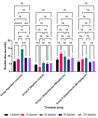
ns signifies no difference observed between the means. ** signifies difference between the means. Figure 1:** Analgesic effect of Phragmanthera usuiensis.
This comparison revealed that no notable difference was observed in the latency time recorded among all the groups after zero, 90-, and 120-minute monitoring periods (p>0.05) Figure 4.1. However, after 30 minutes, the recorded latency time in the negative control group differed from the other groups as it was the lowest (p<0.05) Figure 4.2. Similarly, the positive control group recorded the highest latency time after 60 minutes, which was different from all the other treatment groups (p<0.05) Figure 4.2.
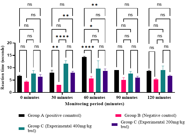
ns = signifies no difference observed between the means. **** = signifies difference between the means. Figure 2: Comparison of the analgesic effect at each monitoring period.
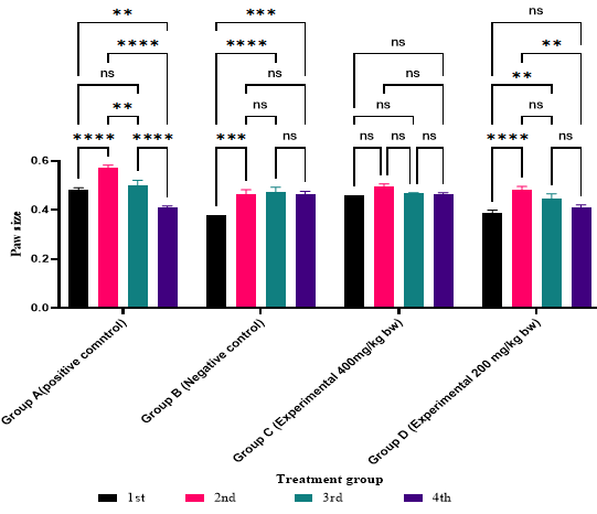
Anti-inflammatory Activity Assessed by Carrageenan-Induced Paw Edema
The anti-inflammatory activity of Phragmanthera usuiensis (Oliv) M. Gilbert was assessed using the carrageenan-induced paw edema animal model. This model is employed to investigate the potential anti-inflammatory effects of the extract in countering acute-phase inflammation induced by subplantar injection of 1% carrageenan into the hind paw of mice. In the negative control group, the injection of 1% carrageenan into the left hind paw of the mice resulted in a progressive increase in paw size. This increase continued up to the third hour of inflammation, with measurements of 0.38±0.00 cm, 0.46±0.02 cm, 0.47±0.02 cm, and 0.46±0.02 cm after the first, second, and third hours, respectively Table 3. Oral administration of diclofenac at a dosage of 10 mg/kg body weight inhibited paw swelling during the second- and third hours following carrageenan injection. The administration of the study plant extract at two different doses resulted in a reduction in swelling following carrageenan injection. One hour after carrageenan injection, the paw size increased from 0.38±0.01 cm to 0.46±0.08 cm in the mice administered the plant extract at a dosage of 200 mg/kg body weight. The reduction in edema formation was observed to continue into the second and third hours in the mice that received the plant extract at a dosage of 200 mg/ kg body weight, with paw sizes measuring 0.44±0.02 cm and 0.41±0.01 cm, respectively. For the mice that received the plant extract at a dosage of 400 mg/kg body weight, a reduction in paw size was observed during the second and third hours, with measurements of 0.49±0.02 cm reducing to 0.46±0.00 cm and 0.46±0.01 cm, respectively.
| Treatment Groups | Paw Size | |||
|---|---|---|---|---|
| 1st | 2nd | 3rd | 4th | |
| Group A (Diclofenac) | 0.48± 0.01 | 0.57± 0.01 | 0.50± 0.02 | 0.41± 0.01 |
| Group B (Normal saline) | 0.38± 0.00 | 0.46± 0.02 | 0.47± 0.02 | 0.46± 0.01 |
| Group C (200 mg/kg) | 0.38± 0.01 | 0.48± 0.02 | 0.44± 0.02 | 0.41± 0.01 |
| Group D(400mg/kg) | 0.46± 0.00 | 0.49± 0.01 | 0.46± 0.00 | 0.46± 0.01 |
Table 3: Data is presented as Mean ± SEM of the triplicate values. SEM: standard error of the mean.
The comparisons of paw sizes in all treatment groups at different monitoring hours were also conducted. Observations from the graph Figure 4.3 revealed that there was no significant difference in paw size for the mice in the experimental group that received the plant extract at a dose of 400 mg/kg body weight (p>0.05) Figure 3. However, the experimental group that was orally administered the plant extract at a dose of 400 mg/kg body weight recorded a significant decrease in paw size after the third hour following the injection of carrageenan.
Acute Toxicity Test
All groups of treated animals did not exhibit any toxic or lethal effect. The present study shows that even at the administration of the limit dose 2000 mg/kg, no sign of toxicity was observed except on group two where two animals showed unusual slow movement after five minutes of dose administration. These effects, however, faded off after 30 minutes. Therefore, it was concluded that the LD50 of extracts was above 2000 mg/kg.
Histopathology of the Mice Liver
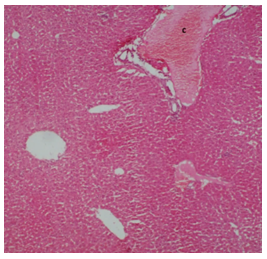
Figure 4 illustrates the histopathology of a normal liver (control) characterized by evenly distributed hepatocytes with a normal architecture separated by hepatic sinusoids.
All the animals that were administered methanolic and aqueous extracts of Phragmanthera usuiensis, did not exhibit significant histopathological changes in the liver when compared to the control group.
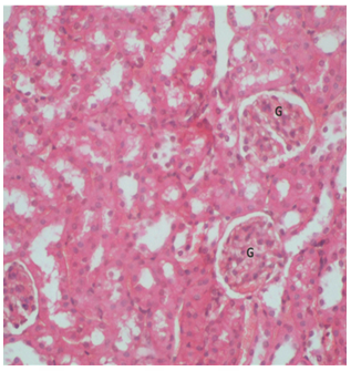
However, all the groups that received both the aqueous and methanol extracts at two dose levels of 300 mg/kg and 2000 mg/kg body weight displayed hepatic portal vein congestion. This congestion, as depicted in the liver section of a mouse dosed with 2000mg of P. usuiensis aqueous extract Figures 5-7, was not observed in the control group, indicating that it was not attributable to the extracts.
Key: C- congestion in the portal center. Figure 4: Photomicrograph showing a liver section from a control mouse showing hepatocyte structure of a normal mouse liver stained with H&E (400X magnification).
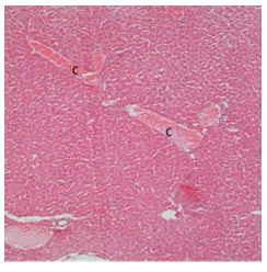
Key: C-Congestion in the portal center. Figure 5: Photomicrograph showing a liver section from a mouse dosed with 2000mg/Kg body weight of aqueous Phragmanthera usuiensis aqueous extract stained with H&E (400X magnification).
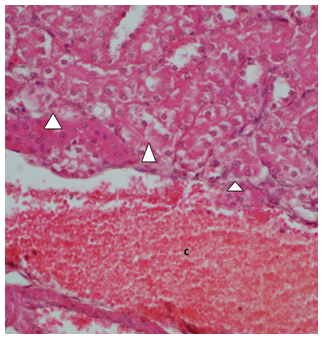
Key: Arrowhead –focal degeneration of tubular epithelium. Figure 6: Photomicrograph showing a kidney section from a mouse dosed with 2000mg/Kg body weight of Phragmanthera usuiensis methanol extract.
Conclusion
Despite the increased use of conventional medicine, this study shows that the use of traditional herbal remedies continues to be embraced by many people in the study region [9, 18]. The active phytochemicals derived from the medicinal plant are devoid of side effects on human health, a reason that has escalated their use as nonprescription medicine [19]. The herbal products today symbolize safety in contrast to the synthetics that are regarded as unsafe to human and environment.
In this study, the oral acute toxicity study of the tested plant extract did not reveal major changes in behavior and mortality of animals in all groups. The extracts seem to be safe at a dose level of 2000 mg/kg, and the LD50 is >2000 mg/ kg. Any pharmaceutical drug or compound with the oral LD50 higher than 1 000 mg/kg could be considered safe and less toxic [20]. The main target organ for drug or bioactive compound is the liver. The liver is exposed to the foreign substances being absorbed in intestines and metabolized to other compounds which may or may not be hepatotoxic to the mice [21]. Despite the congestion of blood vessel observed in the liver of mice that were treated with the extract, these changes were not considered toxicologically significant, as they were not observed in other animals of the same group and that they compared with the control group. However specific assays of toxicity and more biological studies could provide more information regarding the toxic effects of the extracts on liver.
In histopathological studies of the kidneys, congestion of blood vessels and focal degeneration of tubular epithelium with cell swelling was observed in only two mice and only at a dose level of 2000mg/kg body weight. Since no similar changes were observed on other animals of the same groups, the results were judged to be consistent with a normal tissue with a mild kidney injury.
References
-
Heinrich M, Gibbons S (2001) Ethnopharmacology in drug discovery: an analysis of its role and potential contribution. Journal of Pharmacy and Pharmacology 53(4): 425-32.
-
Dröge W (2002) Free Radicals in the Physiological Control of Cell Function. Physiol Rev 82(1): 47-95.
-
Rahman I, Biswas SK, Kirkham PA (2006) Regulation of inflammation and redox signaling by dietary polyphenols. Biochem Pharmacol 2(11): 1439-1452.
-
Wainaina WS, Mucunu MJ, Wakonyu KL, Misonge OJ (2019) Ethnobotanical Survey of Plants Used For Management of Arthritis In Nyamira North Sub-County of Nyamira County, Kenya. The Journal for Haematology 130: 1510-1531.
-
Kaingu CK, Oduma JA, Mbaria JM, Kiama SG (2013) Ethnobotanical Survey of Medicinal Plants Used For the Management of Male Sexual Dysfunction and Infertility in Tana River County, Kenya. The Journal of Ethnobiology and Traditional Medicine.
-
WHO (2002) Traditional Medicine Strategy 2002-2005.
-
(2008) National Coordinating Agency for Population & Development. Seeking Solutions for Traditional Herbal Medicine: Kenya Develops a National Policy, pp: 1-4.
-
Omwenga EO, Hensel A, Shitandi A, Goycoolea FM (2015) Ethnobotanical survey of traditionally used medicinal plants for infections of skin, gastrointestinal tract, urinary tract and the oral cavity in Borabu sub-county, Nyamira county, Kenya. J Ethnopharmacol 176: 508-514.
-
Ondicho J, Ochora E, Matu E, Mutai J (2015) Factors associated with use of herbal medicine among patients in herbal clinics in Gucha. The 2015 JKUAT Scientific Conference: Basic and Applied Sciences, pp: 174-187.
-
Harborne JB (1998) Textbook of Phytochemical Methods. In: Harborne JB (Ed.), A Guide to Modern Techniques of Plant Analysis. 5th(Edn,). Chapman and Hall Ltd, Londan, pp: 21-72.
-
Trease GE (2009) EWC Pharmacognosy. In: Trease GE (Ed.), Baillure Tindall. 16th (Edn.), London, UK, pp: 176- 180.
-
Yimer T, Birru EM, Adugna M, Geta M, Emiru YK (2020) Evaluation of Analgesic and Anti-Inflammatory Activities of 80% Methanol Root Extract of Echinops kebericho M. (Asteraceae). J Inflamm Res 13: 647-658.
-
Vishwakarma S (2017) Tissue Processing In: Techniques in Histopathology and Cytopathology. Jaypee Brothers Medical Publishers (P) Ltd, pp: 57-57.
-
Heinrich M, Mah J, Amirkia V (2021) Alkaloids Used as Medicines: Structural Phytochemistry Meets Biodiversity—an Update and Forward Look. Molecules 26(7): 1836.
-
Heim KE, Tagliaferro AR, Bobilya DJ (2002) Flavonoid antioxidants: Chemistry, metabolism and structure- activity relationships. Journal of Nutritional Biochemistry 13(10): 572-584.
-
Scalbert A, Johnson IT, Saltmarsh M (2005) Polyphenols: antioxidants and beyond. Am J Clin Nutr 81(1 Suppl): 215-217.
-
Kluska M, Woźniak K (2021) Natural polyphenols as modulators of etoposide anti-cancer activity. International Journal of Molecular Sciences 22(12): 6602.
-
Odongo E, Mungai N, Mutai P, Karumi E, Mwangi J, et al. (2017) Antioxidant and anti-inflammatory activities of selected medicinal plants from western Kenya. African Journal of Pharmacology and Therapeutics 6(4): 178- 182.
-
Vaghasiya YK, Shukla VJ, Chanda SV (2011) Acute Oral Toxicity Study of Pluchea arguta Boiss Extract in Mice. Journal of Pharmacology and Toxicology 6(2): 113-123.
-
Adeneye AA, Olagunju JA (2009) Preliminary hypoglycemic and hypolipidemic activities of the aqueous seed extract of Carica papaya Linn. In wistar rats. Biology and Medicine 1(1): 1-10.
-
Rhiouani H, El-Hilaly J, Israili ZH, Lyoussi B (2008) Acute and sub-chronic toxicity of an aqueous extract of the leaves of Herniaria glabra in rodents. J Ethnopharmacol 118(3): 378-386.
- Effects of 5-HTP and Melatonin on the Sleep Cycle of Medical Students
- Adsorption of Bisphenol A on NH4OH- Modified Rice Husk and Sugar Cane Bagasse Biochar
- Comparative Assessment of the Reinforcement Efficiency of Palm Fruit Fibre and Coconut Fibre in High Density Polyethylene (HDPE) Matrix Composite
- Importance of Bio Compounds Naturally Present in Food with Functionality in Animal Metabolism
- Sub-Acute Study on the Cardiotoxic Effects of Monosodium Glutamate Ingestion in Albino Rat
- Weight Management and Its Natural Solutions: A Review