An Overview of Nanocochleates for Improving Oral Absorption Using a Phospolipid-Based Drug Delivery System
A novel approach to oral and systemic drug delivery is nanaocochleate. It is a brand-new lipid-based system that offers a distinct technological platform appropriate for the systemic and oral delivery of numerous compounds with significant biologically therapeutic properties, such as medications, genes, and vaccine antigens. The technology of nanocochleate formulation is especially useful for hydrophobic macromolecules and small molecules with low oral bioavailability, as well as those that are negatively or positively charged. Research on the proof-of-principle of cochleate-mediated oral delivery of macromolecules and small molecule drugs is being conducted in suitable animal models using well-established, clinically significant drugs that are currently only administered by injection. Numerous obstacles that hindered the therapeutic potential of traditional drug delivery systems have been surmounted by phospholipids. Phospholipids, as formulation excipients, have been increasingly important. To address issues with the solubility and permeability of anti-cancer medicines, this study aims to provide an overview of the fundamental properties and applications of phospholipids in oral delivery systems, namely nanocochleates and drug-phospholipid complexes.
Abbreviations
FDA: Food and Drug Administration; GRAS: Generally Regarded as Safe; PS: Phosphatidyl Serine; API: Atmospheric Pressure Ionization; PA: Phosphatidic Acid; DPG: Diphosphatidyl Glycerol.
Introduction
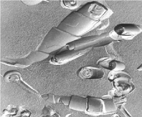
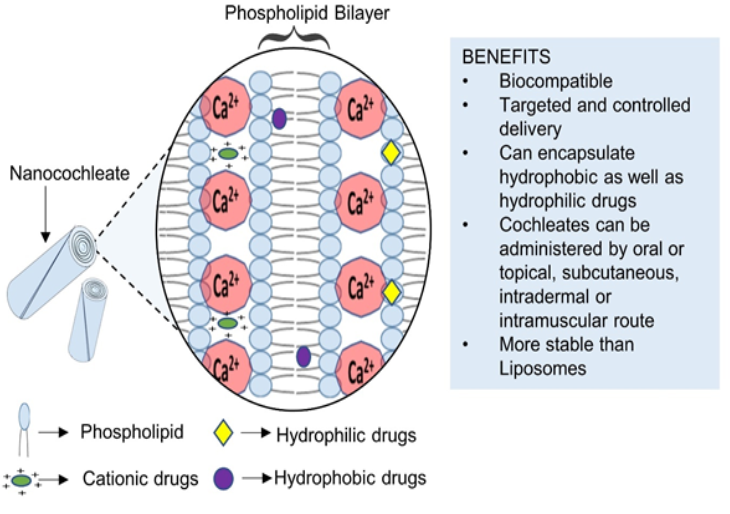
The idea behind the nanocochleate drug delivery vehicle is to potentially deliver drugs in a safe and effective manner by encasing them in a multilayered, lipid crystal matrix called a cochleate. With the use of a special customized method called nanocochleates, particles can be micro-encapsulated and trapped in supramolecular assemblies made of divalent cations and negatively charged phospholipids. A sequence of lipid bilayers makes up the cylindrical (cigar- like) microstructures known as nanocochleates [1]. Stable phospholipid-cation precipitates made of common, naturally occurring substances—phosphatidylserine and calcium, for example—serve as nanocochleate delivery vehicles. Their distinct multilayered structure is made up of a solid lipid bilayer sheet that is either stacked or rolled up in a spiral, with little to no aqueous space inside. This structure shields related “encochleated” molecules from deterioration. Even though the outer layers of the nanocochleate may be exposed to harsh environmental conditions or enzymes, components encapsulated within the interior of the nanocochleate structure remain intact because the entire structure is made up of solid layers [2, 3] due to the hydrophobic and hydrophilic surfaces found in nanocochleates (Figure 1).
One of the biggest causes of mortality in the world, cancer has remained a mystery that has not yet been fully understood. Depending on the type of cancer and its stage, the current treatment options include surgery, radiation therapy, chemotherapy, stem cell therapy, and many more. However, this intricate method is by no means adequate [4]. Despite having all the benefits mentioned above, the low oral bioavailability of chemotherapy drugs reduces the efficacy of oral drug delivery methods. This can cause serious side effects like nausea, abrupt vomiting, severe hair loss, low blood cell count, and anemia by damaging healthy cells and tissues. The pharmaceutical industry places a great deal of importance on the development of effective formulations to support drug delivery through the oral route, even though this is arguably the most preferred method of delivery. Many chemotherapeutic agents are difficult to formulate because they are hydrophobic. Currently available oral dosage forms of various anticancer medications such as capecitabine, cyclophosphamide, 5-fluorouracil, etoposide, and vinorelbine tartrate [4].
Although traditional delivery methods like tablets viz. mucoadhesive tablets [5, 6, 7, 8, 9, 10, 11, 12, 13], Inclusion complex [14, 15, 16, 17]
Sustain release tablets [18] hollow calcium pectinate beads [19], combined tablet dosage form [20], Mouth Dissolving Tablet [21], Solid Dispersion Techniques [22, 23], Tablets for Colonic Drug Delivery [24, 25, 26, 27, 28] Gastro-retentive Drug Delivery System [29], Probiotics [30], Medicated Chewing Gum [31], Nanosuspension Technologies [32, 33], 3D Printing Technology [34] and capsules have been researched, they have drawbacks, particularly low solubility bioavailability as well. This has led to the proposal of several methods, such as the use of prodrugs, drug carriers, or pharmaceutical excipients, to increase the solubility of chemotherapy agents. Various biocompatible nanoparticulate systems derived from oils, lipids, and polymers have been effectively employed to boost the bioavailability of medications when taken orally. Low drug delivery efficiency is one of the main drawbacks of the encapsulation techniques now in use for anti-cancer medication delivery.
Drug leakage and inadequate accumulation at target sites, instability and reticuloendothelial system absorption, encapsulation, and low retention efficiencies. The use of oral chemotherapy has the potential to yield pharmacoeconomic benefits by substantially lowering the overall cost of treatment. These days, phospholipid-based formulations are leading the way because of their superior qualities. They undergo thorough investigation to attain the best possible drug delivery outcomes (Figure 2).
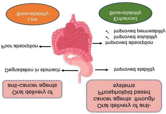
Orally administered anticancer medications with low solubility or bioavailability break down and exhibit low bioavailability. Numerous studies have demonstrated that phospholipid-based drug delivery systems, such as those for paclitaxel, cyclosporine, and curcumin, provide better results.
Sources and Properties of Phospholipids
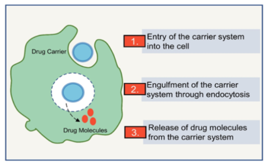
The United States Food and Drug Administration (FDA) has classified phospholipids as “Generally Regarded as Safe” (GRAS). Due to their extremely low toxicity, phospholipids can be administered through all routes of administration [35]. They can be derived from naturally occurring sources like eggs, bovine brain, soybean, sunflower, rapeseed, cottonseed, and so on, or they can be synthetic (both hydrogenated and synthetic phosphatidylcholine) [36]. Nonetheless, naturally occurring phospholipids are advised for oral administration. The component phospholipids are crucial to membrane of a cell. These molecules are amphiphilic, with a hydrophilic head region made up of a negatively charged phosphate group, two hydrophobic tails made up of long-chain fatty acids, and a glycerol or alcohol group connecting the head and tail regions. This allows the molecules to form vesicles or lipidic bilayers in biological systems [37, 38]. Phospholipids are versatile excipients with multiple uses in oral formulations, including emulsifiers, wetting agents, liposome formers and solubilizers. Additionally, they can be incorporated into slow- release tablet formulations and utilized as a matrix material for solid dispersions [39]. Phospholipid-based delivery systems are extensively researched for the delivery of anti- cancer agents because they may offer a solution to the low bioavailability issues. The basic delivery mechanism using phospholipid-based carriers is depicted in Figure 3.
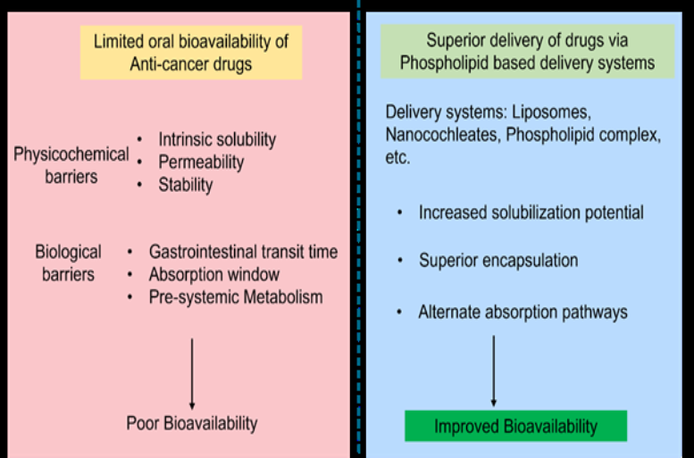
Numerous liposomal formulations based on phospholipids have been introduced to the market, such as Daunoxome®, Myocet®, and Dioxil® [40]. However, the intestinal lipid vesicles’ chemical and enzymatic destabilization limits the use and development of liposomes as oral drug delivery vehicles [41]. Thus, it is advantageous to use various approaches to optimize the therapeutic potential of phospholipids, providing the framework for effectively administering anti-cancer medications orally. Phospholipids and pharmaceutical techniques like nanocochleates and phospholipid complexes can be used to effectively get around the poor biopharmaceutical and physicochemical properties linked with the various anticancer drugs that limit their oral deliverability. Due to their unique qualities, these innovative drug delivery systems can get past several obstacles that prevent drugs from being delivered through the gastrointestinal tract [4, 35, 38, 41]. A visual depiction of enhanced delivery using phospholipid- based systems is presented in Figure 4.
Nanocochleates
In 1975, Dr. Dimitrious Papahadjoupoulos made the initial discovery of cochleates. Liposomes or pre-formed bilayer lipid vesicles with an anionic charge when binding agents are present are the building blocks of cochleates, often referred to as nanocochleates. Bilayer fusion results in morphological transformation that creates big sheets that coil into cochleates, which resemble cigar-shaped cylindrical structures. The Greek word “cochleate” means “a snail with a spiral shell,” which is like the cylinder’s folding pattern (Figure 5). These cochleates can incorporate charged, hydrophilic, and hydrophobic molecules (Figure 6). To prepare cochleates, naturally occurring lipids like lecithins and phospholipids are widely used. Phosphatidylcholine, phosphatidylserine, and other naturally occurring lipids are used to form cochleate [42, 43, 44].
Multivalent cations that are positively charged are typically utilized to interact with the negative charge present on phospholipids. These cations form a noncovalent bond with the lipid. Divalent metal cations such as Ca2+, Ba2+, Mg2+, a Multivalent cation that are positively charged are typically utilized to interact with the negative charge Zn2+ are frequently utilized binding agents [43, 45]. The binding properties of trivalent cations like Al3+ and monovalent cations like Na+ have also been examined [46]. The ability of some cationic drugs, like polylysine and tobramycin, to bind and form cochleate structures has also been investigated [47].
Judeh, et al. recently investigated the characteristics of cochleates bridged with amikacin [48]. Furthermore, Ca2+ in particular is employed as a binding agent due to its capacity to enhance phagocytosis and membrane fusion.
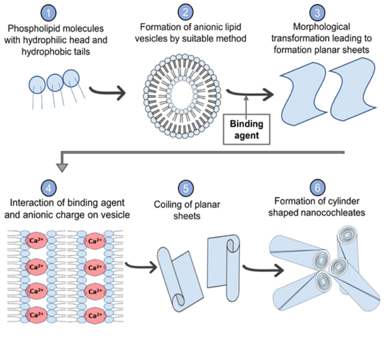
Particle aggregation, which compromises the stability of nanocochleates, is the main barrier to employing Ca2+ as a binding agent, though. Because it functions as a dispersing and stabilizing agent, citric acid can be used to get around this problem [49]. Additional aggregation inhibitors that have been documented include hydroxy cellulose, methylcellulose, casein, milk, and albumin [50]. The increased quantity of tiny molecules causing membrane fusion and the enhanced anti-cancer activity of nanocochleates could be the cause of the effect of enhanced permeability and retention, or EPR. The cylindrical shape of nanocochleates is responsible for their enhanced effect. Particle size and zeta potential ranges of 100–200 nm and ±30 mV, respectively, are advised. This stops the particles from aggregating and strengthens the EPR effect. Additional assessment criteria encompass entrapment effectiveness, medication loading, in vitro release investigations, transmission electron microscopy, and scanning electron microscopy [51, 52, 53].
Advantages of Nanocochleates
The advantages of cochleates are numerous. The nonaqueous construction of the cochleates means that:
- They can be preserved via freeze drying, which offers the possibility of being kept for extended periods of time at room temperatures, which would be beneficial for international shipment and storage before administration.
- They are more stable due to the reduced oxidation of lipids.
- The advantage of these structures over liposome structures is their ability to retain their structure even after lyophilization. Moreover, they show how biological molecules, particularly those with hydrophobic moieties, can be effectively integrated into the lipid bilayer of the cochleate structure. Lastly, when the nanocochleates dissociate or unwind slowly, they can release the biologic molecule in-vivo gradually.
- They have a lipid bilayer matrix that acts as a carrier and is made up of simple lipids that are non-inflammatory, non-immunogenic, and non-toxic because they are present in plant and animal cell membranes.
- They can be made quickly and safely [54].
- They increase the oral bioavailability of a wide range of substances, including those that are poorly soluble in water and protein and peptide biopharmaceuticals, which have proven challenging to use. such as ibuprofen for rheumatism) [55].
- They offer defense against the encochleated medication’s deterioration due to exposure to unfavorable environmental factors like sunlight, oxygen, water, and temperature [5].
Limitations
- They require specific storage condition.
- Sometimes aggregation may occur during storage; this can be avoided using aggregation inhibitor.
- The cost of manufacturing is high.
Structure and Components of Nano-Cochleate Drug Delivery System
The three major components used in the preparation of nanocochleates are atmospheric pressure ionization (API), lipids, and cations.
1. Lipids: Phosphatidyl serine (PS), phosphatidic acid (PA), di-oleoyl PS, phosphatidylinositol (PI), phosphatidyl glycerol (PG), phosphatidyl choline (PC), di-myristoyl PS, phosphatidyl ethanolamine (PE), diphosphatidyl glycerol (DPG), dioleoyl phosphatidic acid, di-stearoyl phosphatidyl serine, di-palmitoyl PG. 2. Cations: Zn+2 or Ca+2 or Mg+2 or Ba+2.
Dosage forms Available for Nanocochleate Drug Delivery • For oral administration: As a granule, powder, emulsion, suspension, pill, tablet, capsule, lozenge, cachet, or tablet form. • For topical or transdermal administration: Powders, sprays, ointments, pastes, creams, lotions, gels, solutions, patches, and inhalants. • For parenteral administration: Sterile isotonic aqueous or nonaqueous solutions, dispersions, suspensions or emulsions, or sterile powders which may be reconstituted into sterile injectable solutions or dispersions just before use.
Mechanism of Nanocochleates
Its unclear what theorizes cochleate’s release and cellular action under in vivo conditions. It is well known that the interior surface of cochleate, which contains a drug molecule encapsulated, is shielded from the harsh conditions found inside the body. Degradation of the outer layer could result in improved absorption and a gradual release of the medication. Two possible mechanisms have been suggested: first, the calcium-rich layer of the cochleate interacts with the cell membrane, rupturing and changing its morphology so that the outer layer of the cochleate fuses with the cell membrane through phagocytosis. The encapsulated medication is released when the cylindrical cochleate structure opens due to a low calcium concentration within the cochleate [43, 55, 56].
Methods of Preparation
Hydrogel Isolation Method: Two polymer solutions are used in the hydrogel method to prepare the cochleate. In short, the drug-loaded pre-formed liposomal dispersion is made. After that, it is combined with polymer A, which includes phosphatidylserine, dextran, and polyethylene glycol. A two-phase aqueous system is created when the prepared liposome and polymer A mixture is added to polymer B (such as polyvinyl pyrrolidone), which is immiscible with polymer A. The biphasic system is then treated to a cation salt solution, which diffuses into polymer B and subsequently into the mixture of liposome particles and polymer A. The polymer’s cationic cross-linkage results in the formation of small size cochleates [13, 57].
Emulsification-lyophilization Method: In this process, multiple emulsions form first, followed by the formation of cochleates. The oil phase is created when the lipid dissolves in a solvent (such as cyclohexane or chloroform). The buffer makes up the outer water phase, and the mixture of a binding and lyophilizing agent makes up the inner water phase. Initially, an oil phase and inner water phases are used to create a primary emulsion with submicron particle size with the help of a probe sonicatorṄ. This emulsion is then added to the outer water phase as a dispersed phase and gently emulsified to create a double emulsion, which is subsequently lyophilized. When lyophilized powder is rehydrated, cochleates are produced [42].
Trapping Method: The calcium chloride solution is added drop wise to the pre-formed liposomes—which are created by combining phospholipids and water—as part of the trapping procedure. It is the most widely used technique for preparing nanocochleates [52, 58].
Dialysis Method: This method involves the use of dialysis to prepare nanocochleates. The initial strategy is known as the “Liposomes before Cochleates dialysis method.” It involves a lipid and detergent mixture. To create lipid vesicles, a two-step procedure entails removing detergents from the mixture. To create cochleates, these vesicles are subsequently subjected to dialysis once more in the presence of a binding agent. The “Direct cochleates dialysis method” is the name of the alternate strategy. With this technique, detergent is directly removed by dialysis against a calcium chloride solution. It is unlikely that the process involves the creation of an intermediate liposome [42].
Delivery of Drugs: Table 1 provides an overview of the medications that have been studied for use in Nanocochleate- based therapy.
| Name of Drug Encapsulated | Biologic Utility | Comments | |
|---|---|---|---|
| 1 | Amikacin | Antibiotic | Better in vitro drug release profile along with increased stability |
| 2 | Artemisinin | Malaria | Higher encapsulation efficiency and controlled release action |
| 3 | Curcumin | Breast cancer | Improved cytotoxicity |
| 4 | Cyclosporine | Immuno-suppressant | Enhanced oral bioavailability by 3-folds |
| 5 | Glipizide | Diabetes | Increased oral bioavailability |
| 6 | Quercetin | Quercetin | Enhanced encapsulation efficiency |
| 7 | Rifampicin | Anti-Tubercular | Increased permeability compared with pure drug |
Table 1: List of Drugs Benefitted by Nanocochleates.
Evaluation and Characterization of Cochleates
Particle Size Determination: The Malvern 2000SM (Malvern, UK) laser diffraction technique was used to determine the mean particle size of the cochleates dispersion and liposomal dispersion. The analysis was performed at a temperature of 30±2°C with a 90° angle of detection. The volume mean diameter D [3, 4]—the average diameter of a sphere whose volume is equal to the particle under measurement—was used to express the mean vesicle size.
Determination of Entrapment Efficiency (EE) of Cochleates: Cochleates were divided into 100 microliter centrifugation tubes. While vortexing, 60 μl pH 9.5 EDTA and 1 ml of ethanol were added to each tube. A translucent, colourless solution is the final result. Equation 3 was used to calculate the entrapment efficiency after the samples were appropriately diluted and their absorbance measured at 289 nm.
Entrapment Efficiency= The drug content in cochleates / total amount of drug present*100.
Fourier Transform Infra-Red Spectroscopy (FTIR): The FTIR measurements of drug-loaded freeze-dried cochleates, DOPS-Na, and KCZ were obtained using a JASCO FTIR 4100 (Japan) that was outfitted with Spectra Manager edition 2. After mixing with KBr, the samples were ready to be put in the sample holder. At room temperature, the spectra were scanned across the wave number range of 3600–400 cm−1. Specific Surface Area: For freeze-dried Nanocochleate, a sorptometer is typically used to measure the specific surface area. The specific surface area can be computed using the following formula. A = 6 / ρd Where ρ denotes the density of the cochleate, A its specific surface area, and d its diameter.
The measured and computed specific surface areas generally agree fairly, but occasionally there may be a discrepancy in the measured values due to residual surfactant [59].
Surface Hydrophobicity: The surface hydrophobicity of nanocochleates affects how colloidal particles interact with the biological environment. Hydrophobicity and hydrophilicity together define the bio-fate of nanocochleates and their contents. Hydrophobicity controls the degree and kind of hydrophobic interactions between nanocochleates and blood components. Surface hydrophobicity has been assessed using a variety of techniques, such as hydrophobic interaction chromatography, two-phase partition, as well as the adsorption of hydrophobic fluorescent or radiolabellled probes. Several advanced techniques for surface chemistry analysis have also been employed recently [60].
Density: Using a gas pycnometer and either air or helium, one can find the density of nanocochleates. Because of the porosity and specific surface area of the structure, the value obtained with air and helium is much more pronounced [61].
In-vitro Release: Utilizing double chamber diffusion cells on a shake stand with phosphate buffer, standard dialysis, diffusion cell, or recently developed modified ultra-filtration techniques can all be used to determine the in vitro release profile of nanocochleates. The two chambers are separated by a hydrophilic, low-protein-binding Millipore membrane. The receptor compartment is assayed for the released drug using standard procedures at various time intervals after the donor chamber is filled with nanocochleates. The in-vitro release behavior of nanocochleates is also ascertained using the modified ultra-filtration technique. In this instance, buffer is added to a stirred ultra-filtration cell directly before the Nanocochleate is added. Aliquots of the dissolution medium are filtered through the ultra-filtration membrane using < 2 positive nitrogen pressure at different time intervals, and the released drug is then measured using standard protocols [31].
Molecular Weight Measurements: Gel permeation chromatography (GPC) with a refractive index detector can be used to determine the molecular weight of the polymer and its distribution within the matrix. It was demonstrated using GPC that the formation of polyalkylcynoacrylate (PACA) nanocochleates is not caused by the rolling up of one or a few long polymer chains, but rather by the entanglement of multiple small oligomeric subunits [62].
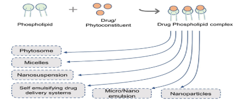
Drug-phospholipid complex
The active ingredient in the drug-phospholipid complex is covalently bound to the phospholipid, resulting in a colloidal dispersion. The drug-phospholipid complex is mostly formed by phosphatidylcholine. In eukaryotic cells, phospholipids containing choline are widely distributed. Phospholipid complexes were first investigated as a means of improving phytoconstituents’ bioavailability. Eventually, it was discovered that they were useful in raising the bioavailability of drugs classified as class II and IV in the biopharmaceutical classification system [38, 63].
Both naturally occurring agents and synthetic drugs, often referred to as phytosomes and pharmacosomes, can be effectively delivered through phospholipid complexation [64]. The resulting complexes can also be added to other delivery systems like nanosuspensions and microemulsions (Figure 6). The dual properties of the drug-phospholipid complex and nanopreparation are present in this kind of nanodrug release system, which may improve the drug’s solubility, stability, and bioavailability while lowering the dosage and harmful side effects. Additionally, it might help achieve targeted drug delivery (Figure 7). Drugs whose absorption is limited in their dissolution or permeation can have more bioavailability through the formation of phospholipid complexes. Differential calorimetric analyses, scanning electron microscopy, transmission electron microscopy, Fourier transform infrared spectroscopy, and nuclear magnetic resonance spectroscopy are a few characterization techniques.
Mechanism of Complex Formation
The phospholipid molecules and the polyphenolic compounds or drug molecule form chemical bonds. A drug- phospholipid complex is created when free carboxylic or active hydrogen atom-like amino, hydroxyl groups esterify with the phospholipid’s hydroxyl group. One possible tool to encourage complex formation is a spacer chain. Both lipophilic and hydrophilic qualities are present in the complex. Through their ability to facilitate the migration of drugs across tissues and cell membranes, they improve the bioavailability of drugs [65, 66].
Stability of Nanocochleate
The structure and characteristics of these stable, lipid- based delivery formulations differ greatly from those of liposomes. This unique structure provides protection from degradation for encochleated, viz, encapsulated molecules. The cochleate structure as a whole is made up of solid layers, thus even though its exterior layers are exposed to severe environments or enzymes, such those found in the stomach, the components inside the structure stay intact. Animal studies demonstrated that nanocochleates cross the digestive epithelium and deliver their cargo molecules into target cells [27, 28]. These unique properties of nanocochleates were used to mediate and enhance the oral bioavailability of broad spectrum of biopharmaceuticals, including compounds with poor water solubility, such as amphotericin B [6, 67, 68]. Before being administered, they can be reconstituted with liquid and are stable against lyophilization. When it comes to the distribution of various medications and delicate substances like proteins and peptides, cochleate technology is the most adaptable.
Safety/Biocompatibility of the Nanocochleate Delivery Vehicles
The main two components of nanocochleate are PS and calcium. PS is a natural constituent of all biological membranes and is most concentrated in the brain. Phospholipids, which are made of anionic lipids and are non-inflammatory and biodegradable, can be synthesised or manufactured from natural sources for use in nanocochleate formulation. Soy PS is available in large quantities and is expensive and suitable for use in humans. These two components which are safe, simple, naturally occurring substances which make nanocochleates safe and biocompatible delivery vehicle. Clinical studies show that PS is very safe which play a role in support of mental functions in the aging brain.
Application of Nanocochleates
- Proteins, peptides, and DNA have all been delivered via nanocochleates for use in gene therapy and vaccination applications.
- Without changing the flavor or aroma of the food, omega-3 fatty acids can be added to cakes, muffins, pasta, soups, and cookies using nanocochleates.
- The benefits of cochleates would be increased bactericidal activity and decreased toxicity.
- Amphotericin B, a possible antifungal agent, has the potential to be administered orally and parentally by nanocochleates.
It has a good safety profile and lower treatment costs. Amphotericin B cochleates that have been prepared exhibit increased stability and efficacy at low dosages. They demonstrate increased patient adherence.
Conclusion
A lipid-based drug delivery method with promise for oral drug administration is nanocochleates. A novel technological platform for oral and systemic drug delivery is represented by cochleate delivery vehicles. They offer a novel technological platform that can be used to administer a wide range of molecules, such as medications, genes, and vaccine antigens, orally and systemically. These molecules have significant biological therapeutic effects. Using themselves as the bridging agents of cochleation, negatively charged liposomes and bivalent cations interact ionically to form cochleates, which makes sense when cationic drugs are microencapsulated. Cochleates and nanocochleate particles can be used as cochleates’ bridging agents to encapsulate hydrophilic and multicationic drugs and peptides. The physico-chemical characteristics of metal ion-bridged cochleates, which include their ability to fuse with the cell membrane and their conversion back to liposomes upon the extraction of the cation bridging agent, are inherent in this new type of cochleate. Because of these special characteristics, charged hydrophilic medications can be delivered across the tissue membrane by cochleates and nanocochleates. As a result, amidocleate has a great therapeutic potential for medication delivery.
Conflicts of Interest
There are no conflicts of interest.
Acknowledgments
The author expresses gratitude to all of his mentors and our principal., Dr. V. V. P. Foundation’s College of Pharmacy, Ahmednagar. The authors would like to thank the academics whose works are referenced and credited in this manuscript for their invaluable assistance. The writers express their gratitude to all the authors, editors, and publishers of books, journals, and articles that served as the source material for the reviews and discussions of the literature included in this work.
References
-
Delmarre D, Tatton N, Krause-Elsmore S, Gould-Fogerite S, Mannino RJ (2004) Formulation of Hydrophobic Drug into cochleate delivery vehicles: A simplified protocol and formulation kit. Drug delivery technology 1: 64-69.
-
Egan WJ, Lauri E (2002) Prediction of intestinal permeability. Adv drug delivery review 54(3): 273-289.
-
Florence AT, Hussain N (2001) Transcytosis of nanoparticles & dendrimers delivery systems: evolving vistas. Adv drug delivery review 50(1): 69-89.
-
Tariq M, T Singh A, Iqbal Z, J Ahmad F, Talegaonkar S (2016) Investigative approaches for oral delivery of anticancer drugs: A patent review. Recent Pat Drug Deliv Formul 10(1): 24-43.
-
Shaikh S, Merekar AN, Godge GR, Gaikwad MR (2016) Formulation and In-Vitro Evaluation of Buccal Mucoadhesive Tablets of Catopril by Using Natural and Synthetic Polymers. World Journal of Pharmaceutical Research 7(5): 1296-1315.
-
Hiremath SN, Kharia AA, Godge GR, Omray LK, Shingai AK (2010) Formulation Strategies for Low Absorption Window Antihypertensive Agents. Research Journal of Pharmacy and Technology 3(1): 113-117.
-
Hiremath SN, Godge GR, Kulkarni PS, Landge RB (2021) Discussion on tablets with special emphasis on formulation and coating defects: A Review. IJCRT 9(7): 124-139.
-
Godge GR, Arjun GM, Balaprasad DA, Machindra DA, Vinayak WD, et al. (2020) Validated Spectrophotometric Method for Simultaneous Estimation of Fenofibrate and Atorvastatin in Synthetic Mixture and in Bulk Tablet Dosage Form. World Journal of Pharmaceutical and Medical Research 6(7): 170-176.
-
Landge RB, Kulkarni PS, Godge GR, Hiremath SN (2022) A Detailed Report on Tablets with Special Emphasis on its Formulation and Manufacturing Technology. International Journal of Creative Research Thoughts 10(3): 624-641.
-
Godge GR, Bharat SC, Shaikh AB, Randhawan BB, Raskar MA, et al. (2023) Formulation Perspectives in Topical Antifungal Drug Therapy: A Review. Journal of Drug Delivery and Therapeutics 13(5): 110-119.
-
Raskar MA, Mungse SS, Kate PA, Godge GR (2023) Validated Simultaneous Derivative Spectrophotometric Estimation of Diflunisal and Lignocaine in Bulk and Pharmaceutical Formulation. Journal of Drug Delivery and Therapeutics 13(8): 51-55.
-
Raskar MA, Kate PA, Mungse SS, Godge GR (2023) Validated Simultaneous Derivative Spectrophotometric Estimation of Azithromycin, Fluconazole and Secnidazole in Bulk and Pharmaceutical Formulation. Journal of Drug Delivery and Therapeutics 13(11): 1-6.
-
Godge GR, Shaikh AB, Bharat SC, Randhawan BB, Raskar MA, et al. (2023) Nasal Drug Delivery System and its Applicability in Therapeutics: A Review. RGUHS Journal of Pharmaceutical Sciences 13(4): 1-12.
-
Kute VC, Godge G (2016) Preparation & In-Vitro Evaluation of Inclusion Complexes of Simvastatin Tablet with Cyclodextrins. World Journal of Pharmaceutical Research 5(2): 1022-1041.
-
Hiremath SN, Godge GR, Sonawale B, Shirsath R (2015) Pharmaceutical Advances in Cyclodextrin Inclusion Complexes for Improved Bioavailability of Poorly- Soluble Drugs. International Journal of Pharmaceutical Sciences and Nanotechnology 8(3): 2894-2905.
-
Hiremath SN, Godge GR (2012) Preparation and in vitro Evaluation of Inclusion Complexes of Nelfinavir with Chemically Modified β-cyclodextrins. Dhaka Univ J Pharm Sci 11(2): 107-116.
-
Hiremath SN, Godge GR, Kharia AA, Vaidya VR (2010) Studies on the Preparation, Characterization and Solubility of Β-Cyclodextrin-Nelfinavir Inclusion Complexes. JPRHC 2(3): 279-284.
-
Godge GR (2016) Formulation development and in-vitro evaluation of sustained release tablets of telmisartan by solid dispersion technology. Asian Journal of Pharmaceutical Technology & Innovation 4(17): 131- 139.
-
Chemate SZ, Godge GR, Pawa KK, Rupnar KA (2016) Preparation and evaluation of hollow calcium pectinate beads for floating-pulsatile drug delivery. Turk J Pharm Sci 13(1): 91-102.
-
Raskar MA, Godge GR, Chitale AB, Giri PD (2015) Validated simultaneous spectrophotometric estimation of telmisartan, hydrochlorthiazide and amlodipine besylate in combined tablet dosage form. Der Pharmacia Lettre 7(11): 120-124.
-
Godge GR, Misal AV, Pawar PY (2015) Formulation and Evaluation of Mouth Dissolving Tablet with Taste Masking Resin. International Journal of Life Sciences and Review 1(7): 253-263.
-
Godge G, Labade S, Misal A (2015) Oral Bioavailability Improvement Using Solid Dispersion Techniques: A Review. International Journal of Life Sciences and Review 1(7): 243-252.
-
Godge GR, Labade SP (2015) Preparation of Solid Dispersion of Poorly Water Soluble Drug Formulation and Consideration. International Journal of Pharma Sciences and Research 6(5): 897-903.
-
Godge GR, Hiremath SN (2014) An Investigation into the Characteristics of Natural Polysaccharide: Polymer Metoprolol Succinate Tablets for Colonic Drug Delivery. Mahidol University Journal of Pharmaceutical Sciences 41(2): 7-21.
-
Godge GR, Hiremath SN (2012) Colon Targeted Drug Delivery System: A Review. Int J Pharm Drug Ana 2(1): 35-48.
-
Godge GR, Hiremath SN (2014) Development and Evaluation of Colon Targeted Drug Delivery System by Using Natural Polysaccharides/Polymers. Dhaka Univ J Pharm Sci 13(1): 105-113.
-
Godge GR, Hiremath SN (2012) Colonic delivery of film coated meloxicam tablets using natural polysaccharide polymer mixture. International Current Pharmaceutical Journal 1(9): 264-271.
-
Hiremath SN, Godge GR (2011) Recent Advances in Pharmaceutical Approaches to Colon Specific Drug Delivery. Pharm Tech 8(4): 1-8.
-
Kharia AA, Hiremath SN, Omray LK, Yadav R, Godge GR (2011) Gastro-retentive Drug Delivery System. Indian Drugs 48(5): 7-15.
-
Sanap SD, Garje MA, Godge GR (2019) Probiotics, their Health Benefits and Applications for Development of Human Health: A Review. Journal of Drug Delivery and Therapeutics 9(S4): 631-640.
-
Sanap DS, Godge GR (2019) Recent Trends in Medicated Chewing Gum Technology: A Review on History. Formulation Strategies and Characterization. Int J Pharm Sci Rev Res 59(2): 72-85.
-
Godge GR, Zarekar SB, Padale PJ (2021) Nanosuspension Technologies: A Innovative Approach in Drug Delivery of Sparingly Soluble Drugs. IJCRT 9(7): 409-420.
-
Godge GR, Garje MA, Dode AB, Tarkase KN (2020) Nanosuspension Technology for Delivery of Poorly Soluble Drugs and its Applications: A Review. Int J Pharm Sci Nanotech 13(4): 4965-4978.
-
Godge GR, Ghume VK, Bidave BB, Golhar AR (2021) 3D Printing Technology in Pharmaceutical Drug Delivery. Int J Pharm Sci Rev Res 68(1): 15-20.
-
Hoogevest PV, Wendel A (2014) The use of natural and synthetic phospholipids as pharmaceutical excipients. Eur J Lipid Sci Technol 116(9): 1088-1107.
-
Singh RP, Gangadharappa HV, Mruthunjaya K (2017) Phospholipids: Unique carriers for drug delivery systems. J Drug Deliv Sci Technol 39: 166-179.
-
Khan J, Alexander A, Saraf S, Saraf S (2013) Recent advances and future prospects of phyto-phospholipid complexation technique for improving pharmacokinetic profile of plant actives. J Control Release 168(1): 50-60.
-
Kuche K, Bhargavi N, Dora CP, Jain S (2019) Drug- phospholipid complex-a go through strategy for enhanced oral bioavailability. AAPS Pharm SciTech 20(2): 43.
-
Hoogevest PV (2017) Review: An update on the use of oral phospholipid excipients. Eur J Pharm Sci 108: 1-12.
-
Teixeira MC, Carbone C, Souto EB (2017) Beyond liposomes: Recent advances on lipid-based nanostructures for poorly soluble/ poorly permeable drug delivery. Prog Lipid Res 68: 1-11
-
Zhang L, Wang S, Zhang M, Sun J (2013) Nanocarriers for oral drug delivery. J Drug Target 21(6): 515-527.
-
Nagarsekar K (2016) Cochleates: New insights into drug delivery system (Doctoral dissertation) 4(2): 55-62.
-
Pawar A, Bothiraja C, Shaikh K, Mali A (2015) An insight into cochleates, a potential drug delivery system. RSC Adv 5(99): 81188-81202.
-
Shuddhodana, Nagarsekar K, Judeh ZMA (2017) Recent advances and developments in cochleate technology. Nanomedicine & Nanotechnology Open Access 2(2): 000119.
-
Shanmugam T, Banerjee R (2011) Nanostructured self- assembled lipid materials for drug delivery and tissue engineering. Ther Deliv 2(11): 1485-516.
-
Ahiwale RJ, Chellampillai B, Pawar AP (2020) Investigation of 1, 2-Dimyristoyl-Sn-Glycero-3- Phosphoglycerol-Sodium (DMPG-Na) Lipid with various metal cations in nanocochleate preformulation: Application for Andrographolide oral delivery in cancer therapy. AAPS Pharm SciTech 21(7): 279.
-
Syed UM, Woo AF, Plakogiannis F, Jin T, Zhu H (2008) Cochleates bridged by drug molecules. Int J Pharm 363(1-2): 118-125.
-
Judeh Z (2021) Insights into the mechanism of formation of nonconventional cochleates and its impact on their functional properties. J Mol Liq 335: 116249.
-
Bozó T, Wacha A, Mihály J, Bóta A, Kellermayer MS (2017) Dispersion and stabilization of cochleate nanoparticles. Eur J Pharm Biopharm 117: 270-275.
-
Mannino R, Gould-Fogerite S, Krause-Elsmore S, Delmarre D, Lu R (2005) Inventors; University of Medicine, Dentistry of New Jersey, Biodelivery Sciences Inc, assignee. Novel encochleation methods, cochleates and methods of use. United States patent application US 10/822,230.
-
Ağardan N, Değim Z, Yılmaz Ş, Altıntaş L, Topal T (2016) The effectiveness of raloxifene-loaded liposomes and cochleates in breast cancer therapy. AAPS PharmSciTech 17(4): 968-977.
-
Poudel I, Ahiwale R, Pawar A, Mahadik K, Bothiraja C (2018) Development of novel biotinylated chitosan- decorated docetaxel-loaded nanocochleates for breast cancer targeting. Artif Cells Nanomed Biotechnol 46(Suppl 2): 229-240.
-
Bhosale RR, Gangadharappa HV, Gowda DV, Osmani RA, Vaghela R (2017) A review on nanocochleates: The inimitable nanoparticulate drug carriers. Adv Sci Eng Med 9(5): 359-369.
-
Fogerite SG, Mannino RJ (1997) Cochleate delivery vehicles, US Patent: 5994318.
-
(2005) Bio delivery sciences International.
-
Asprea M, Tatini F, Piazzini V, Rossi F, Bergonzi MC, et al. (2019) Stable, monodisperse and highly cell-permeating nanocochleates from natural soy lecithin liposomes. Pharmaceutics 11(1): 34.
-
Shende P, Khair R, Gaud RS (2019) Nanostructured cochleates: A multi-layered platform for cellular transportation of therapeutics. Drug Dev Ind Pharm 45(6): 869-881.
-
Yücel Ç, Altintaş Y, Değim Z, Yılmaz Ş, Arsoy T, et al. (2019) Novel approach to the treatment of diabetes: Embryonic stem cell and insulin-loaded liposomes and nanocochleates. J Nanosci Nanotechnol 19(7): 3706- 3719.
-
Tilawat M, Bonde S (2021) Nanocochleates: A potential drug delivery system. J Mol Liq 334: 116115.
-
Uwais MS, Amyf W, Tuo Jina B, Hua Z (2008) Cochleate bridged by drug molecule’s. International Journal of Pharmaceutics 363(1-2): 118-125.
-
Panchagnula R, Sood A (2001) Peroral route: an opportunity for protein & peptide drug delivery. Chem Review 101(11): 3275-3304.
-
Villa AM, Caporizzo E, Papagni A, Miozzo L, Buttero PD (2005) Papagin A Choline & phosphatidylcholine flurescent derivatives localization in carcinoma cells studies by laser scanning confocal fluorescence microscopy. European journal of cancer 41(10): 1453- 1459.
-
Zarif L, Perlin D (2002) Amphoteric B Nanocochleates: Formulation to oral efficacy. Drug Dlivery Technology 4: 34-37.
-
Zarif L, Graybill J, Perlin D, Mannino RJ (2000) Cochleates: new lipid-based drug delivery system. Journal of liposome research 10: 523-538.
-
Gnananath K, Nataraj KS, Rao BG (2017) Phospholipid complex technique for superior bioavailability of phytoconstituents. Adv Pharm Bull 7(1): 35-42.
-
Pandita A, Sharma P (2013) Pharmacosomes: An emerging novel vesicular drug delivery system for poorly soluble synthetic and herbal drugs. ISRN Pharm: e348186.
-
Vaidya VR, Karodi RS, Mohite MT, Hiremath SN, Godge GR (2010) Formulation Optimization of Mucoadhesive Buccal Tablets of Carvedilol using 32 Full Factorial Design. Deccan J Pharmaceutics and Cosmetology 1(2): 7-20.
-
Semalty A (2014) Cyclodextrin and phospholipid complexation in solubility and dissolution enhancement: A critical and metaanalysis. Exp Opin Drug Deliv 11(8): 1255-1272.
- Effects of 5-HTP and Melatonin on the Sleep Cycle of Medical Students
- Adsorption of Bisphenol A on NH4OH- Modified Rice Husk and Sugar Cane Bagasse Biochar
- Comparative Assessment of the Reinforcement Efficiency of Palm Fruit Fibre and Coconut Fibre in High Density Polyethylene (HDPE) Matrix Composite
- Importance of Bio Compounds Naturally Present in Food with Functionality in Animal Metabolism
- Sub-Acute Study on the Cardiotoxic Effects of Monosodium Glutamate Ingestion in Albino Rat
- Weight Management and Its Natural Solutions: A Review