Seroprevalence, Isolation and Molecular Detection of Infectious Bronchitis in Backyard and Commercial Chickens in Central Gondar Zone, Northern Ethiopia
Infectious bronchitis (IB) is a highly contagious disease of the respiratory and urogenital tract of chickens, caused by infectious bronchitis virus (IBV), a member of the family Coronaviridae. Due to the serious infectious and transmission features of the disease mostly in the reproductive and respiratory systems it causes potential economic loss. Hence a cross-sectional study was conducted from February 2022 to June 2022 on chicken serum and tracheal swab samples from backyard and commercial farms in central Gondar zone, Ethiopia, to determine the seroprevalence of IB, associated risk factors and for isolation and molecular detection of the virus. A total of 384 blood samples were collected and tested by an indirect ELISA and Anti-IBV antibody positivity was noted in 92.19% (95% confidence interval (89 % -94.6 %) of the samples. Logistic regression analysis was performed to determine the impact of possible risk factors on seropositivity. Higher prevalence was noted in young chickens than in adults (p< 0.05) and in exotic breeds than in local breeds (p0.05), higher prevalence was obtained in dual purpose chickens (93.75%) than in layers (92.17%) and broilers (90.98%). Higher prevalence was also noted in females (92.7%) than in males (90.98%) and in intensively managed chickens (93.39%) than in extensively managed chickens (90.69%) with p>0.05. Conventional RT-PCR test was also performed for the molecular detection of virus. The test was done on 52 tracheal swab samples collected from intensive and backyard unvaccinated chickens that were pooled in to 26 samples. Accordingly, 3 (11.54%) of the 26 pooled samples were IBV positive. The result showed that this was the first molecular evidence found in the study area. The seroprevalence of the disease in this study was very high for all age groups, breed types, and farm types. The risk factors mentioned and the management methods may have raised the likelihood of infection and the prevalence of the disease. Vaccination and biosecurity measures are advised to manage the disease. Identification and characterization of persistent IBV serotypes that are present in the field is also recommended to manage the disease
Introduction
Background of the Study
The total chicken population in Ethiopia is estimated to be 56.5 million. However, the economic contribution of the sector is not still proportional to the huge chicken numbers, attributed to the presence of many productions, reproduction and infrastructural constraints. Diseases and predators are known to be the major causes of losses in the country. Newcastle disease accounted for the largest proportion of overall flock mortality to be 57.3% followed by fowl pox 31.6%, coccidiosis, 9.4% and predator loss 1.7% [1].
Infectious bronchitis virus causes acute highly contagious viral disease of chicken known as infectious bronchitis, which is characterized by lesions in the reproductive, urogenital, and respiratory organs [2].
Chickens of all ages can contract the disease, although young chickens are more vulnerable because resistance builds of with age. Due to poor performance, lower egg output and quality, and mortality that can be severe in the presence of nephropathogenic strains or when secondary infections emerge, the condition generates significant economic losses in the chicken industry following avian influenza; it is believed that IBV infection causes the third- highest losses among all livestock diseases [3].
The virus is a member of the Coronaviridae family and its 27.6 kb single-stranded positive sense RNA genome has the following genomic arrangement and: fifty untranslated regions 30UTR: 1a/1ab S 3a 3b E M 5a 5b. While the non- structural viral proteins are encoded by two polyproteins, the structural viral proteins include the membrane (M), small membrane (E), nucleoprotein (N), and spike (S).The spike protein, among others, is the subject of extensive research due to its genetic diversity and biological function [4].
Following the discovery of the first IBV serotype in USA in 1931, the virus was detected in other US states, and a large variety of IBV genotypes and serotypes were described globally [5]. Infectious bronchitis is still a problem for the poultry industry despite global attempts to manage it thus far, mostly because of IBV’s large genetic diversity, rapid generation time, and high mutation rate. The effectiveness of natural or vaccination immunity is hampered by the ongoing emergence of genetic diversity and the selection process among IBV variants [6].
In Africa, IBV is one of the most important viral diseases that threaten chicken production. It was first reported in Africa in 1950 in Egypt from birds with respiratory symptoms. The disease is the most prevalent viral respiratory infections of chickens in the continent and is widespread in both vaccinated and unvaccinated poultry birds and is regarded as an epidemic virus [7]. The disease is common in Ethiopia though its prevalence and type of strains has not been well studied in the present study area [8].
Objectives of the Study
General objectives: This study is aimed at determining the seroprevalence of infectious bronchitis virus antibody and to detect infectious bronchitis virus antigen of poultry in central Gondar zone. Specific objectives: To determine the seroprevalence of infectious bronchitis in chickens within the study area
- To isolate and detect infectious bronchitis virus in poultry in the study area
- To study some associated risk factors
Statement of the Problem
Infectious bronchitis is one of the most serious viral infections threatening chicken farming in Africa. Despite reports of respiratory disorders in chicken from all production systems in Ethiopia, there is a paucity of reports on infectious bronchitis (IB). Accordingly, there is only one report about IB seroprevalence which indicates a seroprevalence of 24.6% [9] and no published evidence on molecular detection of the virus in northern Ethiopia in general and the study area in particular. Furthermore, the disease is a poorly studied condition in Ethiopia since it is frequently masked by other infections such as Newcastle disease and other respiratory diseases.
Research Questions
- What is the seroprevalence status of infectious bronchitis in the study area?
- What are the associated risk factors for the occurrence of infectious bronchitis?
- Is infectious bronchitis virus circulating in the study area?
Justification of the Study
Chickens in Ethiopia are not vaccinated against infectious bronchitis especially in rural areas where birds are mostly managed semi-intensively with little or no veterinary care and are left to search for food for most of the day. This practice promotes the rapid spread of the disease. Although
there are some published evidences on the sero-prevalence of the disease with variable figures across the country, there is a limited information on the seroprevalence and there is no information on molecular detection of the virus in both commercial and rural scavenging poultry in Central Gondar Zone. In order to prevent and control the disease by implementing vaccination and biosecurity measures, it is essential to identify the occurrence of the disease in the study area. Therefore; the present study was performed to determine the prevalence of the disease and to detect the existence of infectious bronchitis virus circulating in the study area.
Ethical Approval
Ethical clearance was obtained from the Ethics Committee of the National Veterinary Institute of Ethiopia. Accordingly, handling of the study animals throughout the study period was done based on the guideline of the institute. The poultry owners provided their permission and authorization for the collection of the samples.
Materials and Methods
The Study Area
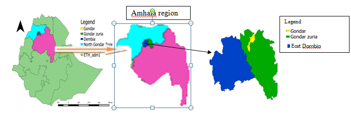
Amhara Regional State is one of the largest state in Ethiopia with a large poultry population. The Central Gondar zone is located in the Amhara National Regional State, and its principal city, Gondar, is 727 kilometers from Ethiopia the capital, Addis Ababa (Figure 1). Geographically, the area lies between 12.3° and 13.38° N latitudes and 35.5° and 38.3° E longitudes. Central Gondar zone is located between 528 and 4620 meters above sea level. The average annual rainfall ranges from 880 mm to 1772 mm, with a monomodal distribution and temperatures ranging from 10°C to 44.5°C [9]. The overall population of central Gondar Zone is 2,642,114, with 2,204,225 (88.66 percent) living in rural villages that rely on agriculture for their livelihood. As a result, the study region is a significant source of livestock that contributes to the country’s growth and domestic production. This area houses around 737,713 sheep, 1,479,366 goats, 3,234,012 cattle, 392,546 donkeys, 12,252 mules, 39,178 horses, and 3,310,498 chickens [10].
Cross-sectional study design was used in this study. For district selection; a simple random sampling was utilized as a sampling strategy. A simple random sampling technique was also used to select poultry farms and individual chickens. Before blood and tracheal swab samples were taken, potential risk factors such as flock history (age, purpose of production, sex, breed, and method of production) and vaccination history in general were noted.
Study Animals
Because maternal antibodies are expected to diminish within three weeks of life, chickens were sampled when
Sample Size Calculation
The sample size for the study was calculated using Thrusfield’s formula [11], with a 95% confidence interval of 5% desired absolute precision and a prevalence of 24.61% from previous studies. Thus, 285 chickens were required for this study. However, to improve the precision of the study the sample size was expanded to 384.
( ) 2 exp exp
2 1.96 P 1 P N d − =
Where N= required sample size, Pexp= expected prevalence d2 =desired absolute precision. z=1.96, p=0.5 and d=0.05
Blood Sample Collection
Two to three ml blood sample was collected from unvaccinated chickens in different farms from the wing vein. To clot the blood, the collection tubes were kept slant for 24 hours at room temperature. Then sera were collected and placed into 2ml tubes and frozen at 20°C until they were processed at National Veterinary Institute of Ethiopia.
Swab Sample Collection and Preparation
Fifty two chickens that appeared to be diseased in total were chosen at random to have tracheal swab samples prepared for PCR analysis. Each swab was placed in a PBS (Phosphate Buffer Saline) containing antibiotic filled sterile tube and transported under cold chain and kept at –20°C in University of Gondar Veterinary Microbiology Laboratory. Then the samples were transported to the National Veterinary Institute of Ethiopia for processing under cold chain and kept under –80°C until processed. Then the samples were taken out from –80°C and thawed for a few minutes. Then in order to make it easier to remove the contents from the swab head, the swab samples from each flock were vortexed and samples from the same farm were pooled into one that makes a total of 26 samples which were then made ready for injection in to specific pathogen-free (SPF) eggs.
Virus Isolation
Specific pathogen-free (SPF) embryonic eggs were used in the viral isolation process. 0.2-0.3 ml of sample supernatant was inoculated into the allantoic cavities of 11day old embryos and no sample were inoculated in to the controls. Eggs were candled every day for four days with mortality occurring within the first 24 hours being regarded as nonspecific. After 96 hours of infection, all of the eggs allantoic fluids were collected and used to infect another egg groups up to three passages. Embryonic deaths after 24 hours of infection were observed and recorded. Infective allantoic fluids were collected from the third passage and kept at –20°C and processed by RT-PCR for molecular detection.
Serological Analysis
According to the manufacturer’s recommendations, an indirect ELISA (ID Screen® Infectious Bronchitis Virus Indirect) was performed to detect antibodies against IBV in chicken serum. The test is designed to detect IBV specific antibodies that are conserved across serotypes, allowing all IBV types to be detected. Four 96-well plates (precoated with purified viral antigen), positive and negative IBV antibody control sera, concentrated conjugate (10 xs), dilution buffer 14, dilution buffer 3, washing buffer (20 x), substrate solution, and stop solution were used in the test in accordance with the manufacturer’s instructions. The microplates were examined using an ELISA micro plate reader at a wavelength of 450 nm. The ELISA sample to positive ratio for each serum determined the interpretation of the data. The percentage of positive sera among the total number of examined sera was used to calculate IBV prevalence.
The unknown test samples were evaluated Parallel to positive and negative controls,. Using a precoated ELISA plate and ready-to-use reagents, all conditions were standardized according to kit manufacturer specifications. The test was valid if the mean OD value of the positive control was greater than 0.250 and when the ratio of the positive and negative control mean values is greater than 3. The ELISA sample-to- positive (S/P) ratio for each serum was used to evaluate the results. Serum samples with an S/P value greater than 0.2 were considered positive.
S Pratio ODsample OD NC ODPC OD NC = − −
Where S/P is the sample to positive ratio; OD is the optical density; ODNC is the optical density of negative control; and ODPC is the optical density of positive control.
RNA Extraction
RNA was isolated by using the Qiagen RNeasy Mini Kit protocol. Based on the this protocol 350µl of RLT lysis buffer was added to 350 µl sample aliquots to bring them to room temperature, and were mixed by pipetting. Next, 350 µl of 70% ethanol was added, and they were again mixed by pipetting. The sample mixture was then poured into an RNeasy spin column and centrifuged for 30 seconds at 13000 rpm. The flow through liquid was then discarded. After centrifuging RNeasy spin column for 30 seconds at 13000 rpm with 700 µl of buffer RW1, flow through liquid was discarded. Then 500 µl of RPE buffer was added in to RNeasy spin column and centrifuged at 13000 rpm for 2 minutes. After discarding the liquid flow, the column was put in a fresh 2 ml tube and centrifuged for 1 minute at 13000 rpm. The column was then put into a fresh 1.5 ml tube. Following the addition of 40 µl of RNAse-free water, the column’s membrane was centrifuged at 13000 rpm for one minute. The water containing RNA was then removed from the tube in an effort to recover as much of it as feasible. The membrane was then re-added to, and it was centrifuged for 2 minutes at 13000 rpm. To keep the RNA until usage, the tube was finally closed.
RT-PCR Analysis
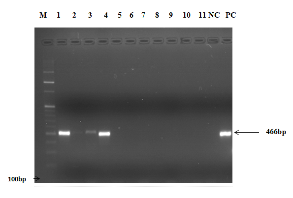
A standard One Step RT PCR Kit was used to test the pools for the presence of IBV by amplifying 466 bp hypervariable region of the S1 gene. Briefly,1X reaction mix, 2μl of forward primer 5’ XCE1(CACTGGTAATTTTTCAGATGG)3’ and 2 µl of reverse primer 3’XCE2 (CCTCTATAAACACCCTTGCA)5’, primer pair, and one step RT PCR 1μl enzyme mix were combined with 5μl of isolated RNA. 5x -Q solution 5 μl, 5x -RT PCR buffer in 5μl, 1μl of 10- mM dNTPs mix, and then 4 μl of RNA-free water were added to make a total volume of 25μl. The thermal protocol that was chosen went as follows: 50°C for 30 min, 95°C for 15 min, 1 cycle, followed by 94°C for 30 sec, 55°C for 1 min, and 72°C for 1 min for 35 cycles. A final extension step lasting five minutes at 72oC was also carried out. Then 1.5 percent agarose gel was used to mix 3 grams of agarose and 200 ml of 1xTAE buffer and kept for two minutes in the microwave then take out of the microwave for cooling. After cooling down to room temperature for 5 minutes, 4 µl of Gel Red was added in to the gel and left for 20 minutes. Then 10 µl PCR product that had 4µl of loading dye added to it was then loaded into each well along with a negative control and a positive control. Finally, 10µl of a molecular marker (ladder) starting at 100 bp was added. After that, electrophoresis was carried out for 1:20 hours at 120 volts, and the result was read under UV light. IBV results that are positive display 466 bp.
Data Analysis
The data collected from field level and laboratory investigation was coded into appropriate variable and entered in to a Microsoft Office Excel spreadsheet. The data was checked for errors entry, coded and then imported to SPSS for descriptive and further analyses. All statistical analyses were performed using SPSS version 26 software. Binary logistic regression was used to study associated risk factors.
Results
Serological Result with Associated Risk Factors
Indirect ELISA test was used to test serum samples from chickens in Gondar city and three districts in northwest Ethiopia for previous exposure to infectious bronchitis virus. 354 of the 384 blood sera tested for infectious bronchitis virus antibodies were positive. The prevalence of IB was found to be 92.19% (89%-94.6% with 95 CI %). Table 1 shows IB seroprevalence in Gondar city and three districts; with no significant difference between them (p > 0.05) .The prevalence of the disease was found to be 96.66% in west Dembya, 90.82% in East Dembya, and 85.39% in Gondar Zuria. A significantly higher prevalence was discovered in young chickens (95.83%) compared to adults (87.5%) with COR 3.286 (95 % CI 1.463-7.378) (p<0.05). The prevalence of IB was also higher in exotic breeds (94.57%) with COR 3.903(95 % CI 1.798-8.471) than local breeds (81.69 %) with a significant difference (P <0.05). The prevalence in females (92.7%) was higher than males (90.98 %) with COR 1.267(95 % CI 0.583-2.753) but the difference was not significant (p>0.05). Though the difference was not statistically significant (p>0.05) the prevalence in intensive farms was higher (93.39%) than the prevalence in extensive farms (90.69%) with COR 1.451(95 % CI.687-3.063). Higher Seroprevalence (93.75%) was observed in dual purpose chickens followed by layers with a prevalence of (92.7%) and broilers (90.98%) but the difference was not statistically significant (p>0.05) (Table 2).
| No. of Chickens Examined | No. of Positive | Prevalence | Univariable COR (95% CI) | p value | |
|---|---|---|---|---|---|
| Origin | |||||
| Gondar City | 108 | 103 | 95.37% | Reference | |
| West Dembya | 89 | 86 | 96.63% | 1.392(0.323-5.990) | 0.657 |
| East Dembya | 98 | 89 | 90.81% | 0.480(.155-1.485) | 0.203 |
| Gondar Zuria | 89 | 76 | 85.39% | 0.284(.097-.830) | 0.21 |
| Total | 384 | 354 | 92.19% (89%-94.7%) |
Table 1: Univariable Logistic Regression Analysis Result in Relation with Study Area.
COR: crude odds ratio; 95% CI: 95% confidence interval. P values <0.05 were statistically significant. Table 1: Univariable Logistic Regression Analysis Result in Relation with Study Area.
| Variables | No. of Chickens Examined | No. of Positive COR (95% CI) | Prevalence | Univariable | p value | Multivariable AOR (95% CI) | p value |
|---|---|---|---|---|---|---|---|
| Age | |||||||
| Young | 216 | 207 | 93.83% | 3.286(1.463-7.378) | 0.004 | 4.122(1.451-11.715) | 0.008 |
| Adult | 168 | 147 | 87.50% | Reference | |||
| Breed | |||||||
| Local | 71 | 58 | 81.69% | Reference | |||
| Exotic | 313 | 296 | 94.57% | 3.903(1.798-8.471) | 0.001 | 3.980(1.147-13.809) | 0.03 |
| Sex | |||||||
| Male | 122 | 111 | 90.98% | Reference | |||
| Female | 262 | 243 | 92.70% | 1.267(.583-2.753) | 0.549 | 1.665(.368-7.528) | 0.508 |
| Production System | |||||||
| Intensive | 212 | 198 | 93.39% | 1.451(.687-3.063) | 0.329 | 1.460(.518-4.117) | 0.475 |
| Extensive | 172 | 156 | 90.69% | Reference | |||
| Production Purpose | |||||||
| Broiler | 122 | 111 | 90.98% | Reference | |||
| Layer | 166 | 153 | 92.17% | 1.166(.504-2.7) | 0.719 | 1.366(.409-4.563) | 0.612 |
| Dual | 96 | 90 | 93.75% | 1.486 (.529-4.176) | 0.452 | 1.665(.368-7.528) | 0.508 |
Table 2: Univariable and Multivariable Logistic Regression Analysis Result in Relation with Associated Risk Factors.
COR: crude odds ratio; AOR: adjusted odds ratio; 95% CI: 95% confidence interval. P values <0.05 were statistically significant. Table 2: Univariable and Multivariable Logistic Regression Analysis Result in Relation with Associated Risk Factors.
RT-PCR Result
Samples that were injected in to SPF eggs were collected after 96 hours and passaged three times and were taken in to molecular biology laboratory for PCR analysis. The test was performed following the standard protocol. Among the
26 pooled samples tested 3 (11.54%) of them were positive during RT-PCR examination. Amplification of an expected DNA band (466 bp) from positive control as well as IBV positive swab samples indicated that the RT-PCR reaction has been performed correctly (Figure 2).
Discussion
A cross-sectional study was undertaken on serum samples from 384 chickens to determine seroprevalence of infectious bronchitis and 26 pooled tracheal swab samples for molecular detection of the virus in central Gondar zone. All of the chickens in this investigation were not vaccinated against infectious bronchitis virus (IBV). Based on the current investigation 92.19% of the selected chickens in the study area were positive for infectious bronchitis. Accordingly this study discovered the highest prevalence of infectious bronchitis as compared to the study carried out by Birhan, et al. [9] in northern Ethiopia who reported a seroprevalence of (24.6%). The result of this study was consistent with those of previous studies by Hutton, et al. [12], who discovered a prevalence of 94.5 % in Debrezeit, Ethiopia and Shiferaw, et al. [13] who reported a seroprevalence of 97.46 % in Bishoftu, Ethiopia. The current study was comparable to those from Nigeria by Owoade, et al. [14], Emikpe, et al. [15] and Mungadi, et al. [16], as well as those from Pakistan by Benazir, et al. [17] who found seroprevalence of 84%, 82.7%, 89%, and 84.40%, respectively.
The finding of this study was higher than the study conducted by Tesfaye, et al. [18] and Yonas, et al. [19] who found 70.6 % and 64.7 % seroprevalence of IBV in Sebeta, Hawassa, and Ada’a Districts of Ethiopia, respectively. The result of the current study was also higher than the studies by Barberis, et al. [20] from Algeria, Thekisoe, et al. [21] from South Africa and Kouakou, et al. [22] from Ivory Coast who reported a prevalence of 78.25%, 43% and 72% respectively. The finding of this study was also higher than the study by Das, et al. [23] and Bhuiyan, et al. [24] from Bangladesh who found a Prevalence of 79.38% and 59.30% respectively. Variations in agro climatic condition and management practices could explain the differences in seroprevalence. Furthermore, the difference in seroprevalence could be linked to a rise in IBV activity among chickens in the research area [25].
The prevalence varied significantly among chickens of different ages (p< 0.05). Young chickens (95.83 %) had the higher prevalence than adults (87.5 %). This result was in line with that of Mungadi, et al. [16], who observed that there was a significant difference between the prevalence in growing and adult chickens. This result was also in line with the study by Cavanagh, et al. [3] who claimed that while all age groups of chicks are susceptible to IBV; young chicks are more vulnerable than older ones. Additionally, as chickens get older, their resistance to diseases is stronger as well. The prevalence of the disease was higher in exotic breeds (94.57%) than the prevalence of in local breed (81.69 %). This might be connected to exotic breeds limited resistance to disease and other environmental stresses [26].
An investigation of the prevalence of IB in chicken farms during the study found that intensive farming had higher seroprevalence (93.39%) than its extensive equivalents (90.69%), although the difference in susceptibility was statistically insignificant (p > 0.005). The higher prevalence rate recorded in intensive farms in this study was consistent with reports from commercial poultry farms in Nigeria (91.3%) by Oyejide, et al. [27] in Jordan (92.9%) by Rousson, et al. [28] and in Ethiopia (94.5%) by Hutton, et al. [12]. The finding of this study was also consistent with the study by Shettima, et al. [29] who indicated an increase seroprevalence in intensive farming. This might be due to the various management protocol types. Given that contact between regions that produce poultry has been linked to the development and maintenance of infectious diseases, this may also be related to the existence of farms in nearby areas [30] and [31].
The current study found higher seroprevalence in layers (93.75%) and dual-purpose chickens (92.17%) as compared with broilers (90.98 %).This finding is in agreement with the study by Shettima, et al. [29] who explains that there was a significant difference in the prevalence among layers and broilers. This could be explained by layer and dual- purpose birds spending more time on the farm than other birds, which leads to their re-infection if there is no effective way to manage it [32]. This study was also in line with the explanation provided by Javed, et al. in [33] who found that the longer the chickens were exposed to the virus, the higher the seroprevalence of IB became.
Though the difference was not statistically significant a higher seroprevalence was obtained in females (92.7%) than in males (90.98 %). This outcome is consistent with the study of local chickens in live bird markets in Sokoto State, Nigeria, who found a higher prevalence in females than in males with insignificant difference. Due to differences in the activity of humoral- and cell-mediated immune responses between the sexes, this may be connected to the presence of a less effective immunological response in males than in females [16].
Isolation of the virus in embryonated eggs, followed by immunological identification of the isolates, is frequently used to make the diagnosis of infectious bronchitis (IB). This is a tedious process and necessitates the employment of particular polyclonal or monoclonal antibodies [34]. Additionally, some isolates can be combinations of various IBV types, which complicate the interpretation of serotyping results. There have been prior descriptions of reverse transcription PCR employing IBV, RNA isolated from allantoic fluid, and tracheal swabs. These methods have been proven to be quite effective for both IBV detection and IBV type identification [35].
As far as molecular detection of the virus is concerned this is the only study we are aware of that describes IBV molecular detection in the study area. The infection is believed to have started in the field since the farms did not use IBV vaccine, as was previously described in this article. This study was in agreement with the study by Hutton, et al. [12] in other research regions of Ethiopia, who reported molecular detection of IBV. The prevalence in this study was higher (11.54%) than the study by Tegegne, et al. [8] who reported a prevalence of 6% in Jimma zone, Ethiopia. This could be due to the difference in implementing biosecurity measures.
Conclusion and Recommendations
The result of the current study shows higher prevalence of the disease (92.19%) and RT-PCR result provides confirmation of the presence of IBV with a prevalence of (11.54%). Accordingly, this is the first molecular evidence that the study area harbors an infectious bronchitis virus.
Based on the above conclusion the following recommendations has been forwarded
- Major method of prevention for the disease like vaccination against the disease should be practiced.
- When new IBV strains appear in a particular area, an IB vaccination program is unsuccessful. In order to select an appropriate virus strain for vaccination, it has been advised to regularly monitor the IBV strains that are currently present in a certain geographic area.
- To lessen the impact of the disease, additional biosecurity measures must be put in place.
- In order to handle an efficient measurement system from the study area in specific and at a country level in general as a future study, sequencing and genotyping of IBV are recommended.
Acknowledgements
The authors extend their appreciation to Pan African university life and earth sciences institute for giving me the fund I needed to complete my project and to the National Veterinary Institute of Ethiopia for providing me with the wonderful opportunity to work in such well-equipped labs with knowledgeable professionals.
Authors’ Contributions
Conceptualization, methodology, formal analysis, investigation, resources, writing-original draft preparation, T.M. and M.B., O.O., data collection, T.M., G.E. and D.W. performing laboratory work T.M. G.D., K.B O.A and A.L.; Writing-review and editing, T.M. and M.B., O.O. All authors have read and agreed to the published version of the manuscript.
Funding
This project was funded by Pan African University Life and Earth Sciences Institute.
Availability of Data and Materials
This article contains all of the data that was created or analyzed throughout the investigation.
Consent for Publication
Not applicable.
Competing Interests
The authors declare that they have no conflicts of interest.
References
-
Roba YT, Bejura W, Teshome T, Tesfaye A, Mengesha N, et al. (2021) Serological Detection of Antibodies Against Gamma Coronavirus Infection in Scavenging Village Chickens in Ada’a District, Ethiopia. Econ Papers 33(4): 26106-26110.
-
Dolz R, Pujols J, Ordonez G, Porta R, Majo N (2008) Molecular epidemiology and evolution of avian infectious bronchitis virus in Spain over a fourteen year period. Virology 374(1): 50-59.
-
Cavanagh, Gelb (2008) Infectious Bronchitis Virus in: Diseases of Poultry. In: 12th (Edn.), Iowa State University Press, USA, pp: 117-130.
-
Laconi A, Van Beurden SJ, Berends AJ, Kühl A, Jansen CA, et al. (2018) Deletion of accessory genes 3a, 3b, 5a or 5b from avian coronavirus infectious bronchitis virus induces an attenuated phenotype both in vitro and in vivo. J Gen Virol 99(10): 1381-1390.
-
Hodgson T, Britton P, Cavanagh D (2006) Neither the RNA nor the Proteins of Open Reading Frames 3a and 3b of the Coronavirus Infectious Bronchitis Virus Are Essential for Replication. J Virol 80(1): 296-305.
-
Belouzard S, Millet JK, Licitra BN, Whittaker GR (2012) Mechanisms of corona virus cell entry mediated by the viral spike protein. Viruses 4(6): 1011-1033.
-
Casais R, Dove B, Cavanagh D, Britton P (2003) Recombinant avian infectious bronchitis virus expressing a heterologous spike gene demonstrates that the spike protein is a determinant of cell tropism. J Virol 77(16): 9084-9089.
-
Tegegne D, Deneke Y, Sori T, Abdurahaman M, Kebede M, et al. (2020) Molecular Epidemiology and Genotyping of Infectious Bronchitis Virus and Avian Metapneumovirus in Backyard and Commercial Chickens in Jimma Zone, Southwestern Ethiopia. Veterinary Sciences 7(4): 187.
-
Birhan M, Temesgen M, Shite A, Berhane N, Bitew M, et al. (2021) Seroprevalence and Associated Risk Factors of Infectious Bronchitis Virus in Chicken in Northwest Ethiopia. Scientific World Journal 13: 4553890.
-
(2021) Central Gondar zone livestock office.
-
Thrusfield M (2007) Veterinary Epidemiology. In: 3rd (Edn.), Blackwell Science Ltd: John Wiley & Sons, London, UK.
-
Hutton S, Bettridge J, Christley R, Habte T, Ganapathy K (2017) Detection of infectious bronchitis virus, avian metapneumovirus, Mycoplasma gallisepticum and Mycoplasma synoviae in poultry in Ethiopia. Trop Anim Health Prod 49(2): 317-322.
-
Shiferaw J, Dego T, Tefera M, Tamiru Y (2022) Seroprevalence of Infectious Bronchitis Virus in Broiler and Layer Farms of Central Ethiopia. BioMed Research International 2022: 1-5.
-
Owoade A, Ducatez MF, Muller CP (2006) Seroprevalence of avian influenza virus, infectious bronchitis virus, reovirus, avian pneumovirus, infectious laryngotracheatis virus, and avian leukosis virus in Nigerian poultry. Avian Diseases 50(2): 222-227.
-
Emikpe BO, Ohore OG, Olujonwo M, Akpavie SO (2010) Prevalence of antibodies to Infectious Bronchitis Virus (IBV) in chickens in southwestern Nigeria. African Journal of Microbiology Research 4(2): 92-95.
-
Mungadi HU, Mera U, Adamu Y, Musa U, Achi C (2015) Seroprevalence of Infectious Bronchitis Virus antibodies in local chickens in live bird markets in Sokoto State, Nigeria. Scientific Journal of Animal Science 4: 53-56.
-
Benazir K, Amjad AC, Nazeer HK, Hidayatullah SA, Nazar K, et al. (2018) Prevalence and clinical pathology caused by infectious bronchitis virus in poultry birds at Sindh, Pakistan. Journal of Veterinary Medicine and Animal Health 10(9): 231-236.
-
Tesfaye A, Kassa T, Mesfin S, Hailu H Teshale S, et al. (2019) Four serotypes of infectious bronchitis virus are widespread in unvaccinated backyard chicken and commercial farms in Ethiopia. World Journal of Veterinary Science 1(1001): 1-4.
-
Yonas TR, Wabi B, Tsedale T, Asamnew T, Naol M, et al. (2021) Serological detection of antibodies against gamma coronavirus infection in scavenging village chickens in Ada’a District, Ethiopia. Biomedical Journal of Scientific & Technical Research 33(4): 26106-26110.
-
Barberis A, Alloui N, Boudaoud A, Bennoune O, Ammar A (2018) Seroprevalence of infectious bronchitis virus in broiler farms in Batna, east Algeria. International Journal of Poultry Science 17(9): 418-422.
-
Thekisoe MMO, Mbati PA, Bisschop SPR (2003) Diseases of free-ranging chickens in the Qwa-Qwa district of the northeastern Free State province of South Africa. Journal of the South African Veterinary Association 74(1): 14-16.
-
Kouakou AV, Kouakou V, Kouakou C, Ducatez MF, Couacy- Hymann E, et al. (2015) Prevalence of Newcastle disease virus and infectious bronchitis virus in avian influenza negative birds from live bird markets and backyard and commercial farms in Ivory-Coast. Research in Veterinary Science 102: 83-88.
-
Das SK, Khan MSR, Das M (2009) Sero-prevalence of infectious bronchitis virus in chicken in Bangladesh. Bangladesh Journal of Veterinary Medicine 7(1): 249- 252.
-
Bhuiyan ZA, Giasuddin M, Khan Z (2018) Seroprevalence of infectious bronchitis virus in different types of chicken in Bangladesh. Asian Journal of Medical and Biological Research 4(1): 132-136.
-
Adene DF (2007) The cornerstones in poultry health and production: concepts, costs and the contemporary applications. University lecture, University of Ibadan, Ibadan, Nigeria.
-
Duguma R, Yami A, Dana N, Hassen HH, Esatu W (2005) Marek’s disease in local chicken strains of Ethiopia reared under confined management regime in central Ethiopia, Revue de Medecine Veterinaire 156(11): 541- 546.
-
Oyejide A, Demangam VL, Akinyemi JO (1988) Serological survey of antibodies to Infectious bronchitis in commercial and indigenous Nigerian chickens using ELISA. Bull. Anim Health Prod Afr 3: 259-262.
-
Roussan DA, Khawaldeh GY, Shaheen IA (2009) Infectious bronchitis virus in Jordanian chickens: seroprevalence and detection. Can Vet J 50(1): 77-80.
-
Shettima YM, El-Yuguda AD, Zanna MY, Abubakar MB, Hamisu TM, et al. (2016) Serological evidence of infectious bronchitis virus among some poultry species in Maiduguri, Nigeria. Alexandria Journal of Veterinary Sciences 51(1): 135-139.
-
Pohjola LK, Ek-Kommonen SC, Tammiranta NE, Kaukonen ES, Rossow LM, et al. (2014) Emergence of avian infectious bronchitis in a non-vaccinating country. Avian Pathology 43(3): 244-248.
-
Wang Y, Jiang Z, Jin Z, Tan H, Xu B (2013) Risk factors for infectious diseases in backyard poultry farms in the Poyang Lake area, China. PLoS One 8(6): e67366.
-
Ayim-Akonor M, Owusu-Ntumy DD, Ohene Asa HE, Oduro Abrokwa A, Hammond P (2018) Serological and molecular surveillance of infectious bronchitis virus infection in free-range chickens and Guinea fowls in the Ga-east district of Ghana. Journal of Veterinary Medicine 6: 4949580.
-
Javed T, Siddique M, Hameed A (1991) Persistence and morpho pathological studies on infectious bronchitis virus in chickens in Pakistan. Assiut Veterinary Medical Journal 25(1): 216-228.
-
Handberg KJ, Nielsen OL, Pedersen MW, Jorgensen PH (1999) Detection and strain differentiation of infectious bronchitis virus in tracheal tissues from experimentally infected chickens by reverse transcription-polymerase chain reaction. Comparison with an immunehistochemical technique. Avian Pathol 28(4): 327-335.
-
Cavanagh D (2007) Coronavirus, avian infectious bronchitis virus. Vet Res 38(2): 281-297.
- Epidemiological Surveillance and Rumors on Social Media
- Awareness and Treatment of Uncontrolled Hypertension in US Overweight/Obese Youths Aged 16–24 Years, NHANES 2021–2023
- Strengthening EPI Through Parental Engagement: Lessons from Dhaka Slums for IA-2030
- Mothers Knowledge of the Prevalence, Causes, Effects, Prevention and Control of Diarrhoea among Children in Ife East Local Government Area, Ile Ife, Osun State, Nigeria
- Covid-19 Reinfections Case Series from October 2023 to October 2024 in A General Medicine Office in Toledo (Spain)
- Water Contact! One Risk Too Many: Risk Factors Associated with Schistosoma haematobium infection in Osun State, Nigeria