The Advantages of Using Mesenchymal Stem Cells in the Treatment of Massive Burns-Case Report
Due to insufficient oxygenation of tissues, cellular and vascular destruction, burns show a different healing process from other wounds. Even if it affects only one organ, namely the skin, it can generate a systematic response in which multiple organs are affected, with high risk of infections and mortality. Therefore it represents a challenging pathology with the involvement of several specialties, which, depending on the way of production, the surface and the depth of the burn, requires special care, long hospitalizations, and multiple surgical interventions. Even if with the evolution of the medicine, the mortality of burns was also improved, the healing process is often unsatisfactory with negative consequences on the functional and physical aspects, which reduce the quality of the patient’s life. This work paper aims to present a different approach in the management of a II-III degree burn, of approximately 20% body surface, at the level of the posterior thorax and the left upper extremity, using mesenchymal stem cells. The procedure involved the collection of skin samples with a diameter of 5 mm each one, match them with saline solution, introduce them in a special device, disaggregate them and inject the resulting suspension solution rich in mesenchymal stem cells into the dermis with the advantage that it can skip over the excisional debridement stage and decrease significantly large skin graft donor areas of the standard approach, and other possible surgical interventions subsequent.
Oproiu AM¹,²* and Grigore A²
¹Department of Plastic Surgery, University of Medicine and Pharmacy, Romania
²Emergency University Hospital of Bucharest, Romania
organs are affected, with high risk of infections and mortality.
patient’s life.
Keywords: Burns; Wounds; Skin; Healing Process; Mesenchymal Stem Cells
Introduction
In USA, approximately 1, 2 million new cases are reported annually, of which 40,000 require hospitalization and 5000 die because of complications, most of them due to sepsis, infections or inhalation [1, 2, 3, 4, 5, 6, 7].
In order to properly manage the burn cases, it is important to evaluate the burn injury correctly and to know the healing process which is different from the other wounds (Table 1).
Even if the phases are the same in every wound, the duration of each step is different in burns [8, 9, 10].
| Inflammatory phase | 1. vascular response |
| Inflammatory phase | vasodilation |
| Inflammatory phase | extravasation of fluid |
| Inflammatory phase | massive burns require fluid replacement because of massive extravasation of plasma due to increased capillary permeability |
| Inflammatory phase | 2. cellular response |
| Inflammatory phase | migration of the neutrophils and monocytes at the site of inflammation |
| Inflammatory phase | later, macrophages replaced the neutrophils |
| Inflammatory phase | role in the phagocytosis and cleaning of death tissue and toxins released by the burn injury |
| Proliferative phase | starts few hours after injury and covers the wound within 5-7 days in partial thickness |
| Proliferative phase | is delayed in deep burns |
| Remodeling phase | the extracellular matrix produce the scar formation |
| Remodeling phase | the process could take years in the deep burn |
Table 1: Burn healing process.
If in the no burn wounds the tissue is fed by underlying blood supplies, the burn injury is characterized by a zone of coagulate necrosis, where the tissue is not sufficiently oxygenated to response to normal healing process. Surrounding this necrosis is a zone of stasis in which is decreased the perfusion of the tissues. Also high capillary permeability, local inflammatory reaction, vasodilation and edema distinguish burns from other trauma injury [11, 12, 13].
An important part of diagnosis is represented by identifying the causative agents (physical, thermal, electrical, radiation, laser, chemical burns) and the depth of burns (Table 2) because local and systematic management is different according to the way of production, the surface and the depth of the burns [14, 15, 16].
| First Degree Burn (epithelial burns) | Skin is erytomatic without vesication |
| First Degree Burn (epithelial burns) | -very painful to the touch |
| First Degree Burn (epithelial burns) | -brisk capillary refill |
| Second-Degree Burns | SUPERFICIAL- vesication and inflammation is seen in the skin |
| Second-Degree Burns | - only papillary dermis is involved |
| Second-Degree Burns | - very painful to the touch |
| Second-Degree Burns | - brisk capillary refill |
| Second-Degree Burns | DEEP- eschar formation |
| Second-Degree Burns | - deep reticular dermis is involved |
| Third-Degree Deep | Full thickness |
| Third-Degree Deep | Presence of eschar |
| Third-Degree Deep | Markers: pain(high to none), color( pink/red to white/brown), capillary refill(brisk to none) |
Table 2: First Degree Burn.
Severe burns, representing by over 20% total body surface area, are characterized by a systematic inflammatory response with the damage of immune system, gastrointestinal system, muscle and hyper metabolism [17]. They require specialized burn centers, special nutrition, control of pain, infection prevention, and rehabilitation is an important part of this process.
If in the first and second superficial degree burns, the healing is by primary intention, in the deep burns the healing is by secondary intention and requires surgical treatments, often multiple surgical interventions, with risk of contraction, hypertrophic scar, late deformities, delaying the patient return to normal.
Regenerative medicine represents a promising approach in the wound healing, but to be able to apply is very important to understand the healing process.
Recent, the medicine paid attention on stem cell because of their capacity to restore damaged tissue. Mesenchymal stem cells are known for their property to be used as autologous, be transplanted for reparing and regeneration the tissue in the clinical practice. It is known that the dermis and the adipose tissue are sources of stem cells [18, 19, 20].
Adipose stem cells have proven to be a superior source of stem cells, even than bone marrow, because of higher quantities and numbers, protective and/or supportive factors which play a role in reducing apoptosis, fibrosis, and inflammation, and also because of the production of a larger number of growth factors [21].
Materials and Methods
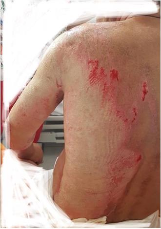
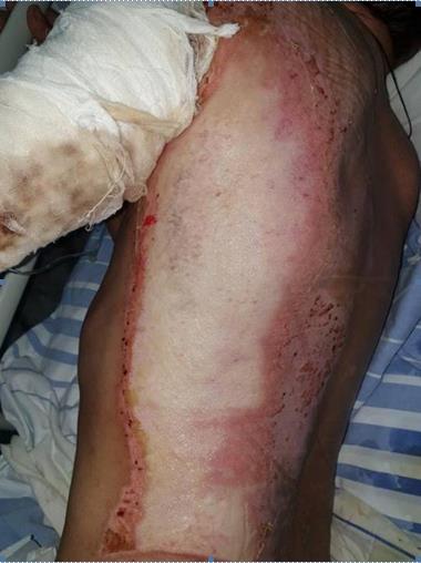
We present a case report of a 51 old male with severe burns, 20% body surface through flammable liquid, grade II-III, which involved left upper extremity and posterior thorax (Figure 1), from whom we applied a different approach from the standard. With the help of an innovative technology called Rigenera, we obtained dermal micro grafts after the tissue was disaggregated by mechanical movements, rich in mesenchymal stem cells.
The technique consists in harvesting the skin using a biopsy punch, introducing it into a special device and disaggregates it by mechanical movements, the result being a micro graft product rich in mesenchymal stem cells, which is injected in the lesion.
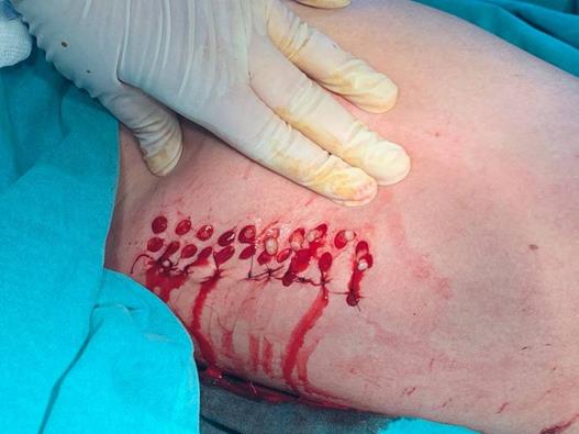
The steps we followed consisted in • Choice of donor site inner anterior right thigh.
• Shaving the donor site gently to remove the epidermis until it starts a superficial bleeding.
• Harvest the tissue with a 5 mm punch biopsy according to the size of lesion, considering that 1mm² of the collected tissue was expected to regenerate 2cm of injury (Figure 2).
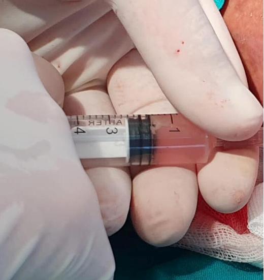
• Introduce the samples in the special device (2 samples/ session) and add 3 ml of saline solution.
• Connecting the device to the machine and letting work for 2 minutes to provide a mechanical disaggregation into a suspension which contains autologous dermal micro graft.
• Introduce the suspension in a sterile syringe.
• Inject the solution in the burn lesion, at a depth of 4mm
(Figure 3).
Results
Clinical observation has documented diminished healing process with good aesthetic results, a better control of pain, without complications and a decreased in hospitalization days up to 50%.
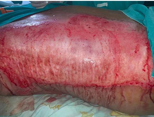
The immediately postoperatively result was impressive. The eschar zone coloured from white to red, was bleeding and marked by streches of mesenchymal stem cell suspension, (Figure 4) being an important tool in the prevent of the power of this suspension.
Also, we noticed that the healing had begun from the periphery of the lesion (Figures 5&6).
The skin is the largest organ of the body with the role of barrier, thermoregulation, avoidance of liquid loss, sensitive, production of vitamin D and any injury disrupts these functions. Using the Rigenera technology, we have been able to isolate mesenchymal stem cells and also a suspension riched in progenitor cells and growth factors easy to apply at the level of injury, which had the effect to accelerate the healing process, avoiding a contracting scar and restoring all the functions of the skin.
Studies showed that dermal micro grafts collected by Rigenera device present mesenchymal stem cell markers such as CD 73, CD 90, CD 105, and CD 34 [22]. Also, it had been demonstrated that approximately 70-80% of the isolated cells are mesenchymal stem cells [23].
During hospitalization, the patient had hyperproteic and hypercaloric diet, and usual scheme of anti biotherapy to avoid infections and didn`t have any complication.
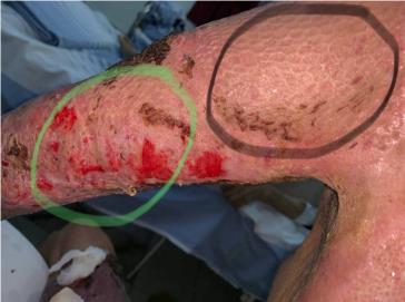
To compare the time and the way of healing between standard method (requires excisional debridement and skin graft) versus Rigenera technique, we chose the standard procedure at the upper extremity and in the third upper arm we also injected the suspension with progenitor cells (Figure 7).
The standard method starts with the excision of necrotic tissue but it has the following disadvantages:
- It requires also healthy tissue excision, the excision should be done until the tissue is bleending,
- It needs large donor sites,
- The risk that the skin graft do not adhere,
- Risk of progressive tissue damage due to unexcused partal-thickness burns and the zone of stasis in excised full-thickness burns.
So, usually, a “wait and see” approach is better to avoid health tissue removal but the risk or infections is increased.
In order to avoid large skin donor areas, rhis technique allowed to collect 30 samples of skin with the diameter of 5mm each one for cover the posterior thorax and 1/3 upper arm. For the rest of the upper extremity, we decided to practice the excision of necrotic tissue and skin graft, because of a more accurate evaluation of those methods. We observed that with the help of micro grafts, the healing process is accelerated and the risk of skin graft to not adhere doesn’t exist.
Another aspect that we managed very well was the dressing. The burn patients require special daily dressing change under sedation due to high pain. The protocol of mesenchymal stem cells requires that the dressings to be changed at the 3 days, not daily, and it wasn`t necessary the sedation. The pain was controlled very well. A big problem of the burns is the pain. The pain is very hard to control, even with a special scheme of pain relievers.
Even if the immunomodulatory function of the mesenchymal stem cells is uncleared, studies promote tissue repair, because of the production of the multiple growth factors, cytokines, collagens and matrix metalloproteinase, promote migrations of keratinocytes and also the differentiation and angiogenesis. The advantage of mesenchymal stem cells is that it can be isolated from a variety of tissue, including not only amniotic membrane, umbilical cord, cord blood, but also bone marrow, adipose tissue, hair follicle dermal papilla and sheath and they enhance process of healing, takeing part in all the phases of healing.
Conclusions
Massive burns represent a different injury which requires many resources, both from the patient and from medical staff and hospital too, with a delayed healing process.
The mesenchymal stem cells represent a promising approach in the wound healing and in the regenerative medicine, with an imunomodulatory function uncompletely elicited, but with many possible clinical applications.
Regarding burns, adipose stem cells are a very accessible source of mesenchymal stem cells, which have demonstrated their role in accelerating the process of healing and minimalizing the complications.
Conflict of Interests
The authors didn’t have any conflict of interests and they had equal contributions.
- Research Progress of Induced Pluripotent Stem Cells and Their Clinical Application Prospects
- Nishan Al-Kamal is the Starting Point of A Feminist Scientist
- Current Concepts and Future Perspectives of Stem Cell Therapy in Peripheral Arterial Disease
- Stem Cell and Oxidative Stress-Inflammation Cycle
- Adipose Derived Mesenchymal Stem Cells Origin, Characteristics and Promises
- Mitochondria Targeted Antioxidants can Improve In Vitro Embryo Production in Buffalo