Successful Surgical Intervention for a Congenital Perineomelia Associated with Atresia Ani and Rectovaginal Fistula in a Cow-Calf
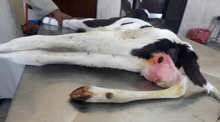
Seven days old cow-calf was admitted to the Veterinary Teaching Hospital, Faculty of Veterinary Medicine, Assiut University, Assiut, Egypt, with congenital malformations. Clinical examination revealed that the calf had a supernumerary ectopic limb at the perineum (perineomelia), atresia ani, and a rectovaginal fistula. The accessory limb was excised from the perineum. Anal opening was created at the base of the tail. Therectovaginal fistula was obliterated transvaginal.
Introduction
In humans and animals, limbs' anomalies are encountered frequently [1, 2]. Polymelia was defined as the congenital malformation characterized by a supernumerary limb (s) at different body regions [3, 4]. Polymelia in symmetrical twins of cattle characterized by the presence of six or eight legs, while in heterotopicpolymelia there are one or two supernumerary limb(s) [5, 6, 7, 8, 9]. Polymelia is classified according to its point of attachment to the body as: notomelia (the back), cephalomelia (the head), thoracomelia (the thorax), and dipygus or pygomelia (the pelvis) [2, 9, 10]. Perineomelia is an extra ectopic limb at the perineum region [11]. Polymelia may develop as a forelimb (cephalomelia, notomelia, and thoracomelia) or as a hind limb (usually as pygomelia). The supernumerary limb may be normal or syndactyl. Usually, these limbs are devoid of muscles and have fused joints [2, 12]. Atresia ani is the congenital abnormality of gastrointestinal tract, characterized bythe absence of anus [13]. Rectovaginal fistula described an abnormal connection between the rectum and vagina. Faeces usually voided by the latter due to imperforate anus [14]. Such congenital deformities present at birth usually result from hereditary factors, environmental conditions, or interactions between the animal's genetic makeup and the environment in which it lives [15]. The present case report presents details of clinical, radio graphical, growth anatomical findings, as well as surgical management of perineomelia, atresia ani, and rectovaginal fistula in a cow-calf.
Case History and Clinical Presentation
Sevendays old, 57 kg body weight cow-calf with a supernumerary ectopic limb at the perineum region (perineomelia) was admitted to the Veterinary Teaching Hospital, Faculty of Veterinary Medicine, Assiut University, Assiut, Egypt. The clinical examination revealed that the accessory limb was undeveloped, didn’t reach to the ground level, and had a point of attachment to the right of the vulvar opening. The limb was loosely attached to the perineal tissues, without a true articulation. On close examination, the calf had also an atresia ani. Bulging at the base of the tail was developed by compressing the abdomen. This was associated with a presence of arectovaginal fistula. It was recognized through voidance of faeces from the vulvar opening. By physical examination transvaginal, there was a communication between the vagina and the rectum, (Figure 1). Nevertheless, the physiological parameters of the calf, including the respiratory and pulse rates (22 breaths/min, 63 beats/min, respectively), and hydration were within normal. The calf walked and suckled normally. The case was decided for surgical intervention.
Surgical Intervention
The calf was sedated by 0.05 mg/kg body weight 2% xylazine HCl (Xyla-Ject, ADWIA Co., SAE, Egypt), intramuscular (IM). The operation was done under theeffect of the caudal epidural anesthesia, at the first intercoccygeal space (Co1 - Co2), using 0.2 mg/kg body weight of 2% lidocaine HCl (Dibucaine, Sigma-Tec Pharmaceutical Industry Co., Egypt), under a strict aseptic condition. The calf was positioned laterally on its right side. The site of the operation was prepared for aseptic surgery. A circumcisional incision was done in the skin,subcutaneous, and fascia, 5 cm from the base of the limb. Excision of the limb from the perineal tissue was performed through a process of sharp dissection. Blood vessels were ligated by chromic cat gut No. zero. The resultant dead space was closed in several layers as in routine closure, using chromic cat gut No. one. The skin was closed in aninterrupted horizontal mattress, using silk No. one. For correction of the atresia ani, a circular excision of the skin was made over the anal bulging at the base of the tail. The rectum was explored after dissection of the perineal muscles. The rectum’s blind end was grasped to the anal sphincter, and then opened. The rectal mucosa was fixed to the circular skin edge by interrupted sutures, using silk No. one, creating a permanent anal orifice. The rectovaginal fistula was closed transvaginal by purse- string suture along its length [16] (Figure 2).
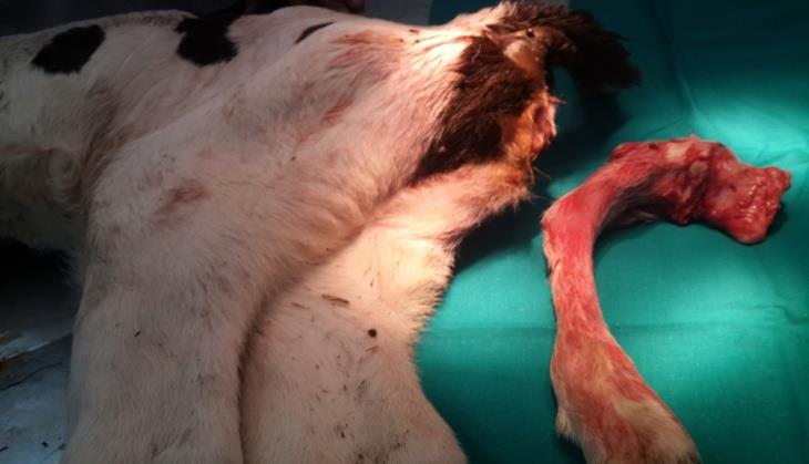
Figure 2: The calf after surgical excision of the accessory limb, the creation of an anal opening, and the closure of the rectovaginal fistula. Post-operatively, the calf was administered 8 mg/kg body weight procaine penicillin and 10 mg/kg body weight dihydrostreptomycin sulphate (Pen & Strep, each ml contains: procaine benzylpenicillin 200 mg and dihydrostreptomycin sulphate equivalent to dihydrostreptomycin 200 mg, Norbrook Laboratories Limited Newry, Co. Down, Northen Ireland), IM, once daily for three consecutive days. Radio graphical as well as, growth anatomical examinations were conducted on the excised supernumerary limb.
Radiographic Examination
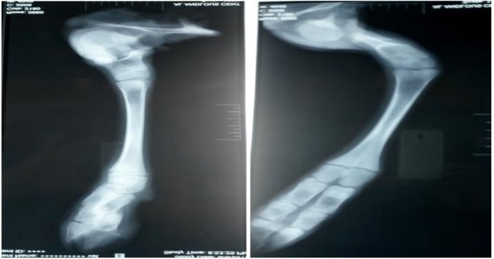
Two views were obtained to the accessory limb; dorsoventral and lateromedial views. The limb presented itself as a pelvic limb. The radiograph of the limb was not informative enough. There was an undeveloped pelvic bone. A short femur with a large rounded distal end was identified. It was attached to a triangular rudimentary tibia-like structure. There was a well-developed calcaneal bone. The metatarsus and the digit were within normal, except for the absence of sesamoid bones (proximal and distal bones). The tarsal joint, and joints distal to it, all were fused. There was a distinct angulation to the limb at the tarsal region. Open growth plates were noted at the distal end of the metatarsus, (Figure 3). This led us to perform growth anatomical examination to the limb for more anatomical details.
Growth Anatomy of the Limb
Growth anatomical examination revealed that the accessory limb had an undeveloped pelvic bone, which was consisted of; a wing of the ilium (Figure 5A), a body of the ilium (Figure 5B), a body of the ischium (Figure 5C), and an acetabulum (Figure 5D). The femur bone was underdeveloped, short, and compact. It had a rudimentary head of the femur (Figure 5E) and a trochanter (Figure 5F) proximally, and an enlarged end distally (Figure 5G). The head of the femur was attached tightly to the pelvic bone at the acetabulum by a fibrous tissue, (Figure 4). A conical, misshapen bony structure, tended to be the articular proximal end of the tibia, was attached firmly to the distal end of the femur (Figure5H). The patella or fibula was not found. Tarsal bones were fused and expressed by a one, large, well developed calcaneal bone (Figure 5I). The metatarsus and the digit appeared to be normal-like. All joints were rigid (arthrogryposis), (Figures 4 & 5).
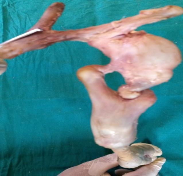
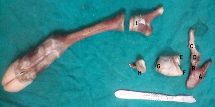
Figure 5: Growth anatomy of the accessory limb: Undeveloped pelvic bone (a wing of the ilium (A), a body of the ilium (B), a body of the ischium (C), with an acetabulum (D). Short femur with a small undeveloped head (E) and trochanter (F) proximally, and enlarged end distally (G). Rudimentary proximal end of the tibia (H). Enlarged calcaneal bone (I). Normal metatarsus and phalanges with tightly fixed joints.
Discussion
A full description; clinically, radio graphically, and growth anatomical features have been given to the congenital perineomelia associated with atresia ani and rectovaginal fistula in the cow-calf. Moreover, the surgical corrections for such multiple malformations in one animal have been detailed in the sequence enabling the veterinarians from the handling of such cases in a correct manner without any confusion. The incidence of congenital malformations in animal livestock ranges from 2% to 3.5% of all births. The musculoskeletal system associated defects constitute about 24% of all malformations [8]. Congenital malformations represent a real threating for animal welfare. These malformations reduce the both; productivity and reproductivity of animal farms, causing severe economic losses. Congenital defects may be due to gene mutations inherited from the parents or caused by new mutations or changes to the DNA [17]. To the best of our knowledge, the only case of congenital perineomelia was reported by Murondoti and Busayi in a Mashona calf [11]. In the present case, the perineomelia was characterized by undeveloped bones, absence, and fusion of some bones, and fixed joints. Similar findings were recorded by Ramadan et al., Chong- sup et al. and Muirhead, et al. [10, 18, 19]. Also, perineomelia in this case, was associated with a gastrointestinal tract defect; in form of an atresia ani, as well as, urogenital tract defect; in form of a rectovaginal fistula. This was in accordance with Murondoti and Busayi, who reported that polymelia may be associated with other body malformations [11]. The limb is developed from an outgrowth of a lateral body wall known as limb bud that consists mainly of mesenchymal cells. Early, these mesenchymal cells developed solely from the mesoderm, from which, the bones and connective tissue of the limb will arise. Later, mesenchymal cells from differentiating somites (myotomes) migrate into the limb bud giving rise to skeletal muscles.Any disruption in these phases may result in such limb abnormalities [20].
Conclusion
The surgical correction for such cases of congenital defects plays a major role in minimizing the economic losses, by rescuing and putting these animals for production in animal farms again. Serious steps should be taken by animal research centers, all over the world to minimize the environmental pollution, which represents the main cause of genetic mutation in such animals, contributing to developing of different congenital anomalies.
References
-
Leipold HW, Dennis SM (1987) Cause, nature, effect and diagnosis of bovine congenital defects. Irish Veterinary News 9: 11-19.
-
Denholm L, Martin L, Denman A (2011) Polymelia (supernumerary limbs) in angus calves. Flock & Herd.
-
Hiraga T, Abe M, Iwasa K, Takehana K, Tetsuka M (1989) Seven-legged calf –dipygus with an extra foreleg at the pelvic region. Nippon Juigaku Zasshi 51(5): 1011-1015.
-
Fourie SL (1990) Congenital supernumerary ectopic limbs in a Brahman-cross calf. Journal of the South African Veterinary Association 61(2): 68-70.
-
Hossain MA, Sen MM, Rahman MA (1980) Teratology– new born calf with a supernumerary limb and atresia ani (case report). Veterinary Medicine Review 2: 178-179.
-
Singh P, Sharma DK, Singh S, Bhel SM, Chandna IS (1989) Polymelia with atresia ani in a calf. The Indian journal of Veterinary & Surgery 10: 62-65.
-
Kim C, S Yeo, Cho G, Lee J, Choi MC (2001) Polymelia with two extra forelimbs at the right scapular region in a male Korean native calf. The Journal of Veterinary Medical Science 63(10): 1161-1164.
-
Leipold HW, Huston K, Dennis SM (1983) Bovine congenital defects. Advances in Veterinary Science and Comparative Medicine 27: 197-271.
-
Rahman MM, Khan MS, Biswas D, Sutradhar BC, Saifuddin AK (2006) Pygomelia or supernumerary limbs in a crossbred calf. Journal of Veterinary Science 7(3): 303-305.
-
Chong-sup KIM, Seong-chan YEON, Gyu-hyen CHO, Joung-hwan LEE, Min-cheol CHOI (2001) Polymelia with two extra forelimbs at the right scapular region in a male Korean native calf. J Vet Med Sci 63(10): 1161-1164.
-
Murondoti A, Busayi RM (2001) Perineomelia, polydactyly and other malformations in a Mashona calf. Veterinary Record 148(16): 512-513.
-
Noden M, De Lahunta A (1985) Limb developmet. In The Embryology of Domestic Animals: Developmental Mechanisms and Malformations. London, Williams and Wilkins, pp: 203-207.
-
Singh J, Singh AP, Patil DB (1993) From Digestive system. In Ruminant Surgery 1st edition, Edited by Tyagi RPS and Singh J, CBS Publishers, New Delhi.
-
Oehme FW, Prier JE (1974) Text book of large animal surgery: Williams & Wilkins, Baltimore/ London, pp: 425-509.
-
Greene HJ, Leipold HW, Huston K, Noordsy JL, Dennis SM (1973) Congenital defects in cattle. Irish Veterinary Journal 27: 37-44.
-
Rahal SC, Vicente CS, Mortari AC, Mamprim MJ, Caporalli EHG (2007) Rectovaginal fistula with anal atresia in 5 dogs. Can Vet J 48(8): 827-830.
-
Albarella S, Ciotola F, D’Anza E, Coletta A, Zicarelli L (2017) Congenital malformations in river buffalo (Bubalusbubalis). Animals 7(2): 9.
-
Ramadan RO, Abdin-Bey MR, Al-Holaibi AK (1988) Notomelia in goata and a calf. Pakistan Vet J 18(1): 47-49.
-
Muirhead TL, Pack L, Radtke CL (2014) Unilateral notomelia in a newborn Holstein calf. Can Vet J 55(7): 659-662.
-
Talamillo A, Bastida MF, Fernandez-Teran M, Ros MA (2005) The developing limb and the control of the number of digits. Clin Genet 67(2): 143-153.
- Psychogenic Erectile Dysfunction in Late Adulthood: A Case Report on Clinical Intervention and Intimacy Restoration
- Clinical Trials on COVID-19 in 2025: A New Chapter in Global Health Research
- Innovations and Challenges in Contemporary Medical Clinical Trials: An Editorial Perspective
- Innovations and Challenges in Contemporary Medical Clinical Trials: A Critical Perspective
- Reimagining Clinical Trials: The Power of Continuous Feedback from Medical Reports
- Factors Influencing Brain Drain: Perspectives from a Medical School in Turkey