Haemato Biochemical Parameters of Goats Experimentally Infected with Trichophyton Verrucosum
This study was designed to investigate the ability of Nubian goat to acquire Trichophyton verrucosum infection. Twenty goats aged 9-12 months were used in this study. Sixteen goats have been successfully infected with the isolated Trichophyton verrucosum isolated from camels naturally infected with dermatophytosis and four goats were used as control. All infected goats showed similar lesions, which were characterized by circumscribed crusty hairless lesions. Haematologically there was significant difference in total white blood cell count in the infected goats; however, no significant alteration was seen in the packed cell volume, haemoglobin, red blood cells count, mean corpuscular volume, mean corpuscular haemoglobin and mean corpuscular haemoglobin concentration. Biochemically, except uric acid, no significant alteration was observed in total serum protein, albumin, globulin, urea, creatinine, sodium and potassium.
Wisal G Abdalla* and Salim MO
Central veterinary Research Laboratories Khartoum, Sudan
Laboratories Khartoum, P.O. Box 8067(Alamarat), Sudan, Tel: +249912614294; E-
mail: wisalgafar8@gmail.com Keywords: Haematological; Biochemical; Experimental infection; Trichophyton verrucosum
Introduction
Dermatophytosis, also called tinea, ringworm, or superficial fungal infection, is one of the most common skin diseases caused by dermatophytes of the genera Microsporum, Trichophyton and Epidermophyton, which show great affinity for the skin and its annexes. It is an infectious disease of animals that causes great economic losses [1]. A lesion observed on affected animals was alopecia and/or circumscribed grayish-white, crusty, raised lesions in head, neck and chest area [2]. Severity and spread of dermatophytosis depend on the immune system of the host, the causative agent’s pathogenicity and environmental factors discussed by Dermatophyte infection may range from mild to severe [3, 4]. Significant decrease in haemoglobin content and packed cell volume percentage due to ringworm was reported by Pasa S, et al. [5]. Serum zinc and vitamin A concentrations were found to be significantly lower in calves with dermatophytosis than those of healthy controls [6]. Concentration of zinc and selenium were found to be lower in cows infected with dermatophytes than healthy ones but the differences were not significant [7].
This study was designed to study experimental infection of Nubian goats with different species of Trichophyton locally isolated from camels and to investigate the haematological and biochemical changes occur that may due to the infection.
Materials and Methods
Animals
Twenty goats (10 males, 10 females) 9-12 months old which were clinically free from dermatophytosis, from Omdurman local market were purchased for the purpose of this experiment. They were ear-tagged, kept in clean disinfected pens and fed dry sorghum hay, concentrates in addition to water.
Skin scraping and Inoculums Preparation
Skin scrapings from infected camels were collected and divided to two portion one used for direct examination with 20% KOH and the second portion cultured on Sabouraud's dextrose plates chloramphenicol and cyclohexamide incubated at 27ºC and 37Cº were done according to AbdelKarim S, et al. [8]. Whole culture of Trichophyton verrucosum, recovered from naturally infected camels with dermatophytosis was used as inoculums for infection of goats.
Infection Procedure
Three areas of each goat (neck, shoulder and hind quarter) were shaved and scarified by using sterile shaving blades. The inoculum was rubbed on the shaved areas of sixteen goats as discussed by Schalm OW, et al. [9]. Four goats were kept as control. In these animals the prepared areas were left without infection. The goats were observed closely for the development of lesions.
Blood Samples
Experimental animals were bled weekly for 12 weeks; whole blood was collected from jugular vein into heparinised vacutainers. Different blood parameters were determined according to Weichselbanm TE [10]. Blood samples in plain vacutainer were allowed to clot at room temperature overnight; serum was separated into clean vials and stored at -20ºC till used for biochemical analysis.
Biochemical Analysis
Total protein was done according to, urea was determined according to and creatinine and uric acid were measured using commercial kits (Spinraect Kinetic Jaffe and Bio Systems) respectively [11, 12].
Statistical Analysis
Statistical analysis was performed using SPSS. Values were expressed as mean ± standard deviation. The One way-ANOVA test was used to compare the parameters between the groups. The significance level was set at P <0.05.
Results
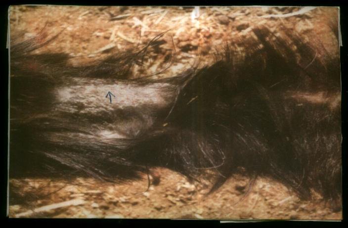
The sixteen experimentally goats age 9-12 months were successfully infected with T. verrucosum. The lesions observed through the experiment of the infected goats were characterized by circumscribed crusty hairless lesion of 1.5-2 cm in diameter surrounded by a zone of minute vesicles. The lesions increased in size and showed profuse exudation matting the hair together. The exudate dried and the annular lesions became covered with thin flakes of light brown coloured scab, which later became thick and brown in colour. Figure 1, the incubation period was 19-21 days; however, the four control goats group didn’t show any lesions.
Blood Analysis
There was significant difference in total white blood cell in the infected goats, however, no significant alteration was seen in the packed cell volume, haemoglobin, red blood cells count, mean corpuscular volume, mean corpuscular haemoglobin and mean corpuscular haemoglobin concentration (Tables 1 and 2).
Biochemical Analysis
Except uric acid, no significant alteration was observed in total serum protein, albumin, globulin, urea, creatinine, sodium and potassium (Tables 3-5).
| PCV% | Hb (g/l) | RBCs (x10^6) | WBCs (x10^3) | |
|---|---|---|---|---|
| 1 | 27±0.82 | 7.35±0.3 | 12.84 ± 0.69 | 10.7 ±0.108 |
| 2 | 26±1.4 | 7.65±0.25 | 12.45±0.41 | 10.76 ±206 |
| 3 | 27.75±0.96 | 8.1±0.42 | 13.26±1.3 | 9.438 ±966 |
| 4 | 26.5±1.29 | 7.75±0.19 | 13.8±0.59 | 8.45 ±0.091 |
| 5 | 27±.82 | 8.1±0.35 | 13.86±2.16 | 8.51 ±0.063 |
| 6 | 26.5±0.58 | 7.6±0.43 | 12.74±0.46 | 8.38 ±.0150 |
| 7 | 27±0.82 | 7.7±0.35 | 13.68±1.3 | 8.86 ±.0450 |
| 8 | 26.75±0.96 | 7.85±0.44 | 12.8±1.87 | 9 ±.0356 |
| 9 | 27±0.82 | 7.65±0.41 | 11.79±1.44 | 8.45 ±.0208 |
| 10 | 27.25±0.5 | 7.85±0.6 | 12.76±1.88 | 8.64 ±0.180 |
| 11 | 27.25±0.96 | 8±0.49 | 11.21±1.11 | 8.78 ±0.132 |
| 12 | 26.75±0.96 | 7.9±0.48 | 12.99±1.44 | 8.78 ±0.126 |
| Control | 26.58±0.67 | 7.73±0.39 | 12.34±1.94 | 10.66 ±0.156 |
| Total mean+ standard | 26.83±0.89 | 7.78±0.42 | 12.75±1.52 | 8.98 ±0.981 |
| P value | 0.44 | 0.43 | 0.39 | 0 |
| MCV (fL) | MCH (pg) | MCHC(g/l) | |
|---|---|---|---|
| 1 | 21.11±1.75 | 5.74±0.42 | 27.24±1.42 |
| 2 | 20.88±0.87 | 6.16±0.37 | 29.5±2.14 |
| 3 | 21.08±2.08 | 6.16±0.68 | 29.1±1.39 |
| 4 | 19.21±0.76 | 5.63±0.22 | 29.31±1.89 |
| 5 | 19.85±3.21 | 5.93±0.8 | 30.04±1.99 |
| 6 | 20.81±0.68 | 5.98±0.43 | 28.68±1.55 |
| 7 | 19.84±1.35 | 5.66±0.5 | 28.55±2.31 |
| 8 | 21.3±3.78 | 6.27±1.22 | 29.35±1.27 |
| 9 | 23.18±3.08 | 6.53±0.53 | 28.39±2.4 |
| 10 | 21.73±3.46 | 6.23±0.8 | 28.81±2.18 |
| 11 | 24.54±3.04 | 7.16±0.34 | 29.41±2.59 |
| 12 | 20.81±2.73 | 6.16±0.95 | 29.53±1.38 |
| Control | 21.92±3.1 | 6.41±1.04 | 29.1±1.55 |
| Total mean+ standard | 21.34±3.69 | 6.19±0.79 | 29.01±1.76 |
| P value | 0.32 | 0.31 | 0.85 |
Table 2: Blood indices of goats experimentally infected with Trichophyton verrucosum.
| Weeks | Na+ (mMol/l) | K+ (mMol/l) |
| 1 | 136±5.16 | 3.45±0.78 |
| 2 | 135.75±3.1 | 3.95±0.13 |
| 3 | 140.5±3.7 | 3.6±0.78 |
| 4 | 135.75±3.1 | 3.9±0.45 |
| 5 | 135.25±4.72 | 4.43±0.26 |
| 6 | 134.5±2.08 | 4.4±0.42 |
| 7 | 137.25±1.5 | 3.53±0.43 |
| 8 | 140.5±1.29 | 3.83±0.57 |
| 9 | 135.5±6.19 | 4.1±0.42 |
| 10 | 134.25±2.06 | 3.65±0.57 |
| 11 | 139±2.58 | 3.75±0.19 |
| 12 | 136.75±2.22 | 3.53±0.46 |
| Control | 136±2.17 | 3.68±0.64 |
| Total mean+ standard division | 136.6±3.43 | 3.8±0.56 |
| P value | 0.1 | 0.17 |
Table 3: Electrolytes of goats experimentally infected with Trichophyton verrucosum.
| weeks | Total protein (g/l) | Albumin (g/l) | Globulin (g/l) |
| 1 | 6.5±0.21 | 2.7±0.28 | 3.78±0.13 |
| 2 | 6.45±0.47 | 2.77±0.29 | 3.68±0.5 |
| 3 | 6.85±0.17 | 3.0±0.14 | 3.85±0.21 |
| 4 | 6.73±0.32 | 3.15±0.17 | 3.58±0.33 |
| 5 | 6.48±0.34 | 2.95±0.45 | 3.53±0.5 |
| 6 | 6.65±0.44 | 3.13±0.25 | 3.53±0.22 |
| 7 | 6.83±0.52 | 2.78±0.22 | 4.05±0.42 |
| 8 | 6.73±0.01 | 3.03±0.39 | 3.7±0.47 |
| 9 | 6.7±0.18 | 3.3±0.17 | 3.68±0.01 |
| 10 | 6.6±0.21 | 2.95±0.25 | 3.63±0.05 |
| 11 | 6.43±3.1 | 2.95±0.3 | 3.48±0.05 |
| 12 | 6.4±0.16 | 2.8±0.01 | 3.58±0.15 |
| Control | 6.31±0.25 | 2.92±0.25 | 3.4±0.31 |
| Total mean+ standard division | 6.54±0.33 | 2.93±0.27 | 3.620,34 |
| P value | 0.07 | 0.41 | 0.19 |
Table 4: Total protein &albumin and globulin of goats experimentally infected with Trichophyton verrucosum.
| Weeks | Urea (mmol/l) | Creatinine (mmol/l) | Uric acid mg/100ml |
| 1 | 5.43±0.28 | 63.43±2.43 | 1.68±0.096 |
| 2 | 5.64±0.27 | 63.43±2.64 | 1.53±0.13 |
| 3 | 5.19±0.21 | 64.31±2.64 | 1.25±0.058 |
| 4 | 5.56±0.32 | 61.22±1.97 | 1.03±0.05 |
| 5 | 5.31±0.23 | 63.43±3.49 | 1.83±0.13 |
| 6 | 5.48±0.30 | 62.32±2.34 | 1.53±0.1 |
| 7 | 5.4±0.09 | 61.88±3.15 | 1.4±0.08 |
| 8 | 5.4±0.39 | 63.21±1.14 | 1.58±0.22 |
| 9 | 5.48±0.24 | 64.09±2.55 | 1.6±0.18 |
| 10 | 5.6±.21 | 62.32±1.84 | 1.33±0.17 |
| 11 | 5.65±.36 | 64.97±1.69 | 1.63±0.13 |
| 12 | 5.48±0.3 | 62.54±1.96 | 1.78±0.01 |
| Control | 5.48±.25 | 62.69±1.53 | 2.66±0.15 |
| Total mean±SD | 5.47±0.27 | 63.01±2.20 | 1.24±0.48 |
| P value | 0.55 | 0.56 | 0 |
Table 6: Nitrogen metabolites of goats experimentally infected with Trichophyton verrucosum.
Discussion
Dermatophytosis is the common name given to diverse superficial infections of the keratinized skin layers and its appendages (hair, feathers and horns). In this study Nubian goats are susceptible to dermatophytosis infection it agree with Khamiev S Kh, who were able to infect Nubian goats with Microsporum canis [13]. The sixteen experimental goats were successfully infected with Trichophyton spp isolated from naturally affected camels. The lesions produced showed a similar pattern of development to those observed in natural field cases. Similar lesions were seen in both male and female goats. No lesions were seen in control goats throughout the experiment. The incubation period in our study was shorter than those reported by Khamiev S Kh and Kumar R, et al. who reported an incubation period of 8-30 days this may be due to different seasons of the experiments [14, 15]. No significant alteration was seen in total red blood cells, the blood indices (MCV, MCH and MCHC) and the packed cell volume in the infected goats this result is in agreement with Anindita D, et al. and disagreement with Nisbet C, et al. who found significantly low PCV [16, 17]. None of the infected goats showed anaemia so no significant difference was observed in haemoglobin our findings in agreement with Anindita D, et al. Karapehlivan M, et al. although it is disagreed with Nisbet C, et al. who reported that infected sheep with T.mentagrophytes had anaemia due to significantly low haemoglobin Significant reduction of white blood cells in the infected goats this result is in agreement with Anindita D, et al. who reported significant reduction in total erythrocyte count, total leukocyte count and the percentage of neutrophils in calves with dermatophytosis, also Nisbet C, et al. found significant reduction in total erythrocyte count while Karapehlivan M, et al. reported no significant alteration in total erythrocyte count [16, 17, 18].
No significant alteration was observed in total serum protein, albumin, globulin, urea, creatinine, potassium and sodium the same result was found by other authors [17]. Significant alteration in uric acid levels this result in accordance with Karapehlivan M, et al. who reported that uric acid was 5.95 ± 0.73 in calves infected with dermatophytosis and 8.93 ± 0.69 in the control [18]. Uric acid is an antioxidant molecule, like serum albumin and bilirubin. It is normally present in all tissue compartments and changes in its level may suggest pathology.
Conclusion
The present investigation showed the ability of Nubian goats to experimental infection with Trichophyton verrucosum. Some haematological and biochemical parameters showed significant changes during the study.
References
-
Osman S, Amer S, Abou Rawash A, Mhrz A (2002) Ringworm in cattle: Clinical, Histopathological and Therapeutic Studies. Assiut Veterinary Journal 47: 264-278.
-
Weitzman I, Summerbell R (1995) The Dermatophytes. Clin Microbiol Rev 8(2): 240–259.
-
Bourdzi Hatzopoulou E (1978) Zooanthroponoses. Epidemiology of dermatophytosis. Scientific yearbook of the veterinary Greece Faculty 19: 195-258.
-
Abo El Foutah E, Gehan A, Soad M, Soha A (2012) Some pathological and mycological studies on ringworm in camels alocality in Sharkia government. Benha Veterinary Medical Journal 23: 26-33.
-
Pasa S, Kiral F (2009) Serum zinc and vitamin A concentration in calves with dermatophytosis. Kafkas Üniv Vet Fak Derg 15: 9-12.
-
Gholam AK, Azizollah E, Mahbobch Z (2009) Zinc and selenium status in cows with dermatophytosis. Comperative Clinical Pathology 18(3): 283-286.
-
Evans EVG, Richardson MD (1989) Medical mycology a practical approach IRL press at Oxford university press.
-
Abdel Karim S, Fadl Elmula A, Abdalla EAD, Fagiri IM (1988) Ringworm in Sudanese goats caused by Trichophyton verrucosum a case report. Journal Veterinary Research 8: 15-20.
-
Schalm OW, Jain NC, Carrol EJ (1975) Veterinary Haematology 3rd (Edn.), Pub Febiger, Philadelphia.
-
Weichselbanm TE (1946) An accurate and rapid method for the determination of protein in small amounts in blood serum and plasma. American Journal Clinical Pathology 10: 40-43.
-
Evans RT (1968) Manual and automated methods for measuring urea based on a modification of its reaction with diacetyl monoxime and thiosemicarbazide. Journal Clinical Pathology 21(4): 527-529.
-
AbuSmara MT, Hago BED (1980) Experimental infection of goats and guinea pigs with Microsporum canis and trials on treatment with canesten cream and neguvon solution. Mycopathologia 72(2): 79-84.
-
Khamiev SKh (1982) Camel ringworm. Vet Bulltein.
-
Khamiev SKh (1983) Epidemiology of ringworm (Trichophyton infection) among camels in Kazakhstan. Veterinariya 9: 42.
-
Kumar R, Khurana R (2002) Effect of dermatophytosis on haematobiochemical parameters in cattle. Haryana Veterinary 41.
-
Anindita D, Bhowmik MK, Biswas P (2006) Dermatophytosis in sheep due to _Trichophyton_ _mentagrophytes_; occurrence, haemato-biochemical and pathomorphological changes. Indian Journal Veterinary Pathology 30: 47-52.
-
Nisbet C, Yarim GF, Ciftci G, Arsian HH, Ciftci A (2006) Effect of trichophytosis on serum zinc levels in calves. Biology Trace Element Research 113(3): 273-80.
-
Karapehlivan M, Erdogan U, Mehmet C (2007) Study of biochemical parameters an antioxidant system in calves with dermatophytosis. Turkey Journal Veterinary Animial Science 31: 85-89.
- Psychogenic Erectile Dysfunction in Late Adulthood: A Case Report on Clinical Intervention and Intimacy Restoration
- Clinical Trials on COVID-19 in 2025: A New Chapter in Global Health Research
- Innovations and Challenges in Contemporary Medical Clinical Trials: An Editorial Perspective
- Innovations and Challenges in Contemporary Medical Clinical Trials: A Critical Perspective
- Reimagining Clinical Trials: The Power of Continuous Feedback from Medical Reports
- Factors Influencing Brain Drain: Perspectives from a Medical School in Turkey