Restoration of a Broken, Endodontically Treated Maxillary Central Incisor Using One-Piece Metal Ceramic Post-Crown: A Case Report
<p style="text-align: justify;">The fracture of anterior teeth can be traumatic to the patient both psychologically and socially. A fractured anterior short tooth that requires complete coverage restorations needs a metal post to ensure retention. The successful treatment of an anterior root canal treated fractured tooth depends not only on good endodontic therapy, but also on good prosthetic reconstruction of the tooth.</p>
Introduction
In the late 19th century, the “Richmond crown,” a single-piece post-retained crown with porcelain facing, was introduced to function as a bridge retainer [1]. It was modified to eliminate the threaded tube and was redesigned as a 1-piece dowel and crown, and actually it was replaced by custom cast post-and core and full veneer crown as superstructure [2]. This procedure required casting a one piece as an integrated or unified components (post and core) which constitutes the framework followed by ceramic stratification although one-piece post-crowns were once made, and according to Rosenstiel and all. Are of only historical interest, certain clinical cases are still candidates to post crown [3].
A 23 years old female patient presented to the department of fixed prosthodontics at the dental clinic of Monastir (Tunisia) for restoring her maxillary central incisor tooth with a chief complaint of endodontic treated fractured right maxillary central incisor (Figure 1). She had no medical history. She complained about fractured and stained maxillary central incisor related to a trauma and depulpation. She asked for an aesthetic, pleasing and natural appearing like life smile with harmony between anterior teeth. She also requested a functional restoration during phonation and mastication.
![Figure 1: The right central incisor was fractured and stained. Clinical examination revealed that this tooth had short clinical crown and endodontically treated (the periapical X-ray is unfortunately not given in this article). It was asymptomatic. , and was planned for one piece post crown. Before treatment is rendered, all facts and findings related to the patient’s disease state should be recorded. First, the patient was recommended to take up initial periodontal preparation and to intensify oral hygiene before starting the rehabilitation process [4,5]. Tooth preparation for endodontically treated teeth can be considered a three-stage operation: removal of the root canal filling material to the appropriate depth, enlargement of the canal and preparation of the coronal tooth structure. The aim of post space preparation is to determine the axis of seating of the metal ceramic post crown .the common used methods to remove gutta-percha are the use of a warmed endodontic plugger or using a rotary instrument, sometimes in conjunction with chemical agents. The post length is equal to two –third the length of the root and at least equal to the height of the anatomic crown, and an amount of 5mm of apical gutta percha should be left. Nevertheless, on short teeth an absolute minimum of 3mm of apical fill is needed. Regarding post diameter, it should not exceed one third of the cross sectional root diameter; when a post is to be used, further reduction is needed to accommodate a complete crown and to remove undercuts from the chamber and internal walls. If more than 2mm of coronal tooth structure remains, the post design probably has a limited role in the fracture resistance of the restored tooth. For post space preparation, gutta percha was removed from the pulp chamber using a thin straight fissure, then a Peeso Reamers (sizes 1 to3), or a twist drill can be used and this is due t to the circular cross-section of the central incisor root [6]. The prepared space was cleaned with mouth wash. The coronal tooth structure was prepared with rotary instruments to provide a sufficient clearance. Every effort should be made to save as much of the coronal tooth structure as possible, because this helps reduce stress concentrations at the gingival margin Care must be taken so that part of the remaining coronal tissue is prepared perpendicular to the post and facial structure of the tooth is adequately reduced for good esthetics [7]. The remaining walls often are thin and weakened because tooth structure has been removed internally and externally, but a width of 1mm is mandatory to avoid jeopardizing the resistance form and an antirotation groove should be placed in the facial preparared wall (Figure 2).](/fulltextimages/1901/fig_1.jpeg)
Figure 1: The right central incisor was fractured and stained. Clinical examination revealed that this tooth had short clinical crown and endodontically treated (the periapical X-ray is unfortunately not given in this article). It was asymptomatic. , and was planned for one piece post crown. Before treatment is rendered, all facts and findings related to the patient’s disease state should be recorded. First, the patient was recommended to take up initial periodontal preparation and to intensify oral hygiene before starting the rehabilitation process [4, 5]. Tooth preparation for endodontically treated teeth can be considered a three-stage operation: removal of the root canal filling material to the appropriate depth, enlargement of the canal and preparation of the coronal tooth structure. The aim of post space preparation is to determine the axis of seating of the metal ceramic post crown .the common used methods to remove gutta-percha are the use of a warmed endodontic plugger or using a rotary instrument, sometimes in conjunction with chemical agents. The post length is equal to two –third the length of the root and at least equal to the height of the anatomic crown, and an amount of 5mm of apical gutta percha should be left. Nevertheless, on short teeth an absolute minimum of 3mm of apical fill is needed. Regarding post diameter, it should not exceed one third of the cross sectional root diameter; when a post is to be used, further reduction is needed to accommodate a complete crown and to remove undercuts from the chamber and internal walls. If more than 2mm of coronal tooth structure remains, the post design probably has a limited role in the fracture resistance of the restored tooth. For post space preparation, gutta percha was removed from the pulp chamber using a thin straight fissure, then a Peeso Reamers (sizes 1 to3), or a twist drill can be used and this is due t to the circular cross-section of the central incisor root [6]. The prepared space was cleaned with mouth wash. The coronal tooth structure was prepared with rotary instruments to provide a sufficient clearance. Every effort should be made to save as much of the coronal tooth structure as possible, because this helps reduce stress concentrations at the gingival margin Care must be taken so that part of the remaining coronal tissue is prepared perpendicular to the post and facial structure of the tooth is adequately reduced for good esthetics [7]. The remaining walls often are thin and weakened because tooth structure has been removed internally and externally, but a width of 1mm is mandatory to avoid jeopardizing the resistance form and an antirotation groove should be placed in the facial preparared wall (Figure 2).
![Figure 2: Extracoronal preparation parallele to the long axis of prepared canal. Note the antirotation groove placed in the facial preparared wall. An impression of the post space was taken with the tooth preparation using vinyl polysiloxane impression material where light body was injected into the sulcus by a syringe after removing the displacemet cord, and inside the prepared post space by a lentulo and a reinforcement wire, as metal post, was inserted to support the material. Additional rubber base materials were then injected around the preparation and the tray filled with heavy body impression material and seated on the light body. The framework was casted in a Nickel - chromium alloy (Figure 3) following standard laboratory procedures [8]. It was finished and polished, returned to the master cast, and tried intraorally to confirm fit, insertion retention , marginal integrity, and mostly the space left for cosmetic ceramic (Figure 4) [9].](/fulltextimages/1901/fig_2.jpeg)
Figure 2: Extracoronal preparation parallele to the long axis of prepared canal. Note the antirotation groove placed in the facial preparared wall. An impression of the post space was taken with the tooth preparation using vinyl polysiloxane impression material where light body was injected into the sulcus by a syringe after removing the displacemet cord, and inside the prepared post space by a lentulo and a reinforcement wire, as metal post, was inserted to support the material. Additional rubber base materials were then injected around the preparation and the tray filled with heavy body impression material and seated on the light body. The framework was casted in a Nickel - chromium alloy (Figure 3) following standard laboratory procedures [8]. It was finished and polished, returned to the master cast, and tried intraorally to confirm fit, insertion retention , marginal integrity, and mostly the space left for cosmetic ceramic (Figure 4) [9].
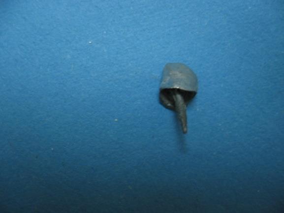
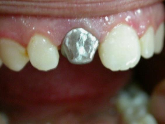
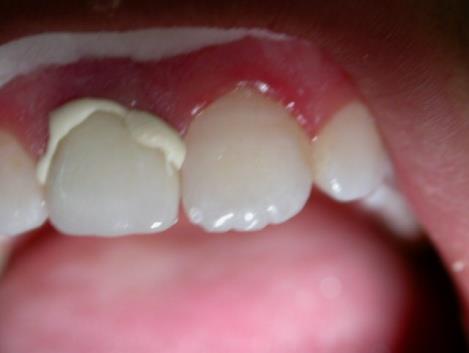
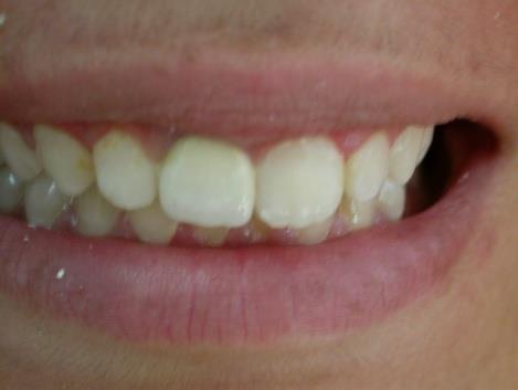
Figure 4: Try in framework for post crown. After shade selection the stratification of ceramics was performed. In the clinic, the metal ceramic post crown was tried again to verify esthetic, static, functional occlusion and phonation. It was cemented using adhesive cement (Figures 5 & 6). The cement was spinned into the canal with a Lentulo Spiral and the inner surface of the post crown and post are coated with the cement and placed in the canal.
Discussion
Introduced in 1878, the Richmond crown has incorporated a threaded tube in the canal with a screw retained crown. It was later modified to eliminate the threaded tube and was redesigned as a 1-piece dowel and crown (Hampson EL, et al. and Demas NC, et al.), which lost its popularity quickly because they were not practical because of divergent paths of insertion of the post space and remaining tooth structure. Endodontically treated teeth not crowned after obturation were lost 6 times more often than teeth crowned after obturation [10]. A 17-year controlled prospective study showed that the type of core restorations under the crowns had no effect on the survival rate of 307 endodontically treated teeth [11]. However, the longevity of a post-and-core restoration was influenced by the amount of remaining dentin height after preparation [12]. Proper restoration of endodontically treated teeth requires a sound knowledge of the endodontic, periodontal, restorative, and occlusal principles [13]. Post space preparation requires good understanding and knowledge of tooth anatomy to avoid unnecessary mishaps [14]. One piece restoration is indicated for the management of mutilated tooth requiring post-core restorations where there is reduced length crown compared to adjacent teeth with or no deep bite. Different coefficients of thermal expansion of the various components of post and core (foundation) and separately fabricated crown may have a harmful effect on the bonds between the tooth-post-core-cement-crown complex and increase the likelihood of decementation during functional loading. This procedure required casting a post-and-core as a separate component from the crown; this 2-step technique improved marginal adaptation and allowed for a variation in the path of insertion of the crow. By decreasing the number of interfaces between components, the single unit restoration helps to achieve a monoblock effect [8]. Regarding corrosion resistance several reports have linked root fracture to corrosion of base metal prefabricated post and core systems [15]. In one study a report on 468 teeth with vertical or oblique root fracture, investigators attributed 72% of these failures to electrolytic action of dissimilar metals used for the post and the core [16]. Endocrowns may perform similarly or better than the conventional treatments using intraradicular posts, direct composite resin or inlay/only restorations and constitute a reliable approach to restore only severely damaged molars and premolars [17, 18]. In the cases discussed in this article, special attention was given towards the path of withdrawal of the post for preparing the tooth to receive the post crown. This path is exactly the axis of the peripheral preparation destined for the one piece post crown .Thus; it is usually used as unit crown and not indicated for abutments of bridges. One piece dowel crown restorations also presented problems when the crown or FPD required removal and replacement. These difficulties led to development of a post and core restoration as a separate entity with an artificial crown cemented over a core and remaining tooth structure.
Conclusion
In the restoration of traumatized endodontically treated anterior teeth, both esthetic and mechanical considerations should be taken into account. If a crown is necessary, due to the loss of external tooth structure, then a post is usually required for anterior teeth, due to the predominantly shearing forces present and the narrow tooth dimensions. Despite, its only historical interest, one- piece metal ceramic post-crown can be indicated for short crown and especially for restoration of anterior teeth with deep bite.
References
-
Smith CT, Schuman N (1998) Prefabricated post-and- core systems: an overview. Compend Contin Educ Dent 19(10): 1013-1020.
-
Smith CT, Schuman NJ, Wasson W (1998) Biomechanical criteria for evaluating prefabricated post-and-core systems: a guide for the restorative dentist. Quintessence Int 29(5): 305-312.
-
Rosenstiel Stephen F, Land M, Fujimoto J (2006) Contemporary fixed prosthodontics. 78635 (Edn.), Mosby, Inc., an affiliate of Elsevier Inc, pp: 337.
-
Armitage GC, Research, Science and Therapy Committee of the American Academy of Periodontology (2003) Diagnosis of periodontal diseases. J Periodontol 74(8): 1237-1347.
-
Bradley J Olson (2012) Restoration of a fractured central incisorv. Compend Contin Educ Dent 33(3): 196-200.
-
Assif DF, Oren E, Marshak BL, Aviv I (1989) Photoelastic analysis of stress transfer by endodontically treated teeth to the supporting structure using different restorative techniques. J Prosthet Dent 61(5): 535-543.
-
Henry PJ (1977) Photoelastic analysis of post core restorations. Aust Dent J 22(3): 157-159.
-
Fattouh M, Abdulrahman A Almalki (2015) Treatment of endodontic treated molar using Richmond crown: case report. Pakistan Oral & Dental Journal 35(2): 343-345.
-
Saafi J, Debbabi I, Alremthi HA, Hajjami H, Cherif M (2017) A modified functionally generated path technique (FGP) for making posterior unit metal- ceramic crown. Journal of Oral Health and Dental Science 2(1): 1-8.
-
Aquilino SA, Caplan DJ (2002) Relationship between crown placement and the survival of endodontically treated teeth. The Journal of Prosthetic Dentistry 87(3): 256-263.
-
Fokkinga WA, Kreulen CM, Bronkhorst EM, Creugers NH (2007) Up to 17-year controlled clinical study on post-and-cores and covering crowns. Journal of Dentistry 35(10): 778-786.
-
Creugers NH, Mentink AGM, Fokkinga WA, Kreulen CM (2005) 5-year follow-up of a prospective clinical study on various types of core restorations. International Journal of Prosthodontics 18(1): 34-39.
-
Silverstein WH (1964) The reinforcement of weakened pulpless teeth. J Prosthet Dent 14(2): 372- 381.
-
Stern N, Hirschfeld Z (1973) Principles of preparing endodontic treated teeth for dowel and core restorations. J Prosthet Dent 30(2): 162-165.
-
Rud J, Omnell KA (1970) Root fractures due to corrosion: diagnostic aspects. Scand J Dent Res 78(5): 397-403.
-
Kakehashi Y, Lüthy H, Naef R, Wohlwend A, Schärer P (1998) A new all-ceramic post and core system: clinical, technical, and in vitro results. Int J Periodontics Restorative Dent 18(6): 586-593.
-
Augusto Sedrez-Porto J, Wellington L, Fernandesda Silva A, Aldrighi Münchow E, Pereira-Cenci T (2016) Endocrown restorations: A systematic review and meta-analysis. Journal of Dentistry 52: 8-14.
-
Belleflamme MM, Geerts SO, Louwette MM, Grenade CF, Vanheusden AJ, et al. (2017) No post-no core approach to restore severely damaged posterior teeth: An up to 10-year retrospective study of documented endocrown cases. J Dent 63: 1-7.
- Psychogenic Erectile Dysfunction in Late Adulthood: A Case Report on Clinical Intervention and Intimacy Restoration
- Clinical Trials on COVID-19 in 2025: A New Chapter in Global Health Research
- Innovations and Challenges in Contemporary Medical Clinical Trials: An Editorial Perspective
- Innovations and Challenges in Contemporary Medical Clinical Trials: A Critical Perspective
- Reimagining Clinical Trials: The Power of Continuous Feedback from Medical Reports
- Factors Influencing Brain Drain: Perspectives from a Medical School in Turkey