Acute Hemorrhagic Edema of Infancy in a 9 Months Old Child: A Case Report
<p style="text-align: justify;">Acute Haemorrhagic edema of infancy (AHEI) is a benign cutaneous condition. It typically presents as low grade fever, purpuric or macular rash and non pitting edema of limbs and face. Its cutaneous manifestations are a result of small-vessel vasculitits demonstrated histologically by leukocytoclastic vasculitis. It has a benign course with complete recovery in most of reported cases. We report a 9 months old child who presented with typical features of AHEI and who has a complete recovery of his illness without any intervention.</p>
Introduction
Acute Haemorrhagic edema of infancy (AHEI) is a benign cutaneous condition. It is one of the least described conditions in the literature. It was first described in 1913 in United States [1]. Its cutaneous manifestations are a result of small-vessel vasculitits demonstrated histologically by leukocytoclastic vasculitis [2, 3]. AHEI patient typically presents with triad of fever, edema and rosette-shaped purpura [4]. It is more common in male children (Male: Female ratio is 2:1) [5]. AHEI is considered as diagnoses in otherwise non-toxic child with purpuric rash or echymotic skin lesions with asymmetrical edema of the face, auricles, and extremities with or without mucosal involvement, age less than 2 years and with no systemic or visceral involvement followed by spontaneous healing of rash within few days or weeks [6]. This condition ultimately resolves by itself without needs for any medications like steroids or antihistamines [7].
A 9 months old boy previously well. He presented to the Royal Hospital with history of purpuric/petechial rash for 2 days. Rash started to appear suddenly at lower limbs, upper limbs and face at same time. The lesions are multiple, scattered and ranging from 0.5-1 cm in diameter. One day after rash, child started to have left upper limb swelling involving the hand and the forearm.
Then he developed left leg non-pitting edema extending over leg to knee joint (Figure 1). Swelling was red in color and not tender in palpation. Child had history of preceding mild upper respiratory tract infection symptoms for 1 week. History of fever (documented 38 C) for two days. There was no history of recent gastroenteritis and/or vaccination (last vaccination was at age of 6 months). There was no history of trauma, insect bite or bleeding diathesis. Child’s activity and feeding were not affected by illness.
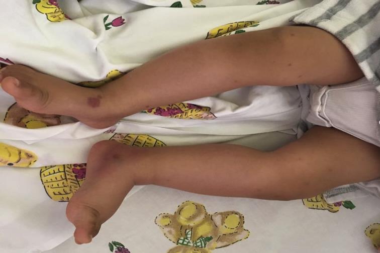
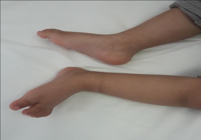
Figure 1: (photo. Taken with parents permission): Initial presentation of the child with purpuric rash and edema of lower limbs. Laboratory workup showed normal complete blood count, coagulation profile, urine microscopy and complements: CBC: Hb 11.5 g/dL, WBC 12.3 109/L, Platelet 509 109/L, ESR 51, C3 1504 (820-1850), C4 345 (150-530), Coagulation profile: PT 9.6 s, APTT 41.4 s, TT15.10s, INR 0.9 The decision was to admit the child with impression of Acute Haemorrhagic edema of infancy. During his admission; the child was doing well, remained afebrile. The rash and edema started to resolve by the second day of admission. He was discharged with minimal edema of upper and lower limbs. Patient was reviewed two weeks after discharge as outpatient visit. He was asymptomatic, active and all skin lesions and limbs edema has resolved (Figure 2).
Discussion
Acute Haemorrhagic edema of infancy (AHEI) is an acute onset self-limiting cutaneous condition [7]. Typical features include fever, purpuric rash and edema [4]. Several forms of rash can present. This may include purpura, ecchymosis, annular lesions, and cockade or targetoid lesions [8]. Skin eruptions usually follow presence of edema which is non-pitting and tender [9]. Edema initially starts to affect the palms and feet in asymmetrical manner. Then it can progress to affect the face [8]. It has been noticed that less 10 % of AHEI cases have extracutaneous manifestations which include glomerulonephritis, abdominal pain, arthralgia, testicular torsion, and intussusception [10, 11]. Preceding infections, vaccination and antibiotics use have been reported as triggers for AHEI [12]. Infections that are reported in literature include Rota virus induced gasteroenteritis and other viruses like herpes simplex virus, and adenovirus [8, 11, 13, 14]. It has been found that the average period of AHEI manifestation following triggering event ranges from two days to one month [12, 15]. Laboratory studies are usually not significant. Possible abnormal findings include increased erythrocyte sedimentation rate (ESR) and C-reactive protein (CRP), mild thrombocytosis and leukocytosis [16, 17]. Level of serum complements, immunoglobulin (Ig) G, A and M are frequently within normal range or slightly elevated [17, 18]. Acute skin eruptions can point to a worrying condition. So in some cases, clinician might need to exclude more serious conditions if presentation is atypical or there is suspicion of more serious illness. Otherwise AHEI can be diagnosed clinically based on typical history and clinical findings. The confirmatory test for AHEI is skin biopsy. In approximately one-third of patient biopsy shows leukocytoclastic vasculitis with IgA immunofluorescence [4, 11]. There is a list of conditions that can give a picture which is like AHEI. Henoch-Schonlein purpura (HSP) is considered the main important differential diagnosis for AHEI. Both HSP and AHEI have leukocytoclastic vasculitis in histopathology and clinically present with skin lesions [16, 17]. Previously AHEI was believed to be a variant of HSP. So it important first to exclude HSP because in case of HSP systemic corticosteroids should be started as soon as the syndrome is detected [16]. Features which can points to HSP are age of affected children is between 3 and 6 years, presence of pleomorphic lesions in the buttocks and extensor surfaces of legs (face usually spared), presence of extracutaneous manifestations and significant raise of serum IgA levels hence vascular IgA deposition is found [17, 19, 20]. There is still no approved treatment for AHEI. In a review of studies, it has been shown that corticosteroids and antihistamines do not affect the course of the disease [4, 15]. Few studies postulated that steroids might have beneficial effect in AHEI management [4, 20]. Risikesan, et al. has reported a child with AHEI who responded well to methylprednisolone and the discontinuation of steroids led to relapse of the disease [4]. Our patient was managed conservatively and follows up visit after 14 days showed complete recovery of the disease.
References
-
Snow I (1913) Purpura, urticaria and angioneurotic edema of the hands and feet in a nursing baby. JAMA 61(1): 18-19.
-
Saraclar Y, Tinaztepe K, Adaliou G, Tuncer A (1990) Acute hemorrhagic edema of infancy (AHEI)—A variant of Henoch-Schönlein purpura or a distinct clinical entity?. Journal of Allergy and Clinical Immunology 86(4 Pt 1): 473-483.
-
Paradisi M, Annessi G, Corrado A (2001) Infantile acute hemorrhagic edema of the skin. Europe PMC 68(2): 127-129.
-
Risikesan J, Koppelhus U, Steiniche T, Deleuran M, Herlin T (2014) Methylprednisolone Therapy in Acute Hemorrhagic Edema of Infancy. Case Reports in Dermatological Medicine 2014: 1-3.
-
Sorensen E, Matiz C, Friedlander S (2014) An 8- Month-Old Boy with Purpuric Skin Lesions. Pediatric Annals 43(1): e4-e8.
-
Krause I, Lazarov A, Rachmel A, Grunwald M, Metzker A, et al. (1996) Acute haemorrhagic oedema of infancy, a benign variant of leucocytoclastic vasculitis. Acta Paediatrica 85(1): 114-117.
-
Halicioglu O, Akman S, Sen S, Sutcuoglu S, Bayol U, et al. (2010) Acute Hemorrhagic Edema of Infancy: A Case Report. Pediatric Dermatology 27(2): 214-215.
-
Dongre A, Kothari D, Khopkar U, Adhe V, Kardekar S (2012) Acute hemorrhagic edema of infancy: A report of two cases. Indian Journal of Dermatology Venereology and Leprology 78(1): 121.
-
Sorensen E, Matiz C, Friedlander S (2014) An 8- Month-Old Boy with Purpuric Skin Lesions. Pediatric Annals 43(1): e4-e8.
-
(2012) Acute Hemorrhagic Edema of Infancy in an 11-Month-Old Boy.
-
Chesser H, Chambliss J, Zwemer E (2017) Acute Hemorrhagic Edema of Infancy after Coronavirus Infection with Recurrent Rash. Case Reports in Pediatrics 2017: 1-3.
-
Binamer Y (2015) Acute hemorrhagic edema of infancy after MMR vaccine. Annals of Saudi Medicine 35(3): 254-256.
-
Fiore E, Rizzi M, Ragazzi M, Vanoni F, Bernasconi M, et al. (2008) Acute hemorrhagic edema of young children (cockade purpura and edema): A case series and systematic review. Journal of the American Academy of Dermatology 59(4): 684-695.
-
Di Lernia V, Lombardi M, Lo Scocco G (2004) Infantile Acute Hemorrhagic Edema and Rotavirus Infection. Pediatric Dermatology 21(5): 548-550.
-
Fiore E, Rizzi M, Simonetti G, Garzoni L, Bianchetti M, et al. (2011) Acute hemorrhagic edema of young children: a concise narrative review. European Journal of Pediatrics 170(12): 1507-1511.
-
Karremann M, Jordan A, Bell N, Witsch M, Dürken M (2008) Acute Hemorrhagic Edema of Infancy: Report of 4 Cases and Review of the Current Literature. Clinical Pediatrics 48(3): 323-326.
-
Moradinejad MH, Entezari P, Mahjoub F, Ziaee V (2011) Acute Hemorrhagic Edema of Infancy; a Report of Five Iranian Infants and Review of the Literature. Iranian Journal of Pediatrics 21(1): 107– 112.
-
PoyrazoĞlu H, Per H, Gunduz Z, DuŞunsel R, Arslan D, et al. (2003) Acute hemorrhagic edema of infancy. Pediatrics International 45(6): 697-700.
-
Garty BZ, Ofer I, Finkelstein Y (2002) Acute hemorrhagic edema of infancy. Isr Med Assoc J 4(3): 228-229.
-
Da Silva Manzoni A, Viecili J, de Andrade C, Kruse R, Bakos L, et al. (2004) Acute hemorrhagic edema of infancy: a case report. International Journal of Dermatology 43(1): 48-51.
- Psychogenic Erectile Dysfunction in Late Adulthood: A Case Report on Clinical Intervention and Intimacy Restoration
- Clinical Trials on COVID-19 in 2025: A New Chapter in Global Health Research
- Innovations and Challenges in Contemporary Medical Clinical Trials: An Editorial Perspective
- Innovations and Challenges in Contemporary Medical Clinical Trials: A Critical Perspective
- Reimagining Clinical Trials: The Power of Continuous Feedback from Medical Reports
- Factors Influencing Brain Drain: Perspectives from a Medical School in Turkey