Tuberculosis of the Sacrum: Our Experience and Review of the Literature
<p style="text-align: justify;">Tuberculosis of the sacrum is a rare disease, despite spinal tuberculosis being the common form of the extra pulmonary disease. More commonly, tuberculosis involves thoracic or lumbar or the cervical spine. We report a case of sacral tuberculosis with a huge cold abscess over the left lumbar and gluteal region in a male, aged 18 years. CT scan revealed features of significant erosion of the sacrum and large volume abscess in the sacral and lumbar region communicating with each other. Anti-tubercular therapy was started. The patient underwent surgical drainage of the abscess. The purulent fluid was positive for acid-fast bacillus, and CB-NAAT test confirming the diagnosis of sacral tuberculosis. The patient recovered uneventfully and was discharged on anti-tubercular therapy for 9 months with follow-up protocol. The 2-month follow-up is marked with clinical improvement. In developing country like ours, sacral tuberculosis must be considered as first in the differential diagnosis in patients presenting with cold abscess and radiological features of sacral lesions.</p>
Introduction
Sacral form of tuberculosis is a rare presentation, and few cases are reported in literature. Tuberculosis and it’s both forms pulmonary and extra pulmonary are significantly prevalent in India. Tuberculosis of spine is the most common and the most serious form of skeletal tuberculosis. The thoracic spine is the most common site of affection, followed by lumbar and cervical spine in decreasing order of frequency [1]. Isolated tuberculosis of the sacrum is rarely reported in the literature, which is one factor accounting for low index of suspicion leading to a delay in diagnosis and initiation of specific therapy for this potentially curable disease. Osteoarticular tuberculosis (OAT) represents 2% to 5% of all cases of tuberculosis and 11% to 15% of extra pulmonary tuberculosis [2, 3]. The principal location of the OAT is the spinal cord, accounting for 50% of cases. Spinal involvement is usually a result of hematogenous spread of M.tuberculosis into the dense vasculature of cancellous bone of the vertebral bodies. Tuberculosis commonly involves the intervertebral disc and the adjacent vertebral bodies, the “paradiscal” area. Involvement of the posterior elements is very uncommon [1]. The primary infection site being either a pulmonary lesion or an infection of the genitourinary system [4, 5, 6, 7]. Spread occurs via the arterial or the venous route. Lumbo-sacral region accounts for only 2 to 3% of all cases with isolated involvement of sacrum rarely reported. The characteristic clinical features include chronic back pain, local tenderness, stiffness, a cold abscess, prominent spinal deformity with paraplegia the most dreaded complication. Early diagnosis and prompt treatment is necessary to prevent permanent neurological disability and to minimize spinal deformity. Antitubercular drugs from the mainstay of treatment with surgery are reserved for those with neurological symptoms [8].
Case Report
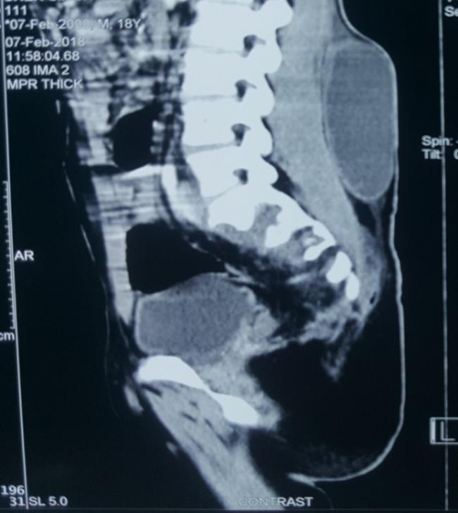
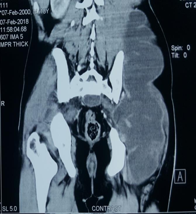
A male patient, 18 years of age presented with swelling in left lumbar region and back since 5 months. Swelling was insidious in onset, gradually progressive, occasionally associated with mild dull aching pain, with no aggravating or relieving factors. He gave history of decline in the appetite and 4-5 kg weight loss. There was no history of fever, or cough. Patient had no predisposing conditions for Potts disease (malnutrition, malignancy, sickle cell anemia, diabetes mellitus). No past history of tuberculosis or any family history. The Patient was thin built, haemodynamically stable, afebrile, but had pallor present. There was no evidence of icterus and lymph nodes were not palpable. There were no neuropathic symptoms (urinary incontinence, constipation, sciatic pain). Local examination revealed 15X10 cm swelling on left lumbar and lower back just overlying the sacral region, crossing the midline, and extending in the left gluteal region. The temperature was not raised and it was non- tender. The swelling was soft, no cough impulse present, non-pulsatile, and non reducible. Radiograph of the lumbo-sacral spine lateral view, and chest PA were within normal limits. The CECT Scan revealed 10 X 2 X 5 cm presacral collection causing significant erosion of the underlying sacrum S2 to S5. A second collection, which was communicating with presacral collection, was seen in the left flanks extending to the gluteal region and upper thigh 28 X 17.6 X 7.7 with continuing upwards in the iliopsoas muscles.
Routine Laboratory investigations: a total leukocytic count of 9300/mm3 with neutrophils (71.5%) and lymphocytes (19.8%), hemoglobin was 9.7 g/dl. Other routine investigations including RFT, RBS were within normal limits. Patient was however positive for HbsAg antigen. Clinical diagnosis of sacral tuberculosis with huge cold abscess was made and patient was started with anti- tubercular therapy category I. Since it was a significantly large size abscess patient was taken up for the incision and drainage under the regional anesthesia. Intra-op around 1.2 L of frank pus was drained, all the loculi were broken cavity was irrigated with betadine solution and normal saline. A drain tube was kept, which was removed on 5th day postoperatively. Pus culture study was sterile after 24 hours incubation. However Zeil Nelson staining was positive for Acid Fast Bacillus. Core body Nucleic Acid Amplification Test (CB- NAAT) was also positive in the abscess fluid for M. Tuberculosis confirming the diagnosis further. Patient was duly started on ATT according to Cat-I. Post op was uneventful with almost complete resolution of swelling and patient was discharged by 5th post op day on ATT for 9 months with regular follow up protocol. The 2 month follow-up is marked with clinical improvement (Figures 1& 2).
Discussion
Isolated tuberculosis of the sacrum is rarely reported in the literature, with a frequency estimated at 5% by Pertuiset, et al. [3]. In a review of 63 cases of spinal tuberculosis by Lindahl, et al., sacral involvement was found in just four cases, while none was involved in 107 patients in the series of Lifeso, et al. [9, 10]. CECT is sensitive for diagnosis as in our case showing significant erosions in sacrum with collection in the fascial and intertmuscular planes extending up to ipsilateral ileopsoas muscle and gluteal region. Confirmation of diagnosis however needs needle aspiration, staining and culture. Tuberculosis is a major health problem in developing countries. OAT represents about 3% of all tuberculosis and 15% of extra pulmonary tuberculosis cases of which Spinal location is seen in at least 50% of cases [2]. Hematogenous route of mycobacterium from primary foci in the lung and/or genitourinary tract is the common mode of spread [11]. It is widely believed that the paravertebral venous plexus of Batson provides the primary pathway for dissemination of the tuberculous bacilli into the vertebral column. The dorsolumbar spine is the seat of choice. Pott’s disease usually presents with back ache (90- 100%), weight loss (58%), neurological involvement (32-76%) [12]. It also spreads into paravertebral spaces, adjacent soft tissue, fascia through subligamentous spread causing different complications including psoas abscess [13]. Psoas abscess can be primary or secondary. Primary is mostly caused by Staphylococcus with Mycobacterium also a common cause. Secondary psoas abscess is associated with contiguous sources like musculoskeletal infections. Two third of the vertebral osteomyelitis is pyogenic and only one third is tubercular. So, Pott’s disease is not a common cause of secondary psoas abscess. Our patient presented with a large swelling, underwent incision and drainage. The purulent fluid drained from the abscess was positive acid fast bacilli on ZN staining and CB-NAAT, thus confirming the diagnosis of sacral tuberculosis. CB-NAAT bears sensitivity and specificity of 95.7% and 99.3% respectively in Dx of TB [14]. Indications for surgical treatments of Pott’s disease include neurological deficit, spinal deformity with instability or pain, no response to medical therapy and large paraspinal abscess [10]. Our patient had a large swelling as primary symptom and mild discomfort. Pt responded well to therapy with no recurrence of swelling with no complications of the disease. The prognosis of sacral tuberculosis is good, if an early diagnosis is made and appropriate treatment is provided.
Conclusion
Sacral tuberculosis is a rare presentation among vertebral tubercular disease. Sacral tuberculosis must be considered in the differential diagnosis among the patients presenting with cold abscess and radiological features of sacral lesions. Early diagnosis and treatment is the key to successful outcome and circumventing its complications.
References
-
Amey P Patankar (2016) Tuberculosis of spine: An experience of 30 cases over two years. Asian Journal of Neurosurgery 11(3): 226-231.
-
Lazrak F, Abourazzak FE, Elouzzani FE, Benzagmout M, Harzy T (2014) A rare location of sacral tuberculosis: A report of three cases. European Journal of Rheumatology 1(2): 78-80.
-
Pertuiset E (2004) Peripheral bone and joint tuberculosis. EMC-Rhumatologie Orthopédie 1: 463- 486.
-
Tuli SM (2004) In: Tuberculosis of the Skeletal System: Bones, Joints, Spine and Bursal Sheaths. 3rd (Edn.), New Delhi: Jaypee Brothers Medical Publication Ltd.
-
Boachie-Adjei O, Squillante RG (1996) Tuberculosis of the spine. Orthop Clin North Am 27(1): 95-103.
-
Schirmer P, Renault CA, Holodniy M (2010) Is spinal tuberculosis contagious? Int J Infect Dis 14(8): e659- e66.
-
Uddin Mohammad, Sultana Nusrat, Rehan Rezwan, Khan Aminuddin A (2014) Pott’s Disease with Psoas Abscess in a Diabetic Patient: A Conservative Approach. Chattagram Maa-O-Shishu Hospital Medical College Journal 13(2): 1-3.
-
Garg RK, Somvanshi DS (2011) Spinal tuberculosis: A review. The Journal of Spinal Cord Medicine 34(5): 440- 454.
-
Lindahl S, Nyman RS, Brismar J, Hugosson C, Lundstedt C (1996) Imaging of tuberculosis. IV. spinal manifestations in 63 patients. Acta Radiol 37(3): 506- 511.
-
Lifeso RM, Weaver P, Harder EH (1985) Tuberculosis spondylitis in adults. J Bone Joint Surg Am 67(9): 1405-1413.
-
Patankar T, Krishnan A, Patkar D, Kale H, Prasad S, et al. (2000) Imaging in isolated sacral tuberculo- sis: a review of 15 cases. Skeletal Radiol 29(7): 392-396.
-
Turgut M (2001) Multifocal extensive spinal tuberculosis involving cervical, thoracic & lumbar vertebra. Br J Neurosurg 15(2): 142-146.
-
Azzam NI, Tammawy M (1988) Tuberculous spondylitis in adults: diagnosis and treatment. Br J Neurosurg 2(1): 85-91.
-
Sharma SK, Kohli M, Yadav RN, Chaubey J, Bhasin D, et al. (2015) Evaluating the Diagnostic Accuracy of Xpert MTB/RIF Assay in Pulmonary Tuberculosis. Plos One 10(10): e0141011.
- Psychogenic Erectile Dysfunction in Late Adulthood: A Case Report on Clinical Intervention and Intimacy Restoration
- Clinical Trials on COVID-19 in 2025: A New Chapter in Global Health Research
- Innovations and Challenges in Contemporary Medical Clinical Trials: An Editorial Perspective
- Innovations and Challenges in Contemporary Medical Clinical Trials: A Critical Perspective
- Reimagining Clinical Trials: The Power of Continuous Feedback from Medical Reports
- Factors Influencing Brain Drain: Perspectives from a Medical School in Turkey