Caverns Sinus Thrombosis
Cavernous sinus thrombosis (CST) is the formation of a blood clot within the cavernous sinus, a cavity at the base of the brain which drains deoxygenated blood from the brain back to the heart. This is a rare disorder and can be of two types– septic cavernous thrombosis and aseptic cavernous thrombosis. Most commonly the form is of septic cavernous sinus thrombosis. The cause is usually from a spreading infection in the nose, sinuses, ears, or teeth. Staphylococcus aureus and Streptococcus are often the associated bacteria. Cavernous sinus thrombosis symptoms include: decrease or loss of vision, chemosis, exophthalmos (bulging eyes), headaches, and paralysis of the cranial nerves which course through the cavernous sinus. This infection is life-threatening and requires immediate treatment, which usually includes antibiotics and sometimes surgical drainage. Aseptic cavernous sinus thrombosis is usually associated with trauma, dehydration, anemia, and other disorders.
Najia al H*, Attia Saleh Al Z, Ibraheem K, Amal Z, Laila SA, liza OS and Hashim N
NICU Consultant at Maternity Children Hospital Makah, Saudi Arabia
Makah, Po Box 8981- Makah, Saudi Arabia, Email: najoo_n1@yahoo.com anemia, and other disorders.
Keywords: Caverns Sinus; Thrombosis; Streptococcus; Cerebral Injury; Sensorimotor Abbreviations: CST: Cavernous sinus thrombosis; ACOG: American College of Obstetricians and Gynecologists.
Introduction
Perinatal stroke may be defined as an acute neurologic syndrome with chronic sequel due to cerebral injury of vascular origin occurring between 20 weeks gestation and 28 days postnatal life [1, 2]. These disorders include focal cerebral injury due to arterial ischemic stroke, cerebral venous thrombosis, and primary intra-cerebral hemorrhage. Perinatal stroke is a common cause of acute neonatal encephalopathy, and may manifest as seizures, altered mental status, and sensorimotor deficits. It is an important cause of chronic neurologic disability.
Sensorimotor Deficits Terminolog
It is a "Neonatal encephalopathy" has emerged as the preferred term to describe central nervous system dysfunction in the newborn period [2, 3]. The American College of Obstetricians and Gynecologists (ACOG) describes neonatal encephalopathy as a clinically defined syndrome of disturbed neurologic function in the earliest days of life in an infant born at or beyond 35 weeks of gestation, manifested by a subnormal level of consciousness or seizures, and often accompanied by difficulty with initiating and maintaining respiration and depression of tone and reflexes.
The terminology does not imply a specific underlying pathophysiology, which is appropriate since the nature of brain injury causing neurologic impairment in a newborn is poorly understood. While neonatal encephalopathy was once automatically ascribed to hypoxia-ischemia, it is now known that hypoxia-ischemia is only one of many possible contributors to neonatal encephalopathy [4]. Whether a particular newborn’s encephalopathy can be attributed to hypoxic-ischemic brain injury is often unclear. Some investigators require stringent criteria for using the term neonatal encephalopathy, such as two or more symptoms of encephalopathy lasting over 24 hours, while others require no more than a low five-minute Apgar score [5, 6]. However, the use of Apgar scores alone is problematic, as Apgar scores may be low due to maternal analgesia or prematurity, or can be normal in the presence of acute hypoxia-ischemic injury, or can be inflated in actual clinical practice.
What Causes Neonatal Stroke
One of the causes of neonatal stroke is hypoxia, an event when oxygen deprivation causes the brain to panic. The health of the mother can sometimes affect whether a child will have a neonatal stroke, especially if she has disorders such as autoimmune disorders, coagulation disorders, prenatal cocaine exposure, infection, congenital heart disease, diabetes, and trauma.
Factors that can affect a neonatal stroke during pregnancy include:
- Placental abruption, placental infection, and chorioamnionitis.
- Other miscellaneous disorders affecting the mother and/or baby’s health and resulting in a neonatal stroke include: blood, homocysteine, and lipid disorders (such as polycythemia, disseminated intravascular coagulopathy, prothrombin mutation, lipoprotein a deficiency, factor VIII deficiency, and factor V leiden mutation).
- A neonatal stroke can also be caused by maternal infection through infections affecting the central nervous system or other systemic infections.
However, several parents and doctors are worried as the cause of a neonatal stroke isn’t always obvious: some healthy children born after an uncomplicated pregnancy and normal labor and delivery may still experience a neonatal stroke.
Case Report
- 5 days old burmawi boy, born FT, SVD, Car delivery and transported to a private hospital, admitted to our hospital at the age of 5 days due to decreased oral intake and decreased activity for 1 day, and 2 episodes of generalized tonic colonic seizures with up rolling eyes, both attacks lasted less than a minute.
- Once the family noticed the seizure they came by their car within 20 minutes and they reported the second seizure in their car with the same character and were aborted in our ER with diazepam, and loading Phenobarbitone.
- The family reported an undocumented fever that night and no other symptoms such as vomiting, diarrhea, cough, and changes in urine or stool or jaundice.
- Regarding his antenatal history, the mother did not suffer from any events or illnesses during her pregnancy, only took folic acid, and followed only twice for ultrasound in the second and third trimester and both the follow ups were normal.
- The child received his Hepatitis B vaccine that morning.
- No consanguinity and no family history of seizures or metabolic or genetic or hematological illnesses.
- No history of abortions or early deaths in family members or cousins.
- So during his admission septic workup was initiated including lumber puncture.
- The first blood culture which came positive on the 3rd day revealed streptococcus pneumonia, however 2 separate tries for lumber puncture came as dry taps so MRI spine was done and it was normal.
- He was started on Meropenem and Ampicillin and we consulted infectious diseases and they advised to continue Meropenem for 14 days.
- The blood culture repeated 3 days later and 1 week later showed no growth.
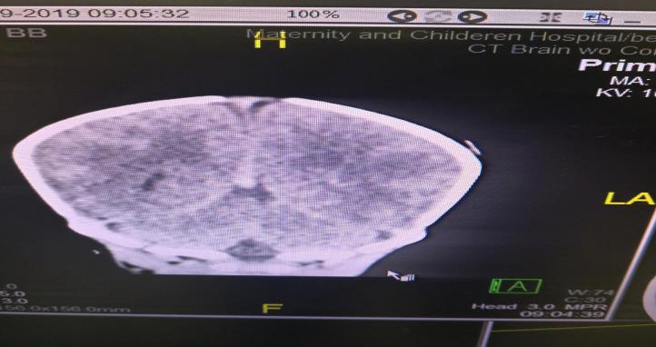
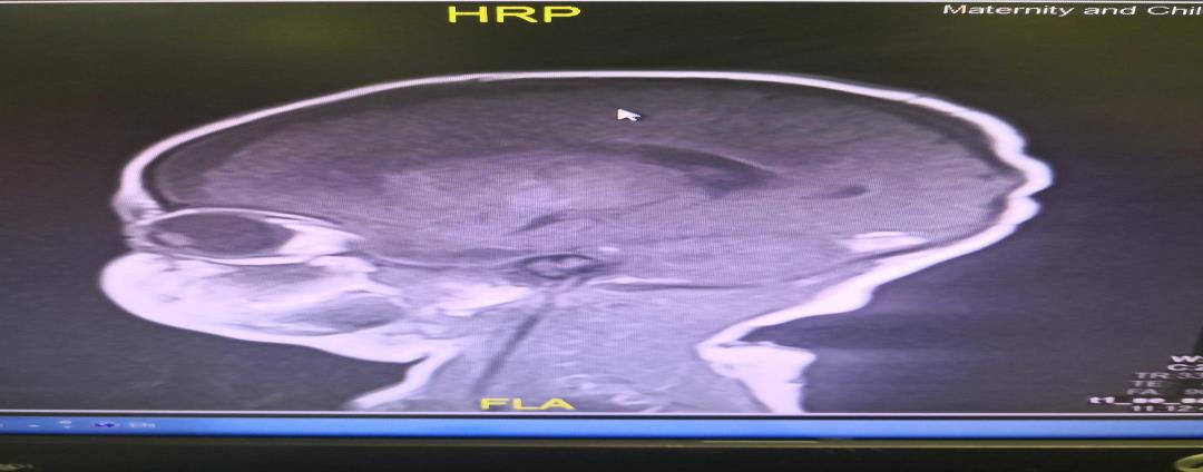
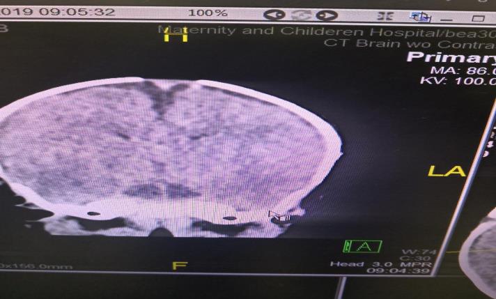
- Neurology consulted and they started him on keppra and on Phenobarbital and advised for an MRI, the MRI showed bilateral acute infarcts and chronic subdural hematoma on the left hemisphere and high signal intensity suggesting acute sinus thrombosis, the radiologist advised for CT venography.
- The CT venography was done 5 days later and showed subdural hematoma seen in the left front parietal region and that no contrast was visualized in the sinuses that could be due to the acute thrombosis.
- The hematology was consulted and they advised for fibrinogen and coagulation profile which were both normal and they ordered protein C and protein S both.
Clinical Presentation in Relation to Imaging Findings
Clinical presentation has previously been described in cohorts of predominantly full-term infants. In the 2 largest studies reported so far, seizures and/or apneas were the presenting symptoms in about 75% of the infants 2, 10 in the study by Berfelo and colleagues, a distinction was made between generalized and focal seizures, with generalized seizures being more common.
Apneas were the presenting symptom in 17 and 19% of cases, respectively, and could well have been of epileptic origin, but this could not be confirmed due to the lack of continuous electroencephalographic monitoring. It has recently been suggested that infants with temporal lobe hemorrhage and possible involvement of the vein of Labbé tend to present with apneic spells. 23, 24 as expected, seizures were almost invariably present in the context of parenchymal lesions (38 of the 42 infants). 2 Cerebral Sino venous thrombosis can also be a chance finding (5% to 13%), diagnosed on a routine cranial ultrasound. It is therefore likely that cerebral Sino venous thrombosis is more common than reported.
Imaging Studies
Sinus films are helpful in the diagnosis of sphenoid sinusitis. Opacification, sclerosis, and air-fluid levels are typical findings. Contrast-enhanced CT scan may reveal Najia al H, et al. Caverns Sinus Thrombosis. Med J Clin Trials Case Stud 2019, 3(3): 000227.
underlying sinusitis, thickening of the superior ophthalmic vein, and irregular filling defects within the cavernous sinus; however, findings may be normal early in the disease course.
Copyright© Najia al H, et al.
A MRI using flow parameters and an MR venogram are more sensitive than a CT scan, and are the imaging studies of choice to diagnose cavernous sinus thrombosis. Findings may include deformity of the internal carotid artery within the cavernous sinus, and an obvious signal hyperintensity within thrombosed vascular sinuses on all pulse sequences. Cerebral angiography can be performed, but it is invasive and not very sensitive. Orbital venography is difficult to perform, but it is excellent in diagnosing occlusion of the cavernous sinus (Figure 2).
Differential Diagnosis
Orbital cellulitis Internal carotid artery aneurysm Stroke Migraine headache Allergic blepharitis Thyroid exophthalmos Brain tumor Meningitis Mucormycosis Trauma
What is the Treatment for Neonatal Stroke?
One of the newest and most experimental forms of treatment is hypothermia. While it doesn’t seemingly make sense that a way to treat a condition is another serious condition, the serious drop in temperatures keeps the infant’s brain and body from overheating and reacting to the increased flow in blood with hypothermia, there is more constriction of blood vessels, and the brain is less likely to react into hyperactive responses that result in brain damage. In fact, doctors have found that stroke patients treated with hypothermia are far more likely to make a rapid recovery.
Najia al H, et al. Caverns Sinus Thrombosis. Med J Clin Trials Case Stud 2019, 3(3): 000227.
Another experimental treatment is Hyperbaric Oxygen Therapy: by putting a child in an environment of 100% oxygen, the important gas floods the body, keeping the blood and the brain from overacting to oxygen deprivation, sometimes preventing permanent brain damage. If your doctor prefers to stay away from experimental forms of treatment, there are other anticoagulant treatments, such as heparin and urokinase. Our patient admitted in NICU not received cooling therapy because he came to hospital 5 days old; restrict fluid to 80ml/kg/day, urgent CT scan done showed infarction. Cerebrospinal fluids unable to take so treat him as meningitis for 21 days MRI brain was done and consult hematology whose started heparin infusion.
What is Prognosis?
It is important to get rapid medical attention to your child if he or she experiences a neonatal stroke: of the children that survive a neonatal stroke, as many as 1 million a year of those survivors may develop cerebral palsy. If, however, you respond quickly and if your doctor is willing to try experimental treatments such as hypothermia or Hyperbaric Oxygen Therapy, your child may quickly recover and may not suffer any long term disabilities. Cavernous sinus thrombosis has a mortality rate of less than 20% in areas with access to antibiotics.
Copyright© Najia al H, et al.
Before antibiotics were available, the mortality was 80– 100%. Morbidity rates also dropped from 70% to 22% due to earlier diagnosis and treatment.
Conclusion
Septic CST most commonly results from contiguous spread of infection from a nasal furuncle (50%), sphenoidal or ethmoidal sinuses (30%) and dental infections (10%). Less common primary sites of infection include tonsils, soft palate, middle ear, or orbit (orbital cellulitis) [3]. The highly anastomotic venous system of the paranasal sinuses allows retrograde spread of infection to the cavernous sinus via the superior and inferior ophthalmic veins. It was previously thought that veins in the area were valve less and that this was the major cause of the retrograde spread, but a recent study has found that the ophthalmic and facial veins are not valveless [7].
Staphylococcus aureus is the most common infectious microbe, found in 70% of the cases. Streptococcus is the second leading cause. Gram-negative rods and anaerobes may also lead to cavernous sinus thrombosis [3]. Rarely, Aspergillus fumigatus and mucormycosis cause CST. Aseptic cavernous sinus thrombosis is much less common and is usually associated with other disorders including trauma, circulatory problems, nasopharynx cancers and other tumours of the skull base, dehydration, and anemia [8, 9].
References
-
(2014) Executive summary: Neonatal encephalopathy and neurologic outcome, second edition. Report of the American College of Obstetricians and Gynecologists' Task Force on Neonatal Encephalopathy. Obstet Gynecol 123(4): 896-901.
-
Ulrika Ådén (2009) Neonatal Stroke Is Not a Harmless Condition. Stroke 40(6): 1948-1949.
-
Renée Shellhaas, Douglas R Nordli, Joseph A Garcia- Prats, John F Dashe (2018) Etiology and prognosis of neonatal seizures.
-
(2016) Cavernous sinus thrombosis - NHS Choices. NHS Choices.
-
(2016) Cavernous sinus thrombosis: Medline Plus Medical Encyclopedia.
-
James Garrity (2019) Cavernous Sinus Thrombosis. Eye Disorders.
-
Guidelines Cavernous sinus thrombosis.
-
Sidhartha Tan, Yvonne Wu, Douglas R Nordli, Leonard E Weisman, John F Dashe (2018) Etiology and pathogenesis of neonatal encephalopathy.
-
Zhang J, Stringer MD (2010) Ophthalmic and facial veins are not valveless. Clin Exp Ophthalmol 38(5): 502-510. Najia al H, et al. Caverns Sinus Thrombosis. Med J Clin Trials Case Stud 2019, 3(3): 000227. Copyright© Najia al H, et al.
- Psychogenic Erectile Dysfunction in Late Adulthood: A Case Report on Clinical Intervention and Intimacy Restoration
- Clinical Trials on COVID-19 in 2025: A New Chapter in Global Health Research
- Innovations and Challenges in Contemporary Medical Clinical Trials: An Editorial Perspective
- Innovations and Challenges in Contemporary Medical Clinical Trials: A Critical Perspective
- Reimagining Clinical Trials: The Power of Continuous Feedback from Medical Reports
- Factors Influencing Brain Drain: Perspectives from a Medical School in Turkey