Novel Vesicular Drug Delivery Systems Proniosomes
These proniosomes, which are dry surfactant-coated particles that form niosomal dispersion in hot aqueous fluids immediately on stirring, are the subject of this review paper. It is easier to transport, distribute, store, and administer dosage forms such as proniosomes thanks to their provesicular structure, which reduces the issues of vesicular system physical stability such as aggregation, fusion, and leakage. Lipophilic or amphipilic medications can be delivered effectively by oral route using proniosome powders, which are suitable dosage forms. Proniosome preparation, characterisation, release profile, applications, advantages, and downsides are all discussed in this review in great detail.
Introduction
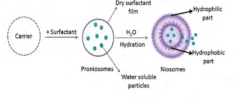
Therapeutic agents can be targeted precisely to the site of action without interfering with other tissues, which is called medication targeting. Immunoglobin, serum proteins, synthetic polymers and liposomes are only some of the carriers that have been utilised to target the medicine [1]. Erythrocytes and niosomes. As particulate carriers, the particulate carriers have special advantages while avoiding the disadvantages of conventional dosage forms. However, the particulate carriers have a number of disadvantages instead of advantages [2]. Using a proniosome-like structure, such as a vesicle, reduces these disadvantages. Niosomal dispersion can be made within minutes of agitation in hot aqueous conditions using proniosomes, a dry formulation of water-soluble carrier particles covered with a surfactant [3].
Hydration causes niosomes to disintegrate or disperse and create an oral or injectable suspension of niosomes that can be delivered in a variety of ways. A more homogeneous size distribution and similar appearance to traditional niosomes are the results of this procedure [4]. Surfactants themselves act as penetration enhancers and are biodegradable, non- toxic, amphiphilic, exhibit the capability of encapsulation and they can encapsulate both hydrophilic and lipophilic medicines. The dry powder form of proniosomes makes them more convenient for transportation, distribution, storage, processing, packaging, and sterilisation, as well as enabling optimal flexibility, unit dosage as a capsule, and a high degree of stability. With all of these advantages, they seem like a good choice for mass manufacturing in the industry. Delivering a wide range of active chemicals is possible with these adaptable systems [5, 6]. There are other advantages to pro-niosomes, including minimal toxicity due to their non- ionic nature, no need for extra precautions, and conditions for formulation and preparations [7]. The approach is very easy to use and does not require the use of unpleasant solvents for the routine and large-scale manufacture of pro- niosomes [8, 9].
Structure of Proniosomes
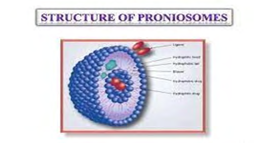
Proniosomes are hypothesised to be microscopic lamellar structures that can be found in a variety of cell types. In order to hydrate the mixture, they utilise a non-ionic surfactant and cholesterol, then add water. Consequently, the hydrophilic and hydrophobic ends of the nonionic surfactant point outward, forming the bilayer. Liposomes, on the other hand, have a phospholipid bilayer, but proniosomes have non-ionic surface-active molecules in their bilayer that separate them from liposomes. Depending on the preparation method, pheniosomes can be unilamellar or multilamellar. Hydrophilic chains on the surface of niosomes face each other, while hydrophobic chains face each other on the inside of the bilayers. Because of this, they may have a mixture of hydrophilic and hydrophobic drugs. In the vesicle, hydrophilic pharmaceuticals are maintained in place, while hydrophobic drugs are enclosed in the container’s bilayer [10, 11].
Advantages of Proniosomes over Niosomes
It is possible to solve the niosome’s issues, such as aggregation, fusion and leakage with the use of proniosomes. The drug-containing proniosomal vesicles serve as a sort of storage facility, allowing the drug’s release to be prolonged. Additionally, proniosome powder in a dry form creates capsules beads, which is a great feature [12, 13]. There are many issues with liposomes, such as oxidation and hydrolysis. Proniosomes solve these issues [14]. Because proniosomes can transport both water-soluble and water- soluble medicines, hydrolysis-resistant medications are not encapsulated.
Formation of Niosomes from Proniosomes
Niosomes can be made by adding different aqueous phases with the medicine to the proniosomes and agitating for a short period of time. Noise is formed from proniosomes. T>Tm Where, T = Temperature Tm = Mean phase transition temperature.
Maltodextrin proniosomes formulation has been shown to promote fast reconstitution of niosomes with minimum residual carrier, according to previous studies If you add warm water to the free-flowing powder of maltodextrin and surfactant, it will come back to life (Figure 1) [3, 5, 6, 15].
Components of Proniosomes
The essential components for the delivery system are as follows
Surfactants
Hlb value: The surfactant’s Hydrophilic Lipophilic Balance [HLB] value plays a critical role in determining the vesicle’s ability to encapsulate drugs. They are surface-active agents that are usually organic compounds with a mixture of water- repellent and water-loving groups in nature. There are both water-insoluble (hydrophobic) and hydrophilic components in surfactants. When used as emulsifying and solubilizing agents, they can also improve the permeability of the material [16]. As vesicles are formed, non-ionic surfactants such alkyl acids, alkyl ethers, alkyl esters and fatty acids are commonly utilised. Any surfactant with an HLB number between 4 and 8 was shown to be compatible with vesicle- forming abilities, as indicated by the Hydrophilic Lipophilic Balance [HLB] number [17, 18]. Among the most commonly employed non-ionic amphiphiles for the creation of vesicles are the following: [19] (Table 1).
| S. No | Non-ionic Amphiphiles | Examples |
|---|---|---|
| 1 | Alkyl ethers and alkyl glyceryl ethers | Polyoxyethylene 4 lauryl ether Polyoxyethylenecetyl ethers Polyoxyethylene stearyl ethers |
| 2 | Sorbitan fatty acid esters | Span 20, 40, 60, 80 |
| 3 | Polyoxyethylene fatty acidesters | Tween 20, 40, 60, 80 |
Table 1: Common non-ionic amphiphiles used for vesicle formation.
CPP (Critical Packing Parameter): Structure of the surfactant, comprising its hydrophilic head group and hydrophobic alkyl chain, can be defined as a relationship between the capacity to produce vesicles and its ability to form vesicles. The type of vesicle formed by a surfactant can be anticipated based on its CPP. The drug’s entrapment efficiency is influenced by the nonionic surfactant’s hydrophilic head group chain length and size.
v CPP=lc×a Where, v = The hydrophilic group volume, lc = Critical hydrophobic group length a = Area of the hydrophilic head group.
In comparison to lauryl (C12)-chained nonionic surfactants, those with stearyl (C18) chains exhibit better trapping efficiency. Surfactants in the Tween family, which contain long alkyl chains and big hydrophilic moiety, had the highest water-soluble drug entrapment effectiveness when combined 1:1 with cholesterol [20, 21]. Phase transition temperature: Surfactant entrapment efficiency increases as the transition temperature of surfactants rises, resulting in a decrease in the permeability of the material. The higher the phase transition temperature, the better the drug entrapment. Due to a high phase transition temperature and limited permeability, the vesicles’ drug leaching can be reduced [22].
Carrier Materials
Allows for a wide range in the amount of surfactant and other ingredients that can be included in the formulation. In addition, the increased surface area allows for more efficient loading, making it more convenient to use. Carriers must be safe and non-toxic, free-flowing, have low solubility in the loaded combination solution, and have good water solubility for easy hydration of the loaded mixture [23, 24]. Table 2 includes a list of common carriers.
| Carrier material investigated | |
|---|---|
| 1 | Maltodextrin |
| 2 | Sorbitol |
| 3 | Spray dried lactose |
| 4 | Glucose monohydrate |
| 5 | Lactose monohydrate |
| 6 | Sucrose stearate |
Table 2: Commonly used carriers.
Solvent
One of the most critical aspects is the choice of solvent, which has a significant impact on vesicle size as well as drug permeability [25]. Research shows that vesicles generated from various alcohol kinds have varying diameters, depending on their water solubility. Solubility in water increases the size of vesicles, which follows the following pattern: 26 Ethanol > propanol > butanol > isopropanol.
A solvent can help a product penetrate more easily. It also has an impact on the spontaneity with which niosomes are formed. As a result of isopropanol and butanol’s rapid phase separation and lesser solubility in water, the formulation comprising these solvents developed more spontaneously than niosomes containing propane or ethanol. Because the branched structure of isopropanol acts as a co-surfactant and may loosen the bilayer packing, research imply that drug penetration is greatest for isopropanol due to the decreased vesicular size due to branching. Skin permeability may be increased by ethanol’s ability to reduce lipid polar head contacts in the membrane [26, 27].
Aqueous Phase
In the aqueous phase of proniosome synthesis, phosphate buffer (pH 7.4), 0.1 percent glycerol, and hot water are the primary ingredients. Additionally, the pH of the hydration medium has a crucial impact in proniosome entrapment efficiency [28]. According to the literature, the pH of the hydration medium has been observed to affect the entrapment efficiency. Drug encapsulation increased as the pH lowered from 8 to 5.5. The ionizable carboxylic group in the chemical structure of the drug may be responsible for the increase in the percentage encapsulation efficiency when the pH is decreased. As pH drops, the unionised flurbiprofen species, which have a higher partitioning to bilayer lipid phase than the ionised species, see an increase in their proportion [29].
Cholesterol
Adding cholesterol to vesicles improves their stability. It is mostly used to stabilise membranes. An essential component of cell membranes is cholesterol, a naturally occurring hormone. Stabilizing the system against aggregate formation due to repulsive steric or electrostatic effects avoids aggregation. It causes the niosome system to change from a gel state to a liquid one. When surfactant monomers are assembled into bilayers to form niosomal membranes, cholesterol molecules accommodate themselves as “vesicular cement” in the molecular cavities formed, and this space filling action results in increased rigidity, decreased permeability, and improved entrapment efficiency for cholesterol-containing membranes compared to cholesterol- free membranes. After a particular level of cholesterol has been reached, it competes with the medicine for vesicular membrane bilayer space, so preventing the drug from entering the membrane and disrupting its regular linear shape [30, 31, 32].
Lecithin
Soya lecithin, which comes from soya beans, and egg lecithin, which comes from egg yolk, are both types of lecithin. So much of lecithin is made up of Phosphatidyl Choline (PC). It serves a variety of functions in the vesicular system. Due to its high Tc (phase transition temperature), it limits the leaking of drugs from the vesicles, helps to raise the percentage of drug entrapment, and results in smaller vesicles due to an increase in hydrophobicity that results in the reduction of vesicle size. The Food and Drug Administration (FDA) has designated lecithin as safe for human consumption. The FDA’s list of inactive ingredients also includes lecithin [33].
Drug
There are a variety of drug options for proniosome formation, but all of them should have the following features [34].
- Low aqueous solubility drugs
- High-dose regularity drugs
- Short half-life
- Controlled medication distribution appropriate drugs
- Higher negative medication responses drugs.
Types of Proniosomes
Depending upon the method of preparation, proniosomes exist in two forms.
Dry Granular Proniosomes
According to the type of carrier and method of preparation of dry granular proniosomes are again divided into:
- Sorbitol-based proniosomes
- Maltodextrin-based proniosomes.
Sorbitol based proniosomes
As a carrier, sorbitol is coated with non-ionic surfactant, creating sorbitol-based proniosomes. Within minutes, niosomes are created by the addition of hot water and agitation. Sorbitol powder is coated with organic solvents, which are subsequently evaporated. Due to the fact that sorbital carrier is soluble in organic solvents, the process must be repeated until the necessary surfactant coating is produced. Sorbitol-based proniosomes have the benefit of homogeneity in their size distribution. When hydrolysis is a concern for the active substance, this can be helpful. Because of the residual sorbitol, the entrapment efficiency of this proniosome is just half as high as it would be without it.
Maltodextrin Based Proniosomes
Fast slurry method is used to make maltodextrin-based proniosomes. There is no effect on the amount of surfactant solution needed to make proniosomes using the slurry approach. Maltodextrin is a water-soluble polysachharide that is utilised in the formulation process as a carrier substance. Hollow-blown maltodextrin particles can be employed to increase surface area by a large amount. Surfactant coatings are thinner because of increased surface area, making the rehydration process more efficient. Hydrophobic and amphiphilic medicines could be delivered using this formulation [35].
Liquid Crystalline Proniosomes
There are three ways in which lipophilic chains of surfactants can be changed into a disordered, liquid state known as lyotropic liquid crystalline state when they are kept in contact with water (neat phase) [36]. In order to dissolve lipids, you can increase the temperature at the Kraft point (Tc), add the solvent that dissolves lipids, or utilise both. Bilayers are stacked on top of one another in an aqueous layer to form the neat phase or lamellar phase. Polarized microscopes reveal thread-like birefringent structures in this type of structure. Proniosomes are created by a ternary lecithin, non-ionic surfactants as monoglycerides and alcohol system, and lamellar liquid crystals are formed at Kraft temperature in the presence of alcohol. In the presence of increased water concentrations, the lamellar crystalline phase is transformed into dispersion of niosomes. For transdermal medication delivery, this lamellar structure of lipids, ethanol, and water can be easily applied. The proniosomes gel and liquid crystalline proniosomes serve as reservoirs for the transdermal administration of the active medicinal ingredient [37].
Preparation of Proniosomes
Slurry Method
Carrier powder and the full surfactant solution are mixed together and added to an evaporator-equipped round- bottom flask, which is then vacuumed to form a dry and free- flowing powder. It may be necessary to add more organic solvent to make a slurry when the amount of surfactant used is less than what is needed. The flask was taken out of the evaporator and held under a vacuum for the duration of the experiment. It is stored at 4°C, sealed in a container, and the dry powder is collected. A steady time frame for proniosome formation has been observed regardless of the surfactant solution/carrier material ratio. Protects active compounds and surfactants against hydrolysis and oxidation using this approach. Surfactants and cholesterol are added to a suitable solvent to make a solution [38, 39, 40, 41]. Evaporation must be carried out at 50-60 RPM, 45 20°C with reduced pressure of 600 mmHg until the bulk in the flask is dry, free-flowing, and no longer contains any solvent. The procedure takes a long time and necessitates the use of specialist vacuum and nitrogen gas equipment. Material typically wasted in small quantities or small dose batches can be tiresome when using the thin film technique, which only allows for predefined lot sizes.
Slow Spray Coating Method
Surfactant in organic solvent is sprayed onto the carrier and the solvent is evaporated, resulting in proniosomes. Carrier can be added to an attached 100ml round-bottomed flask and evaporated in this manner. Surfactant mixtures, cholesterol, and diacetyl phosphate, which should be manufactured and sprayed into the round-bottomed flask on the rotary evaporator, should be added sequentially. The carrier surface should not become saturated throughout the spraying process. As a result, the pace at which the solution is applied is strictly regulated. When using an evaporator, the flask is placed in a water bath at 65-70°C, and the flask is spun under vacuum for 15-20 minutes or until all of the powder has been evaporated, depending on the final aliquots. To apply the surfactant solution, repeat this process until the carrier is dissolved in the organic solvent. As the carrier dissolves, the surfactant coating on the carrier becomes hydrated, allowing multilamellar vesicles to form. Conventional approaches create niosomes with a homogeneous size distribution when comparing the release rates. A desiccator is used to further dry the material overnight at ambient temperature under vacuum. As a result, a dry preparation known as a “proniosome” is created, which is then used in the preparation process and in subsequent research. The niosome dispersion formed from proniosomes is made by hydrating the proniosome preparation with distilled water heated to 80°C for two minutes and vortex mixing the mixture [42, 43, 44, 45]. For hydrophobic drugs, it’s a simple procedure that doesn’t have to worry about the active pharmaceutical ingredient’s stability or sensitivity to hydrolysis [45, 46, 47, 48, 49, 50]. However, because the sorbitol carrier used to make proniosomes is soluble in the solvent used to deposit the surfactant, this process has been observed to be time consuming. The drug’s encapsulation efficiency is hampered by the use of a carrier.
Characterization of Proniosomes
Scanning Electron Microscope
For proniosomes, particle size is of the utmost importance. SEM was used to investigate the surface appearance and size distribution of proniosomes. The proniosomal powder was applied on aluminium stubs using double-sided tape. Using a scanning electron microscope, the aluminium stub was placed inside a vacuum chamber (XL 30 ESEM with EDAX, Philips, Netherlands). A gaseous secondary electron detector (working pressure of 0.8torr, acceleration voltage of 30.00 KV) XL 30 was used to characterise the samples’ morphology Philips, Netherlands [51].
Fourier Transform Infrared (Ft-Ir) Spectroscopy
The infrared spectrum of optimised proniosome powder can be obtained using the potassium bromide pellet method in the 4000-500cm-1 region [52].
Transmission Electron Microscopy
The hydration niosome dispersion’s morphology can be analysed using TEM (transmission electron microscopy). Diluting a niosome dispersion with deionized water results in a 10-fold diluting effect. Carbon coated copper grids are coated with a niosome dispersion and let to dry for one minute to allow some of the niosomes to stick to them. With the corner of the filter paper, the leftover dispersion is absorbed and eliminated. After twice rinsing the grid [deionized water for 3-5 seconds], a drop of 2 percent aqueous solution of uranyl acetate is applied for 1 second. The sample is air dried after the residual solution is absorbed using a filter paper tip. The sample may be seen at 80kV.
Determination of Angle of Repose
The angle of repose of dried proniosomes was measured by the funnel method and cylinder method. Funnel Method: The proniosomal powder was poured into the funnel, which was set in place, and the funnel’s output hole was 10 cm above the surface. For this purpose, powder flowed out of the funnel and formed a cone that could be used to determine the angle of repose. Cylinder Method: To ensure that the opening of the proniosomes powder is 10 cm above the surface, it was placed in a cylinder that had been fastened in place. The powder formed a cone on the surface of the cylinder as it flowed down. In order to determine the angle of repose, we measured the height and diameter of the cone [53].
The angle of repose is calculated by the below equation Tan(Ɵ) = h/r Where, Ɵ = Angle of repose, h & r = height & radius of the pile of powder respectively
Optical Microscopy
The proniosomal powder was examined for the number of vesicles produced following hydration. Niosomes generated after hydration of the proniosomal material were counted using a haemodiameter and an optical microscope. Glass slides are used to mount and magnify the niosomes, which are then examined with 1200X microscopy for morphological analysis. The photomicrograph of the preparation can also be taken from the microscope using a digital SLR camera [54].
Measurement of Vesicle Size
They were diluted 100 times in the same medium they were prepared in. A particle size analyzer was used to measure the vesicle size. He-Ne laser beam 632.8nm with a minimum power of 5Mw is focussed by a Fourier lens (R-5) to a point at the centre of multi-element detector and a small volume sample holding cell. Preliminary measurements on vesicles were made by stirring the samples using an electric stirrer [55].
Determination of Drug Content
A volumetric flask containing proniosomes corresponding to 100mg was used. By shaking for 15 minutes, they were dissolved in 50 ml methanol. It was diluted with methanol to a volume of 100 millilitres (ml). It was diluted to 100 ml by adding 10ml of this solution, which had been concentrated, to the phosphate buffer. The aliquots were taken, and the absorbance at a specific wavelength was measured, and the drug content was estimated [56].
Determination of Entrapment Efficiency
Niosomal suspension was separated from the unentrapped drug using an extensive dialysis technique, followed by centrifugation. Into a dialysis tube, the niosomal suspension was poured, and the cellulose membrane was affixed to one side. A magnetic stirrer was used to keep the dialysis tube submerged in 100 ml of saline buffer at a specific pH. There were two substances separated from each other by use of the osmotic cellulose membrane. A UV spectrophotometric approach was used to estimate the entrapped medication after six hours of rigorous dialysis [57].
Stability Studies
The proniosomes were stored at various temperatures, including refrigeration (2°-8°C), room temperature (25° 0.5°C), and high temperature (45° 0.5°C) for a period of 1 month to 3 months for stability experiments, respectively. Drug content and the average diameter of the vesicles were monitored on a regular basis. As per international climate zones and climatic circumstances, the International Conference on Harmonization (ICH) recommendations recommend that dry proniosomes powders intended for reconstitution be evaluated for accelerated stability at 40 °C/75 percent relative humidity (WHO, 1996). It is 25°C/60 percent RH for countries in zones I and II and 30°C/65 percent RH for countries in zones III and IV for long-term stability studies (RH). The product’s appearance, colour, assay, pH, preservative content, particle matter, sterility, and pyrogenicity should all be examined prior to its use [23].
Assessment of Drug Release from Proniosomes
a) Dialysis Tubing: The dialysis tubing can be sealed hermetically in this system. It is then dialyzed at room temperature against the appropriate dissolving media, and at the appropriate intervals the samples are extracted and centrifuged before being analysed for the drug content using the appropriate method [HPLC, UV, etc]. Maintaining the sink’s condition is critical. b) Reverse Dialysis: This approach utilises a number of 1ml dialysis tubes filled with dissolving medium. After that, the proniosomes are ejected into the dissolving media. The proniosomes can be diluted directly using this technique. The quick release, however, cannot be quantified with this method. c) Franz Diffusion Cell: In-vitro research can be carried out with Franz diffusion cells. They are placed in a donor chamber of the Franz diffusion cell, which has been equipped with a membrane made of cellophane material. The proniosomes are then dialyzed at room temperature in a suitable dissolving media. Samples are taken from the medium at regular intervals and analysed for drug content using a suitable method [HPLC, UV spectroscopy, etc.]. Maintaining the sink’s condition is crucial. d) Zeta Potential Analysis: To ascertain the formulations’ colloidal characteristics, a zeta potential study is carried out. The niosome dispersion resulting from sufficiently diluted proniosomes can be evaluated using a zeta potential analyzer based on electrophorectic light scattering and laser Doppler velocimetry. There’s no going back once you reach 25°C. The charge on the vesicles and their mean zeta potential values, with a standard deviation of five measurements, may be calculated directly from the measurement [58].
Applications of Proniosomes
a) In Studying Immune Response: Proniosomes are utilised to research the immune response because of their increased stability, immunological selectivity, and minimal toxicity. The niosomes are being employed to investigate the nature of the immunological response elicited by antigens. b) In Delivery of Peptide Drugs: Oral peptide medicine administration has been plagued by an enzyme that breaks down the peptide. : Proniosomes may be able to prevent the breakdown of peptides in the digestive tract. One of the most effective and stable methods for drug delivery was found to be oral distribution of a vasopressin derivative that was encased in a proniosome [59]. c) In Anti-neoplastic Treatment: Antineoplastic medicines have a wide range of serious adverse effects. To reduce adverse effects, niosomes can alter the metabolism, increase the drug’s circulation, and extend its half-life. Doxycycline and methotrexate were entrapped in niosomes in two different trials and demonstrated favourable effects on tumour proliferation and plasma levels along with slower clearance for the entrapped medications. Dipalmitoyl phosphatidyl choline proliposomes (PPT-DPPC-PL) for the improvement of the stability of PPT-DPPC are being developed by the researchers [60]. d) In NSAID application: As an NSAID, KT [Ketorolac tromethamine] delivered intramuscularly and orally in divided multiple doses for short-term treatment of postoperative pain. A non-invasive method of drug delivery is needed to ensure that the Ketorolac tromethamine blood levels can be maintained for an extended period of time via the transdermal route, which is an unequivocally attractive method of administration. In patients who have undergone surgery, a non-steroidal anti-inflammatory medicine known as ketorolac is administered either orally or intravenously. Transdermal directed proniosomes are the preferred delivery method due to their non-invasive nature. e) In Hormonal Therapy: Transdermal administration of Levonorgestrel (LN) has been successfully tested using the LN proniosome. Endometrial assays with corpora lutea formation were included in the biological assays [61]. f) Haemoglobin carriers: These hydrogels are confined to the surgical sites near the light source since they develop with difficulty after injection into the body when utilising eosin and visible light. Polymer gelation has been reported for alginates/calcium ions and chitosan/ phosphate ions, for example. A shortage of counter ion concentrations is a common problem when it comes to cross-linking of the polymers discussed above. There are 2 important factors which limit the use of calcium- alginate. Which are as follows:
- Potential immunogenicity
- Longer time in-vivo degradability [62].
- In Drug Targeting: Proniosomes reduce the capacity of the drugs to target RES. Blood serum factors known as poisonings regulate proniosomes uptake. Hepatitis- inflicting parasites can be eradicated with localisation therapy. Drug targeting can also be accomplished using proniosomes that have been conjugated with any carrier system such as antibodies [63].
- In cardiac disorders: Proniosomes can potentially be used to treat heart conditions including hypertension. In the form of proniosomes, Captopril, an anti-hypertensive medication, provides the dug for an extended period of time.
- Sustained release of drug: Because these encapsulated drugs are constantly circulated, proniosomal encapsulation is an ideal method of sustained release drug delivery for drugs with a narrow therapeutic index and poor solubility. .
- To achieve localized drug action: Using proniosomes is one of the finest ways to decrease the adverse effects of a drug while enhancing the drug’s efficacy [3].
- In leishmaniasis: Leishmania parasites attack the liver and spleen cells in leishmaniasis. Treatment with antimonials is the most common method. However, at higher doses, these medications can cause a variety of health concerns, including damage to the liver, kidneys, and heart. Proniosomal formulations, however, have been shown to have minimal side effects when compared to other delivery methods [64] Table 3.
| S. No | Drug | Category | Vesicular type | References |
|---|---|---|---|---|
| 1 | Brimonidine tartarate | Glaucoma treatment | Proniosomal gel | [65] |
| 2 | Budenoside | Anti-asthmatic agent | Dry powder inhaler | [66] |
| 3 | Duloxetine | Anti-depressant | Proniosomal gel | [67] |
| 4 | Lornoxicam | Non-steroidal anti-inflammatory drug | Proniosomal gel | [68] |
| 5 | Curcumin | Neutraceutical | Proniosomal gel | [69] |
| 6 | Letrozole | Anti-cancer agent | Proniosomal powder | [70] |
Table 3: Recent research works that carried out on proniosomes.
Conclusion
BCS Class II/IV drugs can be incorporated into proniosomal powder due to the potency of proniosomes, as well as the capacity to incorporate both hydrophilic and hydrophobic drugs. Other unit dose forms such as beads and capsules can also be made from these powders. There are a variety of ways to administer these proniosomal formulations, including oral, dermal, ophthalmic and transdermal administrations as well as parenteral injection. Proniosomes are commonly employed in both oral and transdermal routes in the current environment. The formulation of numerous drugs, such as anticancer peptides and vaccines, into proniosomes necessitates extensive investigation [71].
Future Perspectives
Research on nutraceuticals, herbal medications, and cosmeceuticals is needed even though proniosomes are a good option for vesicular delivery of drugs. These proniosomes can also be used for medications that undergo enzymatic breakdown, such as peptides and pharmaceuticals that degrade under acidic conditions when taken orally. In addition, this proniosomal method can be used to deliver vaccines to cells that identify antigens. Proniosomes in industry necessitate a wide range of research, based on these findings. The problems encountered with handling proniosomes in an industrial setting must also be solved through numerous pilot scale investigations before a wide range of pharmaceuticals may be conveniently formulated as proniosomes.
References
-
Kamani D, Shah H, Shah R, Solanki J, Chaudhary S, et al. (2012) A Targeted Drug Delivery System. Am J PharmTech Res 2(5): 197-198.
-
Indira U, Shankar U (2012) Proniosomes asa Drug Carrier: A Review, IJPSR 3(12): 4617.
-
Solanki AB, Parikh JR, Parikh RH (2007) Formulation and optimization of Piroxicam proniosomes by 3-Factor, 3-Level Box-Behnken Design. AAPS Pharm Sci Tech 8(4): E86.
-
Hu C, Rhodes DG (1999) Proniosomes: ANovel Drug Carrier Preparation. Int J Pharm 185(1): 23-35.
-
Welsh AI B, Rhodes DG (2001) Maltodextrin-based proniosomes. AAPS PharmSci Tech 3(1): E1.
-
Welsh AI B, Rhodes DG (2001) SEM imaging predicts quality of niosomes from maltodextrin-based proniosomes. Pharm Res 18(5): 656-661.
-
Walve JR, Rane BR, Gujrathi NA (2011) Proniosomes: A surrogate carrier for improved transdermal drug delivery system. Int J Res Ayurveda Pharm 2: 743-750.
-
Schreier H, Bouwstra J (1994) Liposomes and niosomes as topical drug carriers - Dermal and transdermal drugdelivery. J Control Rel 30: 1-15.
-
Baillie AJ, Florence AT, Hume LR, Muirhead GT, Rogerson A (1985) The preparation and properties of niosomes - Non-ionic surfactant vesicles. J Pharm Pharmacol 37: 863-868.
-
Giddi HS, Arunagirinathan MA, Bellare JR (2007) Selfassembled surfactant nano-structures important in drug delivery: A review. Indian J Exp Biol 45(2): 133-159.
-
Comelles F, Sanchez leal J, Gonzalez JJ (2007) Influence of ionic surfactants on the formation of liquid crystals in oleic acid/glycol/water systems. J Surfactants Detergents 10: 137-144.
-
Reddy VS, Mopuri DP, Neelaphar P, Alekhya (2017) A Review ARTICLE on Proniosomes. World Journal of Pharmaceutical Research 6(10): 135.
-
Sadanandan A, George BJ, Samuel J, Praveen Raj (2017) A review on proniosomes: an innovative approach to vesicular drug delivery. World journal of pharmacy and pharmaceutical sciences 6(3): 1039-1053.
-
Radha GV, Rani TS, Sarvani B (2013) A review on proniosomal drug delivery system for targeted drug action. Journal of basic and clinical pharmacy 4(2): 42- 48.
-
Jufri M, Effionora A, Joshita D (2004) Preparation of maltodextrin DE 5-10 based ibuprofen proniosomes. MajalahIlmuKefarmasianI 1:10-2036.
-
Rao R, Kakar R, Anju G, Sanju N (2010) Proniosomes: An Emerging Vesicular System in Drug Delivery and Cosmetics. Der Pharmacia Lettre 2(4): 227-239.
-
Prajapati SK, Kumar S (2012) Proniosomal gel of Flurbiprofen: Formulation and Evaluation. Journal of drug delivery and therapeutics 2(1): 1-5.
-
Gannu PK, Pogaku R (2011) Nonionic surfactant vesicular systems for effective drug delivery - an overview. Acta PharmaceuticaSinicaB 1(4): 208-219.
-
Sulthana AA, George BJ, Samuel J, Thomas N, Daisy PA, et al. (2015) Proniosomes: A Future Revolutionary Drug Delivery System. International Journal of Pharmaceutical. Chemical & Biological Sciences 5(4): 879-882.
-
Uchegbu IF, Florence AT (1995) Non-ionic surfactant vesicles (niosomes): Physical and pharmaceutical chemistry. Adv Colloid Interface Sci 58: 1-55.
-
Arunothayanun P, Bernard MS, Craig DQ, Uchegbu IF, Florence (2000) The effect of processing variables on the physical characteristics of non-ionic surfactant vesicles (niosomes) formed from a hexadecyl diglycerol ether. Int J Pharm 201(1): 7-14.
-
Dahiya NK (2011) Preparation andcharacterization technique in niosomal vesicularsystems - A review. J Pharm Biomed Sci 5: 1-8.
-
Akhilesh D, Faishal G, Kamath JV (2012) Comparative study of carriers used in proniosomes. Int J Pharm Chem Sci 3: 6-12.
-
N Pandey (2011) Proniosomes and Ethosomes: New Prospect in Transdermal and Dermal Drug Delivery System. IJPSR 2(8): 1988-1996.
-
Ishii F, Takemura A, Ishigami Y (1995) Procedure for preparation of lipid vesicles (Liposomes) using coacervation (phase separation) technique. Langmuir 11(2): 483-486.
-
Parikh DK, Ghosh TK (2005) Feasibility of transdermal delivery of fluoxetine. AAPS Pharm Sci Tech 6(2): 144- 149.
-
Annakula D, Errabelli MR, Jukanti R, Bandari S, Veerareddy PR (2010) Provesicular drug delivery systems: An overview and appraisal. Arch Appl Sci Res 2: 135-146.
-
Murdan S, van den Bergh B, Gregoriadis G, Florence AT (1999) Water-in-sorbitan monostearate organogels (water-in-oil gels). J Pharm Sci 88(6): 615-619.
-
Mokhtar M, Sammour OA, Hammad MA, Megrab NA (2008) Effect of some formulation parameters on flurbiprofen encapsulation and release rates of niosomes prepared from proniosomes. Int J Pharm 361(1-2): 104- 111.
-
El-Laithy HM, Shoukry O, Mahran LG (2011) Novel sugar esters proniosomes for transdermal delivery of vinpocetine: Preclinical and clinical studies. Eur J Pharm Biopharm 77(1): 43-55.
-
Venkatesh DN, Priyanka VS, Tulasi K, Kalyani K, Ali SA, et al. (2014) Proniosomes: A superior drug delivery system. Int J Pharm Sci Drug Res 6: 178-182.
-
Nasseri B (2005) Effect of cholesterol and temperature on the elastic properties of niosomal membranes. Int J Pharm 300(1-2): 95-101.
-
Leigh M (2002) Modified Release Drug Delivery Technology. New York: Marshel Decker Inc.
-
Kumar K, Rai AK (2011) Development and evaluation of proniosomes as a promising drug carrier to improve transdermal drug delivery. IRJP 2: 71-74.
-
Alsarra IA, Bosela AA, Ahmed SM, Mahrous GM (2005) Proniosomes as a drug carrier for transdermal delivery of ketorolac. Eur J Pharm Biopharm 59(3): 485-490.
-
Comelles F, leal JS, Gonzalez JJ (2007) Influence of ionic surfactants on the formation of liquid crystals in oleic acid/glycol/water systems. J Surfactants Detergents 10: 137-144.
-
Perrett S, Golding M, Williams WP (1991) A simple method for the preparation of liposomes for pharmaceutical applications: Characterization of the liposomes. J Pharm Pharmacol 43(3): 154-161.
-
Barry BW (2001) Novel mechanisms and devices to enable successful transdermal drug delivery. Eur J Pharm Sci 14(2): 101-114.
-
Mahdi J, Effionora A, Joshita D (2004) Preparation of maltodextrin DE 5-10 based ibuprofen proniosomes. MajalahIlmuKefarmasian I 1: 10-20.
-
Aggarwal D, Garg A, Kaur IP (2004) Development of a topical niosomal preparation of acetazolamide: Preparation and evaluation. J Pharm Pharmacol 56(12): 1509-1517.
-
Shahiwala A (2002) Misra A Studies in topical application of niosomally entrapped nimesulide. J Pharm Pharm Sci 5(3): 220-225.
-
Azeem A, Jain N, Iqbal Z, Ahmad FJ, Aqil M, et al. (2008) Feasibility of proniosomes-based transdermal delivery of frusemide: Formulation optimization and pharmacotechnical evaluation. Pharm Dev Technol 13: 155-163.
-
Thakur R, Anwer MK, Shams MS, Ali A, Khar RK, et al. (2009) Proniosomal transdermal therapeutic system of losartan potassium: Development and pharma cokinetic evaluation. J Drug Target 17(6): 442-449
-
Tiddy GJ (1980) Surfactant - Water liquid crystal phases. Phys Rep 57(1): 1-46.
-
Shahiwala A, Misra A (2002) Studies in topical application of niosomally entrapped nimesulide. J Pharm Pharm Sci 5(3): 220-225.
-
Rishu K, Rekha R, Anju G, Sanju N, Kamal S (2010) Proniosomes: An emerging vesicular system in drug delivery and cosmetics. Sch Res Libr 2(4): 227-239
-
Youan BC, Hussain A, Nguyen NT (2003) Evaluation of sucrose estersas alternative surfactants in micro- encapsulation of proteins by the solvent evaporation method. AAPSPharmSci Tech 5(2): E22.
-
Varshosaz J, Pardakhty A, Baharanchi SM (2005) Sorbitanmonopalmitate-based proniosomes for transdermal delivery of chlorpheniramine maleate. Drug Deliv 12(2): 75-82.
-
Iwai H, Fukasawa J, Suzuki T (1998) A liquid crystal application in skin care cosmetics. Int J Cosmet Sci 20(2): 87-102
-
Ram A, Thakur A, Mittal VK (2012) Proniosomal provesicular system for transdermal delivery of hydralazine for hypertension. Asian J Pharm Clin Res 5(3): 1-7.
-
Chandra A, Sharma PK (2008) Proniosome based drug delivery system of piroxicam. Afr J Pharm Pharmacol 2(9): 184-190.
-
Couvreur P, Fattal E, Andremont A (1991) Liposomes and nanoparticles in the treatment of intracellular bacterial infections. Pharm Res 8(9): 1079-1086.
-
Sankar V, Ruckmani K, Durga S, Jailani S (2010) Jailani S Proniosomes as drug carriers. Pak J Pharm Sci 23(1): 103-107.
-
Kakkar R, Rao R, Kumar DN (2011) Formulation and characterisation of valsartan proniosomes. Maejo Int J Sci Technol 5(1): 146-158.
-
Rani N, Prakash S, Senthamarai R (2010) Formulation and evaluation of rifampicin and gatifloxacinniosomes on logarithmic-phase cultures of mycobacterium tuberculosis. Int J Pharm Biol Sci 1(4): 379-386.
-
Keservani RK, Sharma AK, Ayaz MD (2011) Novel drug delivery system for the vesicular delivery of drug by the niosomes. Int J Res Control Release 1: 1-8.
-
Kapil S, Rao R, Saini V (1012) Preparation and evaluation of lornoxicamniosomal gel. Int Res J Pharm 3: 378-383.
-
Shashikant SU, Inamdar NR (2020) Proniosomes: A Novel Vesicular Drug Delivery System. Am J PharmTech Res 10(02): 267.
-
Shukla ND, Tiwari M (2014) Proniosomal drug delivery systems-Clinical applications. International Journal of Research in Pharmaceutical and Biomedical Sciences 3(3): 275-294.
-
Sudhamani T, Priyadarisini N, Radhakrishnan M (2010) Proniosomes - A promising drug carriers. International journal of pharmatech research 2(2): 1446-1454.
-
Swati G, Ajay P, Vyas SP (2010) Drug Delivery Strategies for Visceral Leishmaniasis. Expert Opin Drug Delivery 7(3): 371-402.
-
Gupta R, Kumar S, Gupta N, Kumar V, Prajapati S K (2014) The proniosomes development and optimization as a surrogated drug carrier for oral delivery of Gliclazide: An-overview. World Journal of Pharmacy and Pharmaceutical Sciences 3(9): 275-294.
-
Alli Malarkodi S, Srilakshmi C, Ganesan G (2013) Proniosome gel: An effective novel therapeutic topical delivery system. Int J PharmTech Res 5(4): 1754-1764.
-
Waghmode M, Shruti (2012) A: Proniosomal drug delivery systems: An overview. IJPCS 1(3): 1045-1056.
-
Eldeeb AE, Salah S, Ghorab M (2019) Proniosomal gelderived niosomes: an approach to sustain and improve the ocular delivery of brimonidine tartrate; formulation, in-vitro characterization, and in-vivo pharmacodynamic study. Drug Delivery 26(1): 509-521.
-
Parthiban S, Pradeepa N, Kumar GPS (2019) Preparation and Charecterization of Proniosomal Dry Powder Inhalerof Budesonide Using Lactose Carrierfor Nasal Administration. World journal of pharmaceutical research 8(7): 1318-1336.
-
Khatoon M, Sohail MF, Shahnaz G, Rehman F, Rehman A (2019) Development and Evaluation of Optimized Thiolated Chitosan Proniosomal Gel Containing Duloxetine for Intranasal Delivery. AAPS PharmSciTech 20(7): 288.
-
Shah H, Nair AB, Shah J, Bharadia P, Al Dhubiab BE (2019) Proniosomal gel for transdermal delivery of lornoxicam: optimization using factorial design and in vivo evaluation in rats. DARU Journal of Pharmaceutical Sciences 27(1): 59-70
-
Aboali FA, Habib DA, Elbedaiwy HM, Farid RM (2020) Curcumin-loaded proniosomal gel as a biofreindly alternative for treatment of ocular inflammation: In- vitro and in-vivo assessment. International Journal of Pharmaceutics 589: 119835.
-
Khudaira N, Agounia A, Elrayessb MA, Najlahc M, Younesd HM (2020) Letrozole-loaded nonionic surfactant vesicles prepared via a slurry-based proniosome technology: Formulation development and characterization. Journal of Drug Delivery Science and Technology 58: 101721.
-
Shruthi PA, Pushpadass HA, Franklin ME, Battula SN, Naik NL (2020) Resveratrol-loaded proniosomes: Formulation, characterization and fortification. LWT- Food Science and Technology 134: 110127.
- Acido Labile or Gastro Irritant Apis and Enteric Release in Galenic Practice: An Overview
- A Study on Knowledge, Attitude and Practice of Hand Hygiene among Healthcare Professionals at a Tertiary Care Hospital, India
- Influence of Inoculum Concentration on In Vivo Incubation Period of Emmia lacerata, Pathogenesis and Management of Wilt in Pepper (Capsicum annuum L.)
- Vanilla’s Chemistry
- Marine Anti-Cancer Compounds and Adverse Effects of Global Warming on Oceans: An Overview
- Serological Investigation of Chikungunya Virus Antibody among Malaria-Suspected Febrile Patients in Some Healthcare Facilities in Rivers State