Measurement of Inhibition Zones, Solvent/Growth Dynamics of Adenanpthera pavonina. Linn. Against Selected Enteric Microorganisms
The aim of this study was to evaluate the inhibition zones, solvent, and growth dynamics of Seed extract of Adenanpthera Pavonina. Linn. Against selected Enteric Microorganisms. Adenanpthera Pavonina.Linn is a Red Sandalwood, (Fabaceae), an unarmed deciduous tree and its seed is traditionally used for treatment of various disease conditions. Five different solvents were used in this study: Ethyl acetate, water, absolute ethanol, fermented palm wine and distilled ethanol from palm wine). In this study, ethanol was distilled from palm wine using fractional distillation method. Palm wine solvent was fermented using barkers’ yeast (Saccharomyces cerevisiae). Isolates used in this study were obtained from Adekunle Ajasin University Akungba- Akoko Health Center (AAUHC) and were re-identified through series of biochemical test and gram staining characteristics. Isolates were identified as Enterobacter aerogenes, Burkhoderia cepacia, Acinetobacter baumanni, Pasteurella multocida, Escherichia coli, Aeromonas hydrophila, Moraxella sp., Burkhoderia pseudomallei, Enterobacter agglomerama. Extensive and series of biochemical tests were carried out to further confirm all the test bacterial strains. Biochemical tests done includes Indole test, Catalase, Citrate, Methyl Red-Voges Proskaeur (MR-VP), Triple Sugar Iron (TSI), Urease, Motility Test, Oxidase Test, to identify and characterized the test isolates, antimicrobial activity of different solvent extracts of Adenanthera pavonina Linn. Seed was tested against enteric bacterial strains by observing the zone of inhibition using agar well diffusion method. The growth dynamic of the test isolate against plant seed extract of Adenanthera pavonina Linn., was done using Ultraviolentvisible spectrophotometer. Ethanolic phytochemical analysis was done qualitatively and quantitatively. In this study, Ethyl acetate and Distilled ethanol from palm wine seed extracts of Adenanthera pavonina Linn., showed the highest antimicrobial activity against all the tested bacteria isolates. Result obtained indicates that the ethanolic, ethyl acetate and distilled ethanol seed extract of Adenanthera pavonina Linn. has antibacterial activities and can be used as antibacterial agent. It was observed that at less than an hour, Pasteurella multocida has the highest growth rate of 0.150λ and Aeromonas hydrophila have the lowest growth rate of 0.102λ. At 24 hours, Pasteurella multocida has the lowest death rate of 0.100λ and Burkhoderia pseudomallei have the highest death rate of 0.068λ Adenanthera pavonina Linn., showed the presence of secondary metabolite and its killing/growth kinetics was very outstanding which was demonstrated during this research work, the plant has a limitless antimicrobial potential, and it can be used for the development of novel antimicrobial agents.
Introduction
Human infections, particularly those involving pathogenic intestinal (enteric) organisms, e.g. bacteria, are the major cause of serious diseases in tropical and subtropical countries of the world [1]. Over the past 20 years, there has been an increased interest in the investigation of natural materials as sources of new antimicrobial agents. Different extracts from traditional medicinal plants have been tested to identify the source of the therapeutic effects. As a result, some natural products have been approved as new antimicrobial drugs, but there is still an urgent need to identify novel substances that are active towards pathogens with high resistance [2].
For a long time, the main focus has been directed to enteric bacteria that are pathogenic to humans, e.g., Streptococcus pyogenes, Bordetella pertussis, Corynebacterium diphtheriae, Clostridium tetani, Salmonella typhimurium, Vibrio cholera, and many others. However, these microbiotas coexist in close association with humans in some useful ways and most of them are not harmful, while others are dangerous to human health. The concentration of microbiota increases steadily along the gastrointestinal tract, with small numbers in the stomach, but very high concentrations in the colon. The stomach and proximal duodenum are exceptionally inhospitable, and very few bacteria are resistant to this acidic condition, to bile or pancreatic enzymes, and they can survive or multiply. However, nowadays multiple drug resistance of these organisms has developed due to the indiscriminate use of commercial antimicrobial drugs commonly used in the treatment of infectious disease [3].
In addition to this problem, antibiotics are sometimes associated with adverse effects on the host including hypersensitivity, immune-suppression, and allergic reactions [4]. This situation forces a search for new antimicrobial substances. Therefore, there is a need to develop alternative antimicrobial drugs for the treatment of infectious diseases from medicinal plants [3].
Adenanthera pavonine (as one of the major medicinal plants) belongs to the family Fabaceae, subfamily Mimosoideae. The tree is known by a host of common names, including red-bead tree, red wood. The tree has been planted extensively throughout the tropics as an ornamental and has become naturalized in many countries. In India its origin is south; it has been cultivated in many parts in southern region. Adenanthera pavonina is an essential representative of the Mimosoideae subfamily [4]. This native Asian tree has been naturalized in many parts of the world. The plant has a long history of traditional medical use for the treatment of many diseases, and its derivatives are used empirically for fever, vomiting, diarrhea, gout, rheumatism, furuncle, hypertension, stomach bleeding, hematuria and cancer [5]. There are reports that Adenanthera pavonina has a wide variety of chemical compounds and biological activities [5].
This plant is a deciduous tree, usually erect, ranging in height between 18-24m and in diameter up to 60cm [6]. It is a fast-growing tree of medium size with smooth bark of brown to greyish color, and many fissures [6]. The spreading crown has few leaves. The leaves are bipinnate, 30-60cm long with numerous oblong leaflets (2-5 x 0.7-2.5cm) that are rounded at both ends and have a small point at the apex. The leaf axis is channeled on the upper surface. The flowers have corolla measured at approximately 4mm.
The fruit is a pod about 22 x 1.6cm with quite hard seeds [7, 8]. The trees produce a large number of red seeds with a single seed weight on average of 0.27g [9]. Based on location, this plant is known as a red sandalwood tree, peacock flower fence, bead tree, coral tree, redwood, red-bread tree, Carolina tree, pigeon’s eye, and dragon’s eye [7] A. pavonina is also called a food tree because its seeds and leaves are often consumed by people.9 It is a species native of tropical Asia, with the first recorded appearances in India. It is common within the tropics of the old world and endemic in Southeast China and India. Moreover, it is an easily adaptable tree to grow on a variety of soils in humid and seasonally humid tropical climates [9].
![Figure 1: Image of Seed of _Adenanthera pavonina_ [10].](/fulltextimages/12233/fig_1.png)
Materials and Methods
Study Area
The study was conducted in Akungba Akoko Community having the coordinates (7.4740N, 5.73790E), this is located in the south-west local government in Ondo State. It has boundary with neighboring on east-Edo and Delta, on west- Ogun and Osun, on the north-Ekiti and Kogi and south- bright Atlantic Ocean. Ondo state is located on the latitude 5ᵒ 45ᵒ and 7ᵒ 52ᵒ and longitude 4ᵒ 20ᵒ and 6ᵒ 05ᵒ E. The antimicrobial evaluation was made in the Microbiological Department laboratory and the phytochemical analysis was made in chemistry Department analytical chemistry laboratory of Adekunle Ajasin University Akungba Akoko, Ondo State, Nigeria.
Plant Sample
Collection and Identification of Samples- Adenanthera pavonina seeds were obtained from different farms areas in Akungba – Akoko community, Ondo state, Nigeria on the 23rd of September 2021 at exactly 3:00pm. The plant’s seed specimen was identified and authenticated by a botanist in the Department of Plant Science and Biotechnology, Adekunle Ajasin University Akungba Akoko, Ondo State [11]. Crystal Distillation of Fermented Palm Wine Process to obtain Ethanol: Collection of fresh bottles of palm wine- Palm wine (Elias Guineansis) was obtained from local farmers in Akungba Akoko area. Palm wine samples were collected immediately after taping from the palm wine tree (Elias Guineansis) so as to avoid any adulteration by addition of water to increase the volume or quantity. This measure was to check any alteration of the physical or chemical composition of the palm wine, hence ensuring greater accuracy of result.
Experimental Procedure
The Fermentation Procedure: Samples of fresh palm wine of approximately 1litre in an enclosed rubber keg were kept in a cupboard while adding equal amount (2g) of barkers’ yeast (Saccharomyces cerevisiae) to the palm wine sample. The fermentation lasted for a period of 3-4 consecutive days at room temperature (260C). Distillation Procedure: The component of the distillation apparatus was assembled by mounting the distillation flask onto an electric heater connected to a power source with a thermometer inserted into the flask to determine the distillation temperature as clamped onto a retort stand with a flow of a coolant connected counter currently to the flow of the vapor coming into the condenser.
A conical flask was also fixed at the mouth of the condenser in order to collect the condensed vapor during boiling. And before any commencement of healthy operation the counter current flow of water in the condenser was tested. Different samples of the fermented palm wine solution (mixtures of ethanol and water) were then heated and, passing water counter-currently in the condenser. The vapor emanating from the flask was condensed and collected in a conical flask. The temperature at which the first drop of the distillate came out was noted as the required distillation temperature and this temperature was held constant through the distillation in each case. The volume of the condensate (distillates noted and recorded [6, 7].
Seed Extraction
The seed extraction process was done by a method described by Kamaliyah SN, et al [7]. Briefly, the ground sample was extracted using (1) Water, (2) Absolute ethanol, (3) Distilled Ethanol, (4) Palm wine and (5) Ethyl acetate solvent. 100g of the powdered sample was extracted with 1000ml of distilled ethanol, 250g of seed powder extracted with 500ml of ethyl acetate, 200g with 1000ml of water, while 100g of the sample was extracted with 1000ml of Absolute ethanol. The sample was soaked overnight for 72hours (~3days) with constant agitation. After 72hours of extraction, the sample was filtered 3 times with muslin cloth and the extract was collected in a round bottom flask, filtered and concentrated using a rotary evaporator and then oven dried at 70%. The crude extract was thus kept in the refrigerator at 40C for further screening [12]. Standardization of Plants Extracts: At aseptic condition, the extracts were reconstituted by adding 1g of each extract to 2.5ml of Dimethylsuphoxide (DMSO) and 7.5ml of sterile distilled water, making it 100mg/ml [12]. Test Isolates: The test isolates used in this study include Klebsiella pneumoniae, Enterobacter aerogenes, Burkhoderia cepacia, Acinetobacter baumanni, Pasteurella multocida, Escherichia coli, Aeromonas hydrophila, Moraxella sp., Burkhoderia pseudomallei, Enterobacter agglomerama. The test enteric isolates were obtained from stock culture of organisms at the Adekunle Ajasin University’s Health center in Ondo state, and was transported to the Microbiology laboratory, Adekunle Ajasin University, Akungba Akoko, Ondo State. Re-Identification of Isolates: The isolates were identified based on their cellular morphological appearances and series of biochemical tests using Bergey’s manual of bacteriological identification. Gram Staining for Identification: A loopful of sterile distilled water was dropped on a clean glass slide by using a sterile inoculating loop after which an inoculum from the culture was mixed with the water on the slide. The smear was allowed to air dry and then heat fixed gently, by passing it quickly over a Bunsen burner flame. The smear was flooded with crystal violet solution for 60seconds (1 minute) and rinsed with water. The smear was again flooded with Lugol’s iodine for 30seconds and rinsed with water, 95% of ethanol was poured on the slides for 15seconds until the crystal violet had been completely washed off. It was then counterstained with Safranin for 60seconds and was rinsed with distilled water and slides were allowed to dry. The slides were then observed under oil immersion objective. Gram positive cells remained purple while Gram negative cells appeared red or pink [8].
Biochemical Identification Tests for Isolates: All slants of test organisms were kept at -40C prior to bioassay of the extracts. Extensive and series of biochemical tests were carried out to further confirm all the test bacterial strains. Biochemical tests done includes Indole test, Catalase, Citrate, Methyl Red-Voges Proskaeur (MR-VP), Triple Sugar Iron (TSI), Urease, Motility Test, Oxidase Test [7].
Standardization of Isolates
Slants of the various enteric isolates (organisms) were reconstituted under aseptic condition. Using a sterile wire loop, approximately one isolated colony of each pure culture was transferred into 9ml of sterile nutrient broth and incubated for 24 hours at 37°C to achieve the 0.5 McFarland standards. After incubation, 0.1ml of the isolated colony was transferred using a sterile needle and syringe into 9ml of sterile distilled water in ten different test tube and then mixed properly, this serves as a source of inoculum containing approximately 106cfu/ml of bacterial suspension [7]. Antimicrobial Assay of Extract Using Agar Well Diffusion Assays: Antibacterial activity was screened by agar well diffusion method [4, 12]. Muller Hinton (MH) plates were prepared and swabbed (sterile cotton swabs) with eight- hours-old broth culture of respective (test) bacteria. Using the sterile cork borer, wells (6mm) were made into each Petri-plate. Various concentrations of Absolute ethanol, distilled ethanol, ethyl acetate and aqueous the plant seed extracts were used to assess the dose dependent activity of the extracts. Simultaneously, the standard antibiotics (as positive control) were tested against the pathogens. Discs of Amoxicillin were used as positive antibacterial controls. Then the plates were incubated at 37°C for 24hours. After the incubation period, the diameter of the inhibition zones of each well was measured. And the values were noted. Triplicates were maintained in each extract and the average values were calculated for the eventual antibacterial activity. Determination of Minimum Inhibitory Concentration and Minimum Bactericidal Concentration of Adenanthera Pavonina Linn Seed Extracts: The Minimum Inhibitory Concentration (MIC) is the lowest concentration of a chemical which prevent visible growth of a bacterium. This is in difference to the Minimum Bactericidal Concentration (MBC) which is the concentration resulting in microbial death as defined by the inability to re-culture bacteria. The closer the MIC is to the MBC, the more bactericidal the compound. MIC and MBC were determined by two-fold serial dilution methods. Plant seed samples were dissolved in DMSO (Dimethyl sulfoxide), which was a widely used commercial solvent derived from trees as a byproduct from the production of paper to a final concentration of 6.25mg/ml and added in increasing concentration such as 12.5, 25mg/ ml, 50mg/ml, 100mg/ml respectively. These concentrations were added to a prepared agar and incubated overnight at
37°C. Growth was observed by visual inspection. The MIC and MCB values were recorded in percentages [4, 12]. Phytochemical Screening of Adenanthera pavonina Seed Extracts (Quantitative and Qualitative): Phytochemical screening was carried out on the obtained plant seed extracts, according to Xu Q, et al. [4] at the chemistry laboratory department of Adekunle Ajasin University [13, 14, 15, 16, 17]. Measurement of Growth dynamic and Death rate of the isolates using Ultraviolet spectrophotometer. Growth dynamic refers to the rate at which cells of microorganism grow at a given time. This test was done to determine the rate of growth of the isolates as well as their killing time in due time. The colony was picked from the stocked culture slant and inoculated into nutrient broth which was incubated for 24 hours at 37°C. A loopful of organism was picked from the broth culture into nutrient broth in three sets which are set A, B, and C, D, E, F, G (seed solvent extracts of Adenanthera pavonina Linn) respectively. Ultraviolet spectrophotometer was set at 620λ wavelength, warmed up for 15 minutes and then the control was first read, the first reading was taken at zero hour, and it continues after every 8hours for 5times. At the 4th reading, which is the 24th hour of set B, Amoxicillin was added to determine up the rate of kill. At the 4th reading, which is the 24th hour of set C, D, E, F, G, extracts of Adenanthera pavonina Linn seed was added to determine the killing time [12, 13, 14].
Results
The test isolate codes, location, time, and date of isolate collection are shown in Table 1a. The isolate codes include T 20, T31, Ik 8, Ik 17, Ok 20, Ok 11, T4, T 19, T27 and Ok 25, which were obtained from Adekunle Ajasin University Akungba Akoko, University Health Center on the 14th of September 2022 at 10:00 AM.
Table1b shows the organoleptic description and location of plant collection. The main plant collected was Adenanthera pavonina Linn seeds, with red color at collection and creamy- red after powdered which was collected at Akungba Akoko Ondo State.
Table 2 shows the colonial morphological characteristic of the bacterial isolates including the color, elevation, surface, and shapes of tests isolates on agar medium for proper confirmatory identifications. It was observed in this Table that isolate T 20, Ik 8, T4 and T 19 gives a grayish colonial pigmentation. Isolate T31 and Ok 20 are grayish brown while Ik 17, Ok 11, T27, and Ok 25 gives are Yellow-brown, pale yellow to grey, white, Pink to red and Greyish to cream colors. T20 and OK25 are flat in elevation where T31, Ik 8, Ik 17, Ok 20, T4 and T 19 are convex and OK11 AND T27 is raised. All isolates are smooth in surface except T27. It was further observed in this Table that all isolates; T31, Ik 8, Ik 17, Ok 20, T4, T 19, T27 and Ok 25 were circular in shape except T 20 and OK11.
| Isolate Codes | Location of Collection | Time of Collection | Date of Collection |
|---|---|---|---|
| Ok 11 | AAUHC | 9.00AM | 10th September, 2022 |
| T31 | AAUHC | 6.00AM | 11th September, 2022 |
| Ik 8 | AAUHC | 6.30.00AM | 12th September, 2022 |
| T 19 | AAUHC | 6.3000AM | 12th September, 2022 |
| Ik 17 | AAUHC | 8.00AM | 13th September, 2022 |
| Ok 20 | AAUHC | 7.00AM | 14th September, 2022 |
| T 20 | AAUHC | 7.00AM | 14th September, 2022 |
| T27 | AAUHC | 8.00AM | 15th September, 2022 |
| Ok 25 | AAUHC | 7.00AM | 16th September, 2022 |
| T4 | AAUHC | 7.00AM | 16th September, 2022 |
Table 1: Morphology Characteristic on Agar Medium of Test Isolates.
Table 1a: Isolate Code, Location and Date/Time of Collection of Sample.
| Botanical Name of the Plant | Plant Part Collected | Color | Appearance | Location |
|---|---|---|---|---|
| Adenanthera pavonina Linn | Seed | Red | Creamy-red when powder | AAUSPC |
| Adenanthera pavonina Linn | Seed | Red | Creamy-red when powder | AAUF |
| Adenanthera pavonina Linn | Seed | Red | Creamy-red when powder | AAUGH |
Table 2: Morphology Characteristic on Agar Medium of Test Isolates.
Table 1b: Evaluation, Description of Medicinal Plant Used and Place of Collection Sample. Key: AAUASPC = Adekunle Ajasin University sport complex, AAUF = Adekunle Ajasin University farm, AAUGH = Adekunle Ajasin University girls’ hostel.
| Color | Elevation | Surface | Shape | |
|---|---|---|---|---|
| T 20 | Greyish to white | Flat | Smooth | Irregular |
| T31 | Grayish brown | Convex | Smooth | Circular |
| Ik 8 | Greyish White | Convex | Smooth | Circular |
| Ik 17 | Yellow brown | Convex | Smooth | Circular |
| Ok 20 | Grayish brown | Convex | Smooth | Circular |
| Ok 11 | Pale yellow to grey, white | Raised | Smooth | Circular |
| T4 | Grayish to white | Convex | Smooth | Circular |
| T 19 | Greyish white | Convex | Smooth | Circular |
| T27 | Pink to red | Raised | Mucoid | Circular |
| Ok 25 | Greyish to cream | Flat | Smooth | Irregular |
Table 3: Morphology Characteristic on Agar Medium of Test Isolates.
Table 3 shows the Gram stain characteristics of test isolates under microscope. It was observed that all isolates; T20, T31, Ik8, Ik17, Ok20, Ok11, T4, T19, T27 and Ok25 were all Gram negative. Isolate T20, T31, Ik17, Ok20, Ok11, T4, T27 were all Rod while Ok 25 while Ik 8 and T19 are Cocci and Bacilli in shape respectively. T20, T31, Ik8, Ik17, Ok20, Ok11, T4, T19, T27 and Ok 25 were observed to be in pairs in arrangement. T20, T31, Ik8, Ik17, Ok20, Ok11, T4, T19, T27 and Ok25 were all observed to be non-spore formers.
| Gram stain | Shape | Size | Arrangements | Spore Formation | |
|---|---|---|---|---|---|
| T 20 | -ve | Rod | Long | In pairs | -ve |
| T31 | -ve | Rod | Long | In pairs | -ve |
| Ik 8 | -ve | Cocci | Short | In pairs | -ve |
| Ik 17 | -ve | Rod | Long | In pairs | -ve |
| Ok 20 | -ve | Rod | Long | In pairs | -ve |
| Ok 11 | -ve | Rod | Short | In pairs | -ve |
| T4 | -ve | Rod | Short | In pairs | -ve |
| T 19 | -ve | Bacilli | Short | In pairs | -ve |
| T27 | -ve | Rod | Short | In pairs | -ve |
| Ok 25 | -ve | Rod | Long | In pairs | -ve |
Table 4: Gram Stain Characteristics of Test Isolates. Key: = negative, + = positive.
Table 4a shows the confirmatory series of biochemical tests for the bacterial test isolate. In this table isolates were tested for their positivity and negativity to Indole test, Catalase, Citrate, Methyl Red (MR-VP), Triple Sugar Iron (TSI), Urease Motility Test, , Oxidase Test. ,It was observed in this table that isolates T 27, Ok 25, Ok 20, Ok 11, Ik 8, T31, and T20 were, indole negative while T 19, T4, Ik 17 are negative. All isolates were catalase positive. All isolates were citrate positive except T 19, T4 and Ik 8. It was observed that all isolates were Methyl red positive except isolate T4 and IK17. All isolates were observed to be urease negative except T27 and OK20. T27, Ok 25, Ik 17 and T 20 were the only isolates positive for Voges Proskaeur while others were negative.
Isolate T27, Ok 11, T 19, Ik 8 were positive for motility test while Ok 25, Ok 20, T4, Ik 17, T31, T 20 were negative. It was observed in this table that isolate T27, Ok 25, Ok 11 and T 20 were oxidase negative and Ok 20, T 19, T4, Ik 17, Ik 8 and T31 were oxidase positive. It was further observed in this table that, isolate T27, Ok 25 and T4 while Ok 20, Ok 11, Ik 8, T31, and T20 T 19 and Ik 17, were able to ferment Lactose. All isolates were good fermenter of sucrose except Ok 11, T4 and Ik 8. All isolates were fermenter of Dextrose and fructose. All isolates were fermenter of maltose except isolate T 19, Ik 8. All isolates were able to ferment glucose except OK20 and IK8.
| Isolate code | Biochemical test | |||||||||||||
|---|---|---|---|---|---|---|---|---|---|---|---|---|---|---|
| Indole | Catalase | Citrate | Methyl -Red | V.P | Urease | Motility | Oxidase | Sugar fermentation | ||||||
| Lactose | Sucrose | Dextrose | Glucose | Maltose | Fructose | |||||||||
| T27 | -ve | +ve | +ve | -ve | +ve | +ve | -ve | - ve | +ve | +ve | +ve | +ve | +ve | +ve |
| Ok 25 | -ve | +ve | +ve | -ve | +ve | -ve | +ve | - ve | + ve | +ve | +ve | +ve | +ve | +ve |
| Ok 20 | -ve | +ve | +ve | -ve | -ve | +ve | +ve | +ve | -ve | +ve | +ve | -ve | +ve | +ve |
| Ok 11 | -ve | +ve | +ve | -ve | -ve | -ve | -ve | -ve | -ve | -ve | +ve | +ve | +ve | +ve |
| T 19 | +ve | +ve | -ve | -ve | -ve | -ve | -ve | +ve | -ve | +ve | +ve | +ve | -ve | +ve |
| T4 | +ve | +ve | -ve | +ve | -ve | -ve | +ve | +ve | +ve | -ve | +ve | +ve | +ve | +ve |
| Ik 17 | +ve | +ve | +ve | +ve | +ve | -ve | +ve | +ve | -ve | +ve | +ve | +ve | +ve | +ve |
| Ik 8 | -ve | +ve | -ve | -ve | -ve | -ve | -ve | +ve | -ve | -ve | +ve | -ve | -ve | +ve |
| T31 | -ve | +ve | +ve | -ve | -ve | -ve | +ve | +ve | -ve | +ve | +ve | +ve | +ve | +ve |
| T 20 | -ve | +ve | +ve | -ve | +ve | -ve | +ve | -ve | -ve | +ve | +ve | +ve | +ve | +ve |
Table 4a: Re-Confirmatory Biochemical Tests of Enteric Isolates. Key: + = positive, - = negative.
Table 5 shows the qualitative phytochemical analysis of Adenanthera pavonine seed extracts using Ethyl acetate, Distilled Ethanol, Absolute Ethanol and Water. In this table it was observed that Tannin, Saponin and Flavonoid were present in all Adenanthera pavonina seed extracts except in water extract. It was also observed that Phenol was absent in all the seed extracts but present in Ethyl acetate extract. It was also observed in this table that Quinines, Cardiac Glycosides and Steroid were not present in all the seed extracts, while Alkaloid was abundant and present in all the seed’s extracts.
| Isolate Code | Isolate Names |
|---|---|
| T27 | Klebsiella pneumoniae |
| Ok 25 | Enterobacteraerogenes |
| Ok 20 | Burkhoderia cepacia |
| Ok 11 | Acinetobacter baumanni |
| T 19 | Pasteurella multocida |
| T4 | Escherichia coli |
| Ik 17 | Aeromonas hydrophila |
| Ik 8 | Moraxella sp. |
| T31 | Burkhoderia pseudomallei |
| T 20 | Enterobacter agglomerama |
Table 4b: Identified Organisms.
Figure 2 shows the measurements of zones of inhibition of Absolute ethanol of Adenanthera pavonina seed. It was observed in the chart that all the organisms have high zone of inhibition to the ethanol seed extract of Adenanthera pavonina at 100mg/ml and 50mg/ml, at 25mg/ml and 12.5mg/ ml concentration the diameter of the zone of inhibition reduced, while at 6.25mg/ml concentration there was no visible zone of inhibition. It was observed that at 100mg/ml concentration, Pasteurella multocida has the highest zone of inhibition with diameter 25mm while Burkhoderia cepacia has the lowest zone of inhibition with 17mm. At 50mg/ml concentration, Klebsiella pneumoniae has the highest zone of inhibition with diameter 19mm.
It was also observed in the chart in Figure 2 that all the organisms have high zone of inhibition to the ethanol seed extract of Adenanthera pavonina at 100mg/ml and 50mg/ml, at 25mg/ml and 12.5mg/ml concentration the diameter of the zone of inhibition reduced, while at 6.25mg/ ml concentration there was no visible zone of inhibition. It was observed that at 100mg/ml concentration, Pasteurella multocida has the highest zone of inhibition with diameter 25mm while Burkhoderia cepacia has the lowest zone of inhibition with 17mm. At 50mg/ml concentration, Klebsiella pneumoniae has the highest zone of inhibition with diameter 19mm while Burkhoderia cepaciahas the lowest zone of inhibition with 14mm. At 25mg/ml concentration, Enterobacter agglomerama has the highest zone of inhibition with diameter 14mm while Aeromonas hydrophila has the lowest zone of inhibition with diameter 9mm. At 12.5mg/ ml concentration, Enterobacter aerogenes and Burkhoderia pseudomallei have the highest zone of inhibition with diameter 9mm. At 6.25mg/ml concentration there was no visible zone of inhibition on most of the organisms.
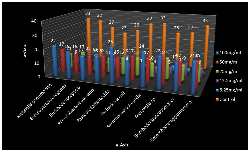
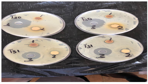
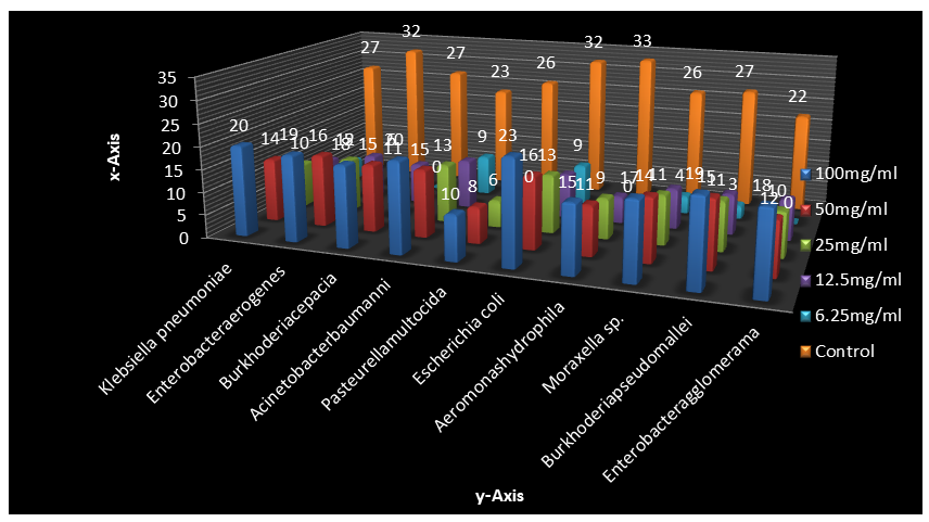
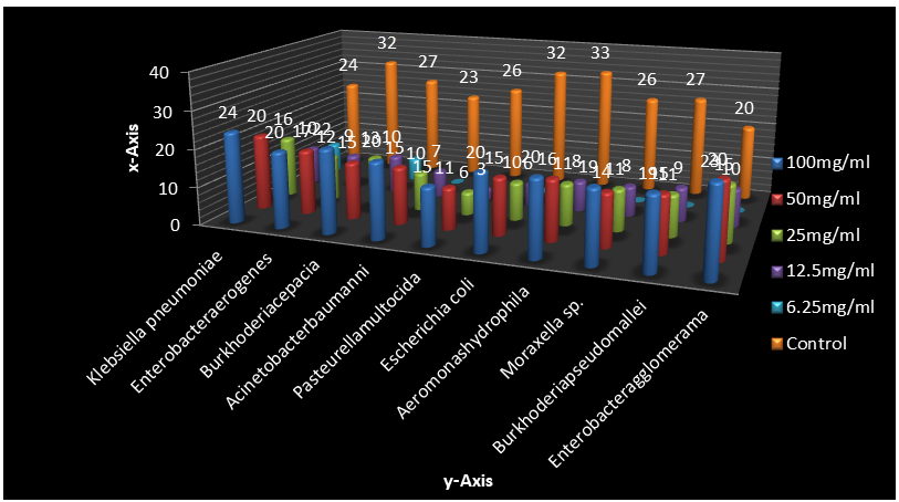
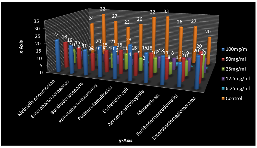
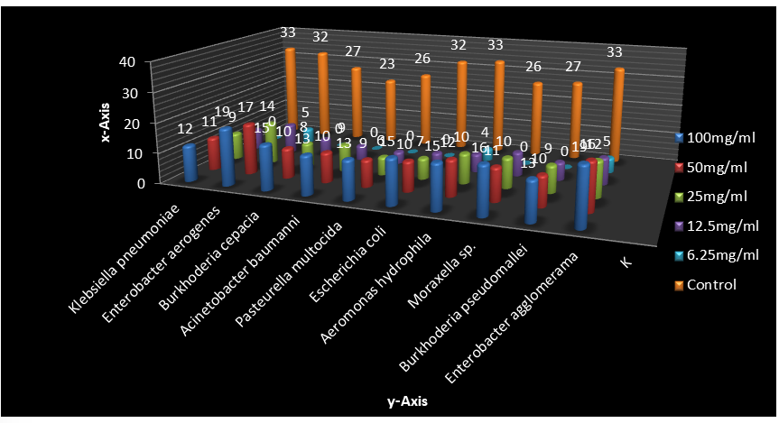
Figure 8 shows the minimum inhibitory concentration and minimum bactericidal concentration of the seed extracts of Adenanthera pavonina seed. It was observed that the value of MIC and MBC were dependent on the bacterial species. The MICs of Aqueous, Distilled ethanol and Ethyl acetate extract of the seeds all falls within the concentration of 6.25mg/ ml and 25mg/ml while the MIC of ethanol seed extract is 12.5mg/ml – 25mg/ml against all the bacteria species. It was observed that the MBC of ethanol seed extract was 25mg/ ml against all the bacterial isolates, while MBCs for Aqueous and Distilled ethanol seed extracts were between 50mg/ml- 100mg/ml. It was further observed that the MBC of Ethyl acetate seed extract was 25mg/ml, -50mg/ml. The MIC of the palm wine extract against the entire test isolates were 50mg/ml and MBC were 100mg/ml.
Key: EMIC = Ethanol seed extract of Adenanthera pavonina Minimum inhibitory concentration, EMBC = Ethanol seed extract of Adenanthera pavonina Minimum Bactericidal concentration, WMIC = Aqueous seed extract of Adenanthera pavonina Minimum inhibitory concentration, WMBC = Aqueous seed extract of Adenanthera pavonina Minimum Bactericidal concentration, DMIC = Distilled Ethanol seed extract of Adenanthera pavonina Minimum inhibitory concentration, DMBC = Distilled Ethanol seed extract of Adenanthera pavonina Minimum Bactericidal concentration, ETMIC = Ethyl acetate seed extract of Adenanthera pavonina Minimum inhibitory concentration, ETHMBC = Ethyl acetate seed extract of Adenanthera pavonina Minimum Bactericidal concentration, PMIC = Palm wine seed extract of Adenanthera pavonina Minimum inhibitory concentration, PMBC = Palm wine seed extract of Adenanthera pavonina Minimum Bactericidal concentration, Mg/ml = milligram/mole.
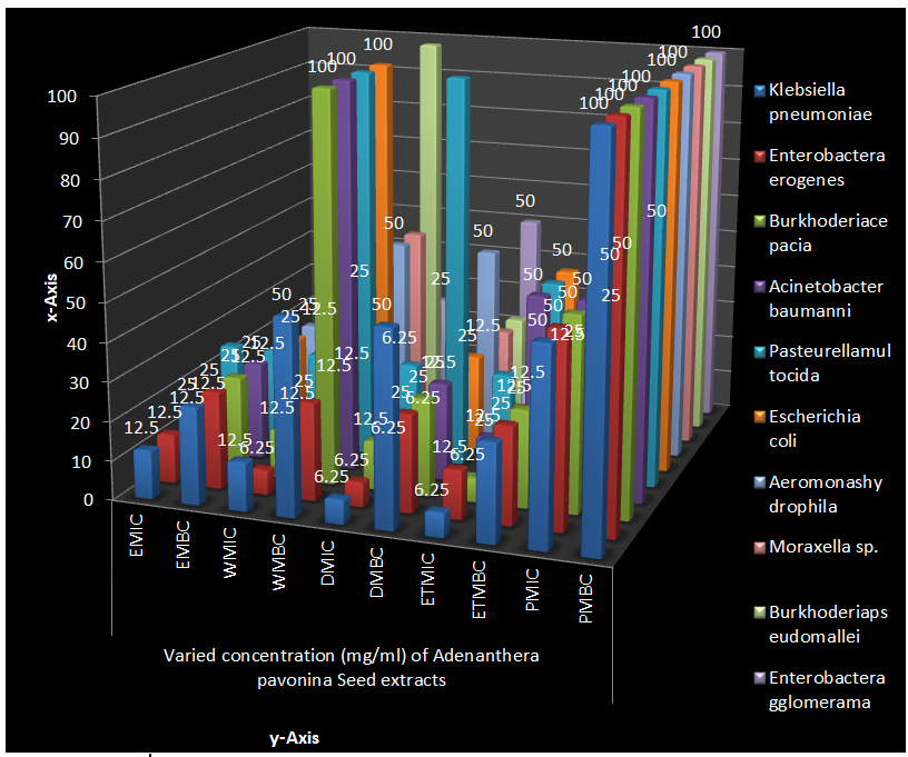
Figure 9 show the growth dynamic of bacteria isolates using ultraviolet spectrophotometer with the wavelength 620λ. It was observed that at 0 hour, Burkhoderia pseudomallei has the highest growth rate of 0.300λ and Pasteurella multocida have the lowest growth rate of 0.190λ. At 24th hour, Escherichia coli have the lowest death rate of 0.195λ and Burkhoderia pseudomallei have the highest death rate of 0.120λ.
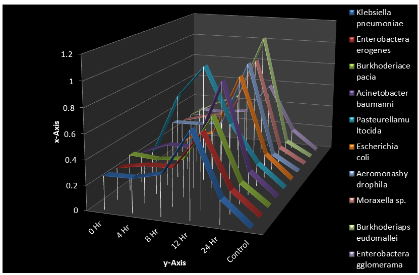
Figure 10 shows the growth dynamic and killing time of bacteria isolates with the addition of absolute ethanol seed extract Adenanthera pavonina using ultraviolet spectrophotometer. In this Figure, it was observed that at 0 hour, Pasteurella multocida has the highest growth rate of
0.142λ and Escherichia coli have the lowest growth rate of 0.056λ. At 24th hour Enterobacter aerogenes has the lowest death rate of 0.097λ and Escherichia coli have the highest death rate of 0.040λ.
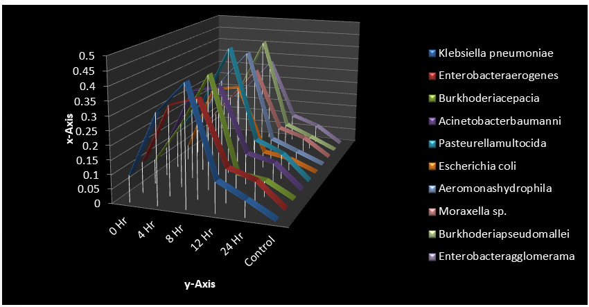
Figure 11 shows the growth dynamic and killing time of bacteria isolates with the addition of distilled ethanol seed extract of Adenanthera pavonina using ultraviolet spectrophotometer. In this Figure, it was observed that at 0-hour Burkhoderia pseudomallei has the highest growth rate of 0.146λ and Escherichia coli have the lowest growth rate of 0.096λ. At 24th hour, Burkhoderia cepacia has the lowest death rate of 0.150λ and Acinetobacter baumanni have the highest death rate of 0.060λ.
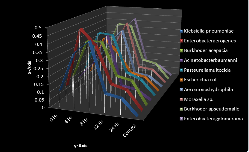
Figure 12 shows the growth dynamic and killing time of bacteria isolates with the addition of aqueous seed extract of Adenanthera pavonina using ultraviolet spectrophotometer. In this Figure, it was observed that at 0 hour, Enterobactera erogenes has the highest growth rate of 0.321λ and Escherichia coli has the lowest growth rate of 0.141λ. At 24th hour Aeromonas hydrophila has the lowest death rate of 0.091λ and Moraxella sp have the highest death rate of 0.048λ.
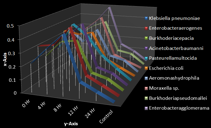
Figure 13 shows the growth dynamic and killing time of bacteria isolates with the addition of Ethyl acetate seed extract of Adenanthera pavonina using ultraviolet spectrophotometer. In this Figure, it was observed that at 0 hour, Pasteurella multocida has the highest growth rate of
0.150λ and Aeromonas hydrophila have the lowest growth rate of 0.102λ. At 24th hour Pasteurella multocida has the lowest death rate of 0.100λ and Burkhoderia pseudomallei have the highest death rate of 0.068λ.
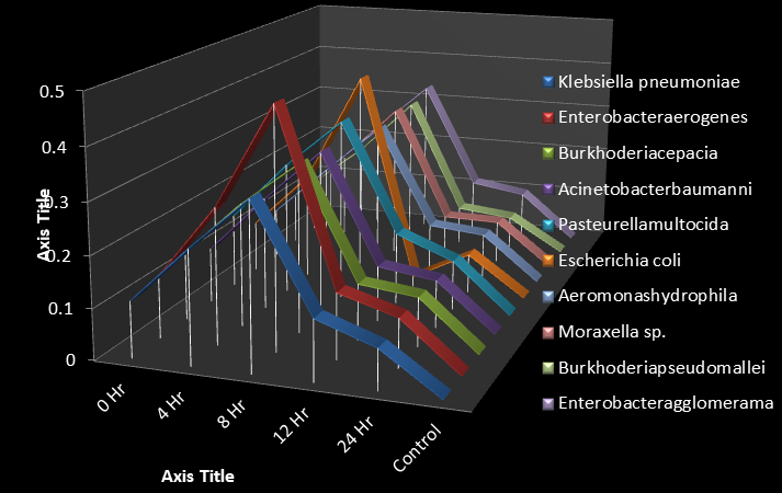
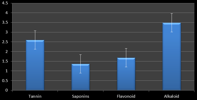
Figure 14 shows the quantitative phytochemical constituents’ analysis of Adenanthera pavonina Seeds extracts. The highest percentage of yield was found in phenol (3.487%) and the lowest is recorded in Cardiac Glycosides with percentage yield of 0.299%.
| Phytoconstituents | ASE | ASD | ASW | ASET |
|---|---|---|---|---|
| Tannin | +ve | +ve | -ve | +ve |
| Saponin | +ve | +ve | -ve | +ve |
| Flavonoid | +ve | +ve | -ve | +ve |
| Alkaloid | +ve | +ve | +ve | +ve |
Table 7: Qualitative Phytochemical Constituents analysis of Adenanthera pavonina Seeds extracts.
Key: +ve = Positive –ve = Negative, ASE = Adenanthera pavonina seed Absolute ethanol extract, ASD = Adenanthera pavonina seed Distilled ethanol extract, ASW = Adenanthera pavonina seed Water extract, ASET = Adenanthera pavonina seed Ethyl acetate extract.
Discussion
The purpose of this research work to measure the zones of inhibition, solvent and growth dynamics of Adenanthera pavonina. Linn. (Seed extract) against selected enteric microorganisms. Microbial infection, mostly those that are associated with intestinal linings are common widespread disease in the world especially in underdeveloped and developed countries where most of its population are always affected by many diseases which may be attributed to different anthropological and natural activities such as drinking of dirty water irrational use of antibiotics, ingestion of contaminated foods and low immunization. Due to rise in resistant to most of currently used medications much effort has been made to subdue and find treatments for these bacterial infections [18, 19].
Seeds of Adenanpthera pavonina.Linn. Plant was extracted using Absolute ethanol, distilled ethanol from palm wine (Elias Guineansis), water, fermented palm wine and Ethyl acetate which were tested for their antibacterial activities against selected enteric microorganism. Ten enteric isolates were tested in this study which includes Klebsiella pneumoniae, Enterobacter aerogenes, Burkhoderia cepacia, Acinetobacter baumanni, Pasteurella multocida, Escherichia coli, Aeromonas hydrophila, Moraxella sp., Burkhoderia pseudomallei, Enterobacter agglomerama.
The use of plant derived drugs for the treatment of different diseases has long been exploited traditionally for decades by herbalists and researcher with a good knowledge of local flora. In the treatment of diseases or infections, medicinal plants are considered as the best alternative to synthetic drugs. Consumed either as a preventive measure or as a viable treatment solution, Phyto-drugs are not prone to certain side effects and easily metabolized by the body system compared with the synthetic drugs. The presence of bioactive compounds in medicinal plants are being studied to help elucidate, which of them individually or collectively are responsible for their antimicrobial potency against selected microbes, thereby confirming the belief that local plants are the platform for traditional African medicine [14]. In Nigeria, native plants are used either in whole or in synergy with other plants as herbal medicine, to develop new treatment to diseases. Like in other developing countries, Nigerian plants are consumed as ‘plants for medicine and plants for food [17]. The therapeutically and dietary needs that specific native plant species meet has been underscored in many developing countries [13].
However, Seed has been used for years in the treatment of infectious diseases. The seed of Adenanpthera pavonina. Possess pharmacologically active ingredients that may be exploited for local development of antimicrobial agents. The phytochemical analysis of the Adenanthera pavonina was also evaluated to discover the phyto-active-chemical components that makes up the plant and responsible for its antimicrobial and therapeutic activities.
The solvent extracts (Absolute ethanolic, distilled ethanol, fermented palm wine and ethyl acetate) extracts of Adenanpthera pavonina. Linn seed examined in this study revealed antimicrobial activities against all the tested bacterial isolates. Absolute ethanol extract shows the highest antimicrobial activity followed by ethanol extracted and distilled from fresh palm wine, fermented palm wine and ethyl acetate in descending order (Absolute ethanol< fresh palm< fermented palm wine< ethyl acetate) The result obtained from this study is similar to a study conducted by Abdu K, Ajayi IA [19, 20] which stated that that the seed Ethanolic extracts of Adenanthera pavonina Linn seed shows excellent antimicrobial potential. Finding of the study reveals that, the activity of seed extracts was better antimicrobial potentials than the plant’s leaf extract [19]. In a similar study, it was reported that ethyl acetate extract of Adenanthera pavonina seeds exhibit significant growth inhibition potential against the selected microorganisms [20]. However, there are differences in the antimicrobial activities as observed in this study, which might be attributed to the amount of the secondary metabolite components extracted by the solvents. It was observed that the palm wine extract has a promising activity of microbial solvent dynamics compared to other solvents used during the course of this research work. The palm wine contains natural acetic and lactic acid which function as a natural acid which is the product of microbial fermentation. This accounts for the activity of palm wine over other solvents. The palm wine has better organoleptic properties compared to other solvent, with is research, palm wine is a good solvent extractor. In this study, it was discovered that, Pasteurella multocida, was the most antimicrobial susceptible organism to the absolute ethanol extract of Adenapthera pavonina Linn seed followed by Aeromonas hydrophila, Klebsiella pneumoniae, and Moraxella sp. while Acinetobacter baumanni and Burkhoderia cepacia were the least antimicrobial susceptible at high concentration which is 100mg/ml and 50mg/ml. The measurements of zones of inhibitions and positive control used in this study which include a commonly used antibiotic agent (Amoxicillin) for the bacterial isolates data are recorded.
The antibacterial activities of the aqueous seed extract of Adenanthera pavonina_Linn seed against the tests bacterial isolates studied were discovered, that most of the isolates were moderately sensitive where _Enterobacter agglomerama and Moraxella sp., has the highest zone of inhibition and Klebsiella pneumoniae, Acinetobacter baumanni, Pasteurella multocida and Burkhoderia pseudomallei were least susceptible.
There is a great antimicrobial activity discovered in this study in distilled ethanol from palm wine extract of Adenanthera pavonina_Linn seed. Escherichia coli isolate was discovered to be the most antimicrobial susceptible isolate while _Aeromonas hydrophila shows the least susceptibility to this extract. Klebsiella pneumoniae was susceptible to ethyl acetate extract of Adenanthera pavonina Linn seed and Pasteurella multocida was least susceptible. Test isolates showed moderate susceptibility to palm wine seed extract of Adenanthera pavonina Linn. At the highest concentration (100mg/ml and 50g/ml) Klebsiella pneumoniae has the highest zone of inhibition value while Burkhoderia pseudomallei have the least zone of inhibition. However, has discovered in this study the zones of inhibition on all of the isolates were higher at the highest concentration of each solvent extract of the plant and became lesser as the concentration of the extract is reduced, a progressive reduction in antimicrobial activity as the concentration reduces from 100mh/ml to 25mg/ml.
In this study, the growth and killing dynamics of the Adenanthera pavonina Linn seed plant against the test isolates was also measured using Ultraviolet spectrophotometric signatures. The addition of antibiotic (Amoxicillin) to the test organism at the 24th hour speed up the death rate of the organisms and also, the addition of solvent seed extract of the plant at the 24th hour also speed up the death rate of the test organisms. The measurement of an exponential bacterial growth curve in batch culture was traditionally a part of the training of all microbiologists; the basic means requires bacterial enumeration (cell counting) by direct and individual (microscopic, flow cytometry) [4], direct and bulk (biomass), indirect and individual (colony counting), or indirect and bulk (most probable number, turbidity, nutrient uptake) methods. In the study, ultraviolet spectrophotometer is used to determine the lag phase, log phase or exponential phase, stationary phase, and death phase.
The qualitative and quantitative phytochemical analysis for the Adenanthera pavonina Linn seed extract was obtained from this study. The presence of medically important and active constituent of plants such as Tannin, Saponin, Flavonoid and Alkaloid was discovered in the Absolute ethanol, distilled ethanol, and ethyl acetate seed extract of Adenanthera pavonina Linn. Also, the presence of Alkaloid was discovered in aqueous extract of Adenanthera pavonina Linn. Seed where Tannin, Saponin, Flavonoid was not detected.
Hence the antimicrobial activity of Adenanthera pavonina Linn Seed was due to the phytochemical constituents present in relatively large proportion. The biological function of flavonoids includes protection against allergies, inflammation, free radicals, platelet aggregation, microbes, ulcers, hepatoxins, viruses and tumors [11, 12]. This may be the reason behind the use of the extracts of this plant in the treatment of intestinal troubles in herbal medicine. Some of the general characteristics of saponin include formation of foam in aqueous solutions, hemolytic activity cholesterol binding properties and bitterness [4]. Alkaloids ranked the most efficient therapeutically significant plant substance. Pure isolated plant alkaloids and their synthetic derivatives are used as basic medicinal agents for their analgesic, antispasmodic and bactericidal effects. They exhibit marked physiological activity when administered to animals. Tannin has a stringent property; it hastens the healing of wounds and inflamed mucous membrane [4]. The presence of tannin in this plant strongly supports its use in treating wounds, burns and hemorrhoids in herbal medicine.
The quantitative phytochemical screening of Adenanthera pavonina Linn seed was also observed. This study revealed that, alkaloid is the most abundant phytochemical in the seed, with 3.487 while Saponins was the least with 1.367. The phytochemicals analysis of explains the constituent of the plant that is killing or inhibiting the growth of tested enteric pathogenic organisms causing diseases. Thus the zone of inhibition shows that the plant (Adenanthera pavonina. Linn. seed) antimicrobial activity and the phytochemicals show the particular component of the plant that is active.
In Conclusion, based on the results obtained in this study, all the solvent extracts showed varying degrees of antimicrobial activity against the test enteric bacterial isolates. This in vitro study demonstrated that some of the medicinal plant extracts like Adenanthera pavonina Linn seed can be as effective as modern medicine to combat the test of the isolates. According to results of this study, it can be concluded Adenanthera pavonina Linn. Seed extract showed broad spectrum of antibacterial activity as evidenced by high zone of inhibition against the test isolates. These results provide evidence that some of the secondary metabolites present in the studied medicinal plants were confirmed as they contained multiple bioactive compounds which might have synergetic impact on inhibition of pathogenic microbes tested. The finding of this study also indicated that Adenanthera pavonina Linn seed extracts for antimicrobial activity it can and may the used for new drug development programs and further study can be developed and isolation of the most potent active compound especially from the most active solvent extracts based on this research work.
Further studies should be done on isolating compounds and fractions of the plant for antimicrobial activity and toxicological screening, in-vivo studies should also be conducted in order to confirm the safety and efficacy of this plant’s secondary metabolites. Awareness should be made concerning this plant to people in the community where it planted to consume moderately and plant new ones to prevent the extinction of the plant.
Acknowledgements
The authors wish to express their appreciation to all the technical staff of the laboratory unit of the Department of Microbiology, Faculty of Science, Adekunle Ajasin University, Akungba-Akoko, Ondo State, Nigeria, for their technical support.
References
-
Ara A, Saleh MM, Hashem MA, Mesbahuddin A, Choudhury M (2019) Phytoconstituents of Adenanthera pavonina Linn from the bark extracts. Beni-Suef Univ J Basic Appl Sci 8: 20.
-
Aprelia HD (2020) Pengaruh skarifikasi Asam Sulfat (H2SO4) dan Giberelin (GA3) terhadap pematahan dormansi biji saga pohon (Adenanthera pavoninaL.) Undergraduate thesis, Universitas Islam Negeri Maulana Malik Ibrahim. 13620117.
-
Mohammeda A, Gbonjubola VA, Koorbanallyc NA, Islama MS (2017) Inhibition of key enzymes linked to type 2 diabetes by compounds isolated from Aframomum melegueta fruit. Pharmaceutical Biology 55(1): 1010- 1016.
-
Xu Q, Qu J, Song B, Liu F, Chen P, et al. (2019) Lathyrus sativus originating from different geographical regions reveals striking differences in Kunitz and Bowman-Birk inhibitor activities. Journal of Agricultural and Food Chemistry 67 (29): 8119-8129.
-
Kuruppu PP, Anchala I, Charitha L, Goonasekara (2019) Medicinal plants commonly used against cancer in traditional medicine formulae in Sri Lanka. Saudi Pharmaceutical Journal 27(4): 565-573.
-
Jaganathan GK, Yule KJ, Biddick M (2018) Determination of the water gap and the germination ecology of Adenanthera pavonina (Fabaceae, _Mimosoideae_); the adaptive role of physical dormancy in mimetic seeds. AoB Plants 10(5): 1-11.
-
Kamaliyah SN, Ifar S, Kusmartono, Chuzaemi S (2019) Effect of Cutting Interval and Cutting Methods on Adenanthera pavonina L. Annual Forage Yield. Journal of Global Biosciences 8(12): 6642-6654.
-
Krishnan HB, Kim S, Pereira AE, Jurkevich A, Hibbard BE (2022) Adenanthera pavonina, a potential plant- based protein resource: seed protein composition and immunohistochemical localization of trypsin inhibitors. Food Chemistry 12(13): 100253.
-
Nayara MPA, Gustavo P, Henrique S, Fábio NS, Lívia GP, et al (2019) Enzymatic treatment improves the antioxidant and antiproliferative activities of Adenanthera pavonina L. seeds Biocatalysis and agricultural biotechnology 18: 101002.
-
Sharma A, Goyal R, Sharma L (2016) Potential biological efficacy of Pinus plant species against oxidative, inflammatory, and microbial disorders. BMC complementary and alternative medicine 16(1): 35.
-
Osuntokun OT (2021) Gram Negative Bacteria (GNB) Isolated from Used Home-made and Surgical Nose/Face Mask by Local Residents of Akungba Akoko, Ondo State, a Threat to Life and False Sense of Protection against SARS-CoV-2 (COVID-19). Asian Journal of Advanced Research and Reports 15(10): 1-17.
-
Olumekun VO, Osuntokun OT, Ajayi AO, Omotuyi IO, Olonisakin A (2022) Assessment of Antibacterial and Gas Chromatography/Mass Spectrophotometric Analysis (GC-MS) Profile of Purified Volatile Compounds from Multi-phases Solvent Extraction Method from Stem Extract of Aframomum melegueta [Roscoe] K. Schum. South Asian Research Journal of Natural Products 5(2): 68-102.
-
Osuntokun OT, Azuh VO, Adejoro BF, Akele EO (2021) Antimicrobial Spectrum, Growth/ killing kinetics, Conventional/ Molecular assay and Ultraviolet Spectrophotometer Signatures of Characterizing Shigella Flexneri and Enterococcus Faecalis and Isolated from Swine House isolates”, International Journal of Pharmacy and Infections Therapy Int J Phar Inft Thrp 2(10): 1018-1034.
-
Osuntokun OT, Olumekun VO, Ajayi AO, Omotuyi IO, Olonisakin A (2020) Assessment of In-vitro antioxidant/ enzymes inhibitory potentials of Aframomum melegueta [Roscoe] K. Schum (grains of paradise) leaf, stem bark, seed bark and seed extracts. Archives of Current Research International; 20(2): 40-57.
-
Wechsler S, Smith D (2018) Has resistance taken root in US corn fields? Demand for insect control. American Journal of Agricultural Economics 100(4): 1136-1150.
-
Mohammeda A, Gbonjubola VA, Koorbanallyc NA, Islama MS (2017) Inhibition of key enzymes linked to type 2 diabetes by compounds isolated from Aframomum melegueta fruit. Pharmaceutical Biology 55(1): 1010- 1016.
-
Susilowati A, Ahmad AG, Rangkuti AB, Harahap MM, Sujarwo, et al (2021) The health status of saga tree (Adenanthera pavonina) in campus area of Universitas Sumatera Utara (USU). inference Series: Earth and Environmental Science 782(4): 042029.
-
Uzama D, Bwai MD, Sunday AT (2012) The phytochemicals proximate and elemental analyses of Securinega virosa leaf extracts. Research Journal in Engineering and Applied Sciences 1(6): 351-354.
-
Abdu K, Adamu M (2020) Isolation, Bioactivity and Charaterisation of 3-Ethynyl-5-(2, 3 dehydropyr role) Pyridine from the Stem Bark of Adenanthera pavonina. Chem Sci Int J 28(4): 1-10.
-
Ajayi IA, Ajibade O, Oderinde RA (2011) Preliminary Phytochemical Analysis of Some Plant Seeds. Research Journal of Chemical Sciences 1(13): 58-62.
- Acido Labile or Gastro Irritant Apis and Enteric Release in Galenic Practice: An Overview
- A Study on Knowledge, Attitude and Practice of Hand Hygiene among Healthcare Professionals at a Tertiary Care Hospital, India
- Influence of Inoculum Concentration on In Vivo Incubation Period of Emmia lacerata, Pathogenesis and Management of Wilt in Pepper (Capsicum annuum L.)
- Vanilla’s Chemistry
- Marine Anti-Cancer Compounds and Adverse Effects of Global Warming on Oceans: An Overview
- Serological Investigation of Chikungunya Virus Antibody among Malaria-Suspected Febrile Patients in Some Healthcare Facilities in Rivers State