Immobilization of Heparin on Bacterial Nanocellulose Hydrogels Induces Tubulogenesis of Human Endothelial Cells
Models that mimic the angiogenesis initial processes, as adhesion, migration, proliferation and tubulogenesis, are extremely valuable for investigating the action of new anti-cancer drugs. There still is a need for an angiogenesis model that reflects in vivo environment. To address this challenge, we developed a 3D matrix well-defined based on covalently immobilization of heparin (HEP) on bacterial nanocellulose (BNC) hydrogels. Successful immobilization was confirmed by qualitative and quantification analysis. Human umbilical vein endothelial cells (HUVECs) were seeded on bottom and top surfaces of BNC and BNC-HEP hydrogels and cells behavior were analyzed. The bottom surfaces of BNC-HEP hydrogels were able to support cell adhesion and promote proliferation and tubulogenesis formation. Results here presented indicate that the tubulogenesis process could be controlled by physico-chemical properties of the developed hydrogel. The interaction between the bioactive molecule, heparin, and the particular microstructure of BNC induced tubulogenenic behavior of HUVECs in vitro.
AC and Porto LM
Chemical and Food Engineering Department, Federal University of Santa Catarina,
Brazil
Catarina, Brazil, Email: emily_reis@intelab.ufsc.br
tubulogenenic behavior of HUVECs in vitro.
Keywords: Angiogenesis; Bacterial Nanocellulose; Heparin; Huvecs; Tubulogenesis; 3d Matrix
Introduction
One of the main phases of the anti-tumor drugs pre- clinical-trial have been related to the quantification of angiogenesis process by in vitro or in vivo assays. Moreover, pre-clinical trial phases also investigate the endothelial cells behavior analyzing their potential to adhere, migrate, proliferate and form tubulogenic networks [1]. Actually, tubulogenesis assays have been Immobilization of Heparin on Bacterial Nanocellulose Hydrogels Induces Tubulogenesis of Human Endothelial Cells performed on 3D commercial matrices that reproduce tumor environment. However, the use of those matrices for cancer drugs screening produce inconclusive results due to their chemical variability from batch-to-batch which difficults the standardization of pre-clinical tests [2]. Consequently, there is an urgent demand regarding to the development of 3D matrices with a well-established chemical (minimal complexity) and microstructural Ann Adv Biomed Sci
proprerties that mimics the extracellular matrix (ECM) in cancer development or in a healthy tissue.
Bacterial nanocellulose (BNC) is a hydrogel synthesized during fermentation of bacteria from Gluconacetobacter genus, that resembly ECM due to its nanofibrous 3D microstructure. On static Fermentation the bacteria secrete BNC hydrogels containing two distinct surfaces. A surface characterized by a high density of fibers (top surface) and another (opposite) surface presents higher porosity (bottom surface) [3].
Structural ECM proteins, growth factors and glycosaminoglycans (GAGs), as heparin, have been associated as components responsible for the induction of neovascularization by the recruitment of endothelial cells [4]. The use of heparin on the development of tissue engineering scaffolds has shown that GAGs enhance significantly angiogenesis independent of the addition of other exogenous growth factors [5]. Thus, in this study we propose to perform the chemical immobilization of heparin, on BNC hydrogels as an alternative to induce endothelial cells adhesion, proliferation which could provide an ideal microenvironment to endothelial cells organize themselves as tubulogenenic networks assuming there in vivo behavior.
Experimental
Materials
Reagents and chemicals were purchased from Sigma- Aldrich do Brasil Ltd. (São Paulo, SP). Culture media and supplements were purchased from Thermo Fisher Scientific do Brasil Ltd. (São Paulo, SP). Heparin (HEPAMAX-S® sodium heparin - 5000 IU/mL) was supplied by Blau Farmacêutica S.A. (Cotia, SP).
BNC-HEP Hydrogels Synthesis
Gluconacetobacter hansenii (strain ATCC 23769) was cultured in a sterile medium composed of mannitol (25 g), yeast extract (5.0 g), and bactopeptone (3.0 g), diluted in 1 L of water and pH adjusted to 6.5. Mixture was stirred and transferred to 24 wells plates (1 mL/well). After four days, on static culture conditions at 26°C, hydrogels were removed and purified in 0.1 M NaOH solution at 50°C for 24 h and then rinsed with distilled water to pH 6.5. Finally, the hydrogels were sterilized by autoclaving (121°C for 20 min).
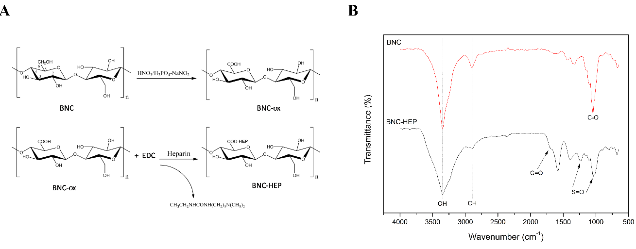
BNC hydrogels were oxidized, with HNO3/H3PO4- NaNO2, to convert free hydroxyl groups of BNC in carboxyl groups [6]. Those carboxyl groups were activated with EDC (1-ethyl-3- (3-dimethylaminopropyl) carbodiimide) allowing the covalent immobilization of heparin (Figure 1a). Briefly, oxidized BNC were immersed in 0.1 M citrate buffer solution (pH 4.8) containing 2 mM of EDC. Those hydrogels were shaking during 24 h at 4°C and after washed with deionized water. Samples were placed in citrate buffer (pH 4.8) containing 1000 IU heparin (v/v) for 24 h at 4°C with shaking. Then, BNC- HEP hydrogels were washed with alkaline phosphate buffered saline and rinsed with distilled water. Hydrogels were lyophilized at -50°C for 24h to perform the chemical and microstructural characterization and they were sterilized by UV before to perform in vitro tests.
Characterization of BNC-HEP
The functional groups on BNC and BNC-HEP were analyzed by Fourier Transformed Infrared Spectroscopy (FTIR). Infrared spectra were recorded on an Agilent spectrophotometer (model Carry 600), with a resolution of 4 cm-1, in a range of 4000-600 cm-1, and using attenuated total reflectance. Scanning electron microscope (SEM) was performed to analyze the microstructure of top and bottom surfaces of BNC and BNC-HEP, using a JEOL JSM-6390LV microscope at 10 kV. Toluidine blue colorimetric assay was used to determine the amount of heparin immobilized on BNC-HEP. The amount of heparin immobilized on BNC-HEP hydrogels was quantitated in comparison with the calibration curve prepared [7].
Citotoxic potential of BNC-HEP hydrogels was evaluated following the direct contact assay. For that, immortalized murine fibroblast cells (L929) were cultured in DMEM (Dulbecco’s Modified Eagle’s Medium) supplemented with 10% of FBS and 1% penicillin/streptomycin. L929 cells were seeded in each well of 24 wells culture plate at 9.0 × 10³ cells/cm2 for 24 h in a humidified atmosphere (5% CO2) at 37°C. BNC hydrogels, non-cytotoxic material, were used as control group [8]. After that, BNC and BNC-HEP hydrogels were added into each well. After 1, 3 and 7 days of in vitro culture, the metabolic activity of L929 cells were determined by MTS [3-(4,5-dimethylthiazol-2-yl)-5- (3carboxymethoxyphenyl)-2-(4-sulfophenyl)-2H- tetrazolium] colorimetric assay from Promega Biotecnologia do Brasil, Ltda. (São Paulo, SP), performed according to the manufacturer’s instructions.
Adhesion, Proliferation and Tubulogenesis Induction of Huvecs on Hydrogels
HUVECs were seeded at a density of 105 cells/cm2 on the bottom surface of BNC-HEP hydrogels to evaluate tubulogenesis induction. After 24 h, in a humidified atmosphere containing 5% of CO2 at 37°C, samples were fixed with 4% of formaldehyde for 30 min at room temperature. Phase contrast images were taken using an Olympus BX4 microscope (Olympus America Inc.).
Statistical Analysis
Data were analyzed statistically by one-way analysis of variance (ANOVA) with Tukey test. Values represent the mean ± standard medium error, with p< 0.05. All experiments were performed in triplicate at two separated experimental times.
Results
Functionalization of BNC Hydrogels
Characteristic bands of BNC appeared at 3345 cm-1 and 2898 cm-1, where the absorption band assigned to hydroxyl groups and hydrogen bonds, respectively [9] (Figure 1b). BNC-HEP showed a broad absorption at 3100–3500 cm-1 which was assigned to N–H stretching absorption and hydrogen bonded hydroxyl groups; Other heparin characteristic bands were observed at 1701 cm−1 (C=O) and 1248 cm-1 (S=O) [9, 10].
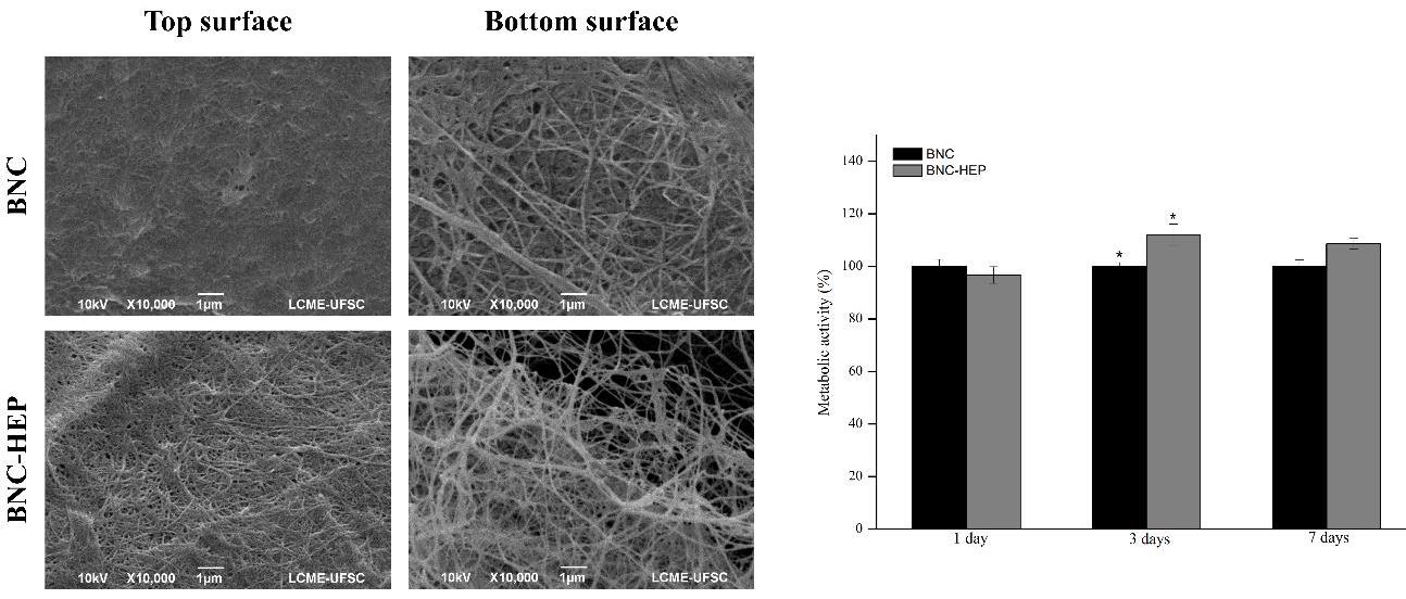
SEM micrographs related to the top and bottom surfaces of BNC and BNC‒HEP hydrogels were shown in Figure 2a. BNC hydrogels were characterized by an entangle arrangement of BNC fibers on top surface and with a porous arrangement of fibers on the bottom surface. The top surface of BNC-HEP kept the same microstructure observed on the top surface of BNC. Moreover, the bottom surface of BNC-HEP showed a similar microstructure observed on the bottom surface of BNC, however within more pores. In addition, results showed that BNC and BNC-HEP hydrogels did not induce any citotoxic effect on L929 cells during 7 days of in vitro culture Figure 2b. The amount of heparin immobilized on BNC hydrogels was quantified by toluidine blue dye assay which revealed a density of HEP immobilization corresponding to 4.7 ± 0.2 μg⋅cm-2 approximately 1% of BNC dry weight.
Tubulogenesis Induction
After 24 hours of culture, HUVECs were aligned and organized as tubulogenic networks on the bottom surface of BNC-HEP. In detail, Figure 3a showed the formation of endothelial tubulogenic networks containing loops and branch points. Figure 3b showed the presence of endothelial tip cells, characterized by cell protrusions called filopodia followed by endothelial stalk cells.
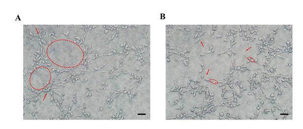
bars represent 100 µm.
Discussion
Tubulogenesis assays have been usually carried out by seeding endothelial cells on a natural commercial ECM (from animal origin, such as Matrigel®) [11]. These commercial ECM contain many pro-angiogenic factors which make it difficult to assess the real influence of drugs on the tubulogenesis process [12]. In this context, we propose the development of a novel 3D tubulogenesis matrix (BNC-HEP hydrogel) with no chemical variation to perform the screening of anti-tumoral drugs and also to study the behavior of endothelial cells (adherence, proliferation and morphology).
SEM micrographs revealed that microstructural differences between bottom and top surface of BNC agree with the observations already published [3]. Yizao Wan and collaborators adsorbed heparin on BNC hydrogels, however they did not observed any microstructural difference between BNC and BNC immersed in heparin solution [10]. Those results suggested that the oxidation process used in our work to immobilize chemically heparin may change the microstructure of BNC-HEP. As result, we found that the immobilization of HEP by oxidative reaction became the bottom surface of BNC-HEP more porous than BNC. In addition, it was observed that the BNC surfaces (top and bottom) interfere directly on HUVECs cell behavior, which was evidenced also by Berti and colleagues [3].
In vitro tests HUVEC cells normally organized themselves as tubes, branch points and loops that can be used to quantify tubulogenesis process [12]. When 105 cells/cm2 HUVECs were seeded on the bottom surface of BNC-HEP, they auto-organize themselves establishing endothelial vascular networks (tubulogenesis) with several capillar sprouts, after 24 h. Capillary sprouts have been composed by the presence of endothelial tip, that coordinate cells alignment, and were followed by aligned proliferative cells, denominated stalk cells [13, 14]. Thus, the presence of endothelial stalk cells indicates high cell proliferation on BNC-HEP. Further investigations will be performed to analyze the influence of known anticancer drugs on the tubulogensis formed on BNC-HEP hydrogels. Our results comproved that endothelial vascular networks could be in vitro designed by chemical immobilization of HEP in bottom surface of BNC hydrogels.
Acknowledgements
The authors thank the Brazilian National Council for Scientific and Technological Development (CNPq) and CAPES, Brazil, for the financial support. The authors also thank the Analytical Laboratory units at the Federal University of Santa Catarina (UFSC), LCME, LACERT and EQA.
References
-
Hait WN (2010) Anticancer drug development: the grand challenges. Nat Rev Drug Discov 9(4): 253-254.
-
Prestwich GD (2007) Simplifying the extracellular matrix for 3-D cell culture and tissue engineering: A pragmatic approach. J Cell Biochem 101(6): 1370- 1383.
-
Berti FV, Rambo CR, Dias PF, Porto LM (2013) Nanofiber density determines endothelial cell behavior on hydrogel matrix. Mater Sci Eng C Mater Biol Appl 33(8): 4684-4691.
-
Afratis N, Gialeli C, Nikitovic D, Tsegenidis T, Karousou E, et al. (2012) Glycosaminoglycans: Key players in cancer cellbiology and treatment. FEBS J 279(7): 1177-1197.
-
Rajangam K, Behanna HA, Hui MJ, Han X, Hulvat JF, et al. (2006) Heparin binding nanostructures to promote growth of blood vessels. Nano Lett 6(9): 2086-2090.
-
Kumar V, Yang T (2002) HNO3/H3PO4-NANO2 mediated oxidation of cellulose-Preparation and characterization of bioabsorbable oxidized celluloses in high yields and with different levels of oxidation. Carbohydrate Polymers 48(4): 403-412.
-
Bae JS, Seo EJ, Kang IK (1999) Synthesis and characterization of heparinized polyurethanes using plasma glow discharge. Biomaterials 20(6): 529-537.
-
Pértile RAN, Moreira S, Gil RM, Correia A, Guãrdao L, et al. (2013) Bacterial Cellulose :Long-Term Biocompatibility Studies. J Biomater Sci Polym Ed 23(10): 1339-1354.
-
Wan YZ, Huang Y, Yuan CD, Raman S, Zhu Y, et al. (2007) Biomimetic synthesis of hydroxyapatite/bacterial cellulose nanocomposites for biomedical applications. Materials Science & Engineering 27(4): 855-864.
-
Wan Y, Gao C, Han M, Liang H, Ren K, et al. (2011) Preparation and characterization of bacterial cellulose/heparin hybrid nanofiber for potential vascular tissue engineering scaffolds. Polym Adv Technol 22(12): 2643-2648.
-
Kleinman HK, Martin GR (2005) Matrigel: Basement membrane matrix with biological activity. Semin Cancer Biol 15(5): 378-386.
-
Irvin MW, Zijlstra A, Wikswo JP, Pozzi A (2014) Techniques and assays for the study of angiogenesis. Exp Biol Med 239(11): 1476-1488.
-
Smet DF, Segura I, Bock KD, Hohensinner PJ, Carmeliet P (2009) Mechanisms of vessel branching: Filopodia on endothelial tip cells lead the way. Arterioscler Thromb Vasc Biol 29(5): 639-649.
-
Wang J, Wan Y, Huang Y (2012) Immobilisation of heparin on bacterial cellulose-chitosan nano-fibres surfaces via the cross-linking technique. IET Nanobiotechnology 6(2): 52-57.
- Origin, Evolution, and Functional Impact of Short Insertion- Deletion Variants in Human Genomes: A Review
- Harnessing Molecular Glues for Next-Generation Vaccine, Cancer and Cardiovascular Disease Drug Development: A Comprehensive Review
- Lateral Cervical Epidermal Inclusion Cyst in a Paediatric Patient: A Rare Case Report
- Malarial Plasmodium Falciparum with Hepatitis B and C Virus Infections among Blood Donors in Ife Central Local Government Area, Ile Ife, Osun State, Nigeria
- Withanolides and Withaferin A- What’s next in Ashwagandha Research
- Designing of Dual Pulse Photoacoustic Tomography for Imaging of Drug-Response and Tumor Growth