Effect of Extended Toe Range of Motion on Muscle Stiffness after Exercise
Fatigue and high-intensity training induce muscle strain and increase muscle stiffness. Increased muscle tone and muscle stiffness are factors in injury and disability and require prompt palliative care. However, muscle stiffness is not measured in everyday life, leading to injuries and disabilities in the face of disappointment. In looking at routine prevention, we wondered whether muscle stiffness could be reduced from extended toe range of motion. Walking is a complex, continuous movement, but most common shoes on the market compress the plantar arch cap and toes. We conducted a prospective cohort study in which 20 subjects underwent post-load training and then wore flat shoes to measure muscle stiffness. 20 subjects performed a 10-kilometre run, and 30 subjects performed a 30-kilometre run and 20 subjects performed a 30-kilometre run. km of running and 30 min of walking. The intervention group wore flat shoes, while the control group wore their daily shoes. 30-minute preand post-walking muscle stiffness measurements of the lateral head of the gastrocnemius, quadriceps and lumbo-dorsal fascia were taken to test whether the intervention group’s muscle stiffness could be reduced. The results showed that the increase in muscle stiffness of the lateral head of the gastrocnemius muscle in the intervention group was inhibited. In addition, the toe area was found to be extended by wearing flat-shaped shoes.
Introduction
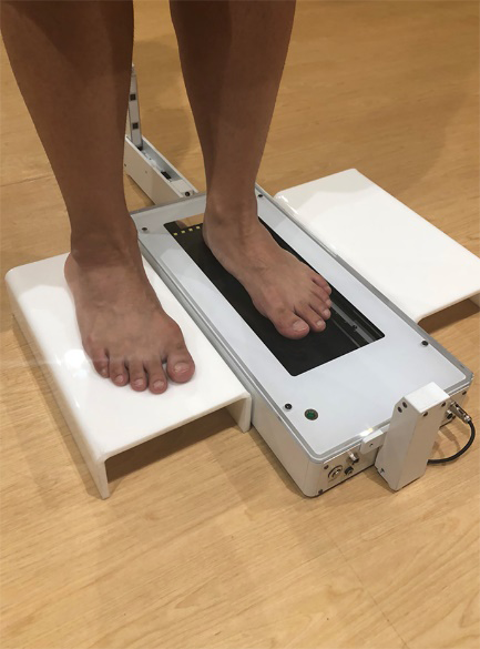
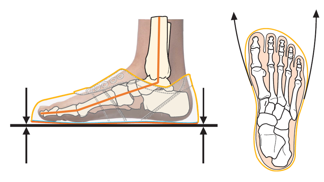
Walking is an action consisting of a series of complex movements. The human leg is intertwined with numerous bones and muscles, and even movements that appear simple can place a high burden on the human body [1]. According to the Ministry of Health, Labour and Welfare’s H30 annual National Health and Nutrition Survey, the average number of steps taken per day by Japanese adults is 6794 steps for men and 5942 steps for women, and for running over 10 km, it is said to exceed 2000 steps, although there are individual differences [2]. This is compounded by a decrease in walking opportunities in Japan, where exercise habits have declined due to Covid19 [3]. Especially in high-impact exercise such as running, shoes, which serve as an interface with the ground, may have a significant impact on the human body. Increased muscle stiffness is an indicator of fatigue, and it is desirable to prevent increased muscle stiffness from inducing injury or disability [4]. Failure to do so may result in separation of the flesh or secondary injury, requiring massage or other care [5, 6]. While investigating shoes worn while walking, we found that many of the common shoes in circulation press on the plantar arch cap and toes [7]. In particular, common shoes are designed with a high toe and plantar arch cap area [8]. This is intended to reduce motion load by covering the foot motion. However, we hypothesized that this restricted foot motion and thus triggered an increase in muscle stiffness. In previous studies, we have found that flat-shaped shoes increase toe movement [9]. Therefore, we wondered if it would be possible to extend the toe area by wearing flat shoes, and to suppress the increase in muscle stiffness by increasing toe movement. Although care therapies have been developed to alleviate the increase in muscle stiffness, it is desirable to be able to prevent it on a daily basis without having to spend time on care. A prototype shoe with a flat shape is shown in Figure 1.
Methods
Twenty subjects underwent high-intensity training, then were divided into 10 intervention and 10 control subjects. A prospective cohort experiment was conducted with the intervention group wearing the prototype shoes. The high- intensity training session consisted of a 10-km run (45 minutes). The intervention group had a mean age of 20.3 years (6 males and 4 females), and the control group had a mean age of 20.2 years (6 males and 4 females). The study was conducted after obtaining approval for “gait analysis using wearable sensors” from the ethical review of human subjects at Maebashi Institute of Technology. The approval number is 22-009.
The intervention group wore prototype shoes, and the control group wore their daily shoes. 30 minutes of walking was performed before and after the 30-minute walk, and muscle stiffness of the lateral head of the gastrocnemius, quadriceps and lumbar dorsal fascia were measured by PEK- 1 to verify whether the increase in muscle stiffness of the intervention group could be suppressed. Muscle stiffness of the lateral head of the gastrocnemius, quadriceps and lumbo- dorsal fascia were obtained at 0, 10, 20 and 30 minutes. A foot scanner was also used to investigate the effect of the prototype shoes on the plantar arch cap region. Toe ground contact surfaces were extracted from the infrared sensor of the foot scanner, and the increase in ground contact surfaces before and after walking was measured.
Results
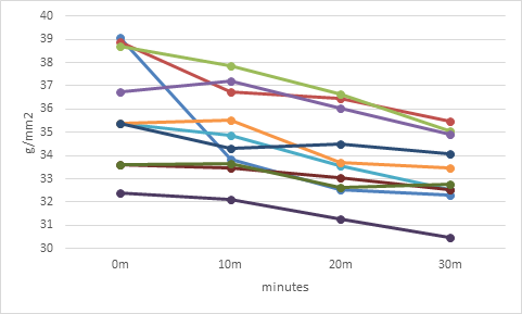
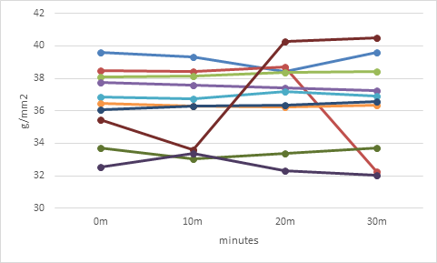
Muscle stiffness of the lateral head of the gastrocnemius, quadriceps and lumbo-dorsal fascia were obtained at 0, 10, 20 and 30 minutes, respectively, and tested using Two-way Factorial Analysis of Variance (TFA). The results showed that the hypothesis of no difference between the two groups was rejected at p-value=0.013 in the intervention and control groups for the lateral head of the gastrocnemius muscle. The hypothesis of no difference between the two groups was rejected at p-value = 0.00000131 for the dorsal lumbar fascia intervention group and the control group. However, the hypothesis of no difference between groups could not be rejected because the p-value exceeded 0.05 for the 0-, 10-, 20-, and 30-minute group comparisons. In terms of muscle stiffness of the lateral head of the gastrocnemius muscle, the intervention group is shown in Figure 3 and the control group in Figure 4.
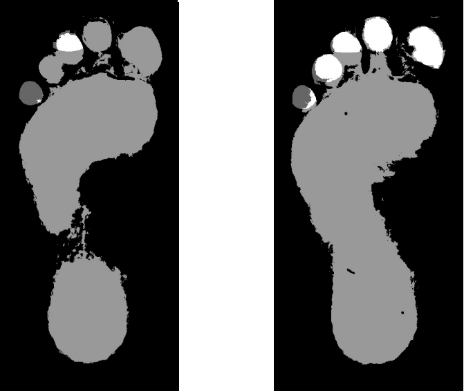
Foot scanner analysis was performed before and after walking in the intervention and control groups. The toes were automatically extracted from the ground surface using an infrared sensor; a normal distribution could not be assumed due to the small number of N. The Wilcoxon signed-rank sum test was used to test for responsiveness of the data. In the intervention group, the hypothesis of no difference before and after walking was rejected with a p-value = 0.0128. In the control group, the hypothesis of no difference before and after walking could not be rejected because the p-value exceeded 5%. Figure 5 shows an example of a scan image before and after walking in the control group.
Conclusion
IEfforts to prevent injury through shoe shape were seen as early as 1986, with Schwellnus using shock-absorbing materials [10]. Fraissler [11] used insoles to treat hallux valgus, demonstrating short-term but effective results. Zhang measured stiffness at various muscle sites and reported correlations with sports injuries [12]. This study focuses on changes during exercise, which differs from the present study, which focuses on daily walking habits. In our study, flexibility in toe movement was found to suppress the increase in muscle stiffness of the lateral head of the gastrocnemius muscle. Since the subjects were only around 20 years old, continued experiments with multiple ages and research conditions are necessary, but the fact that the suppression of muscle stiffness was possible is useful. In the future, we would like to expand the scope of this study to include prevention of frailty, especially by conducting experiments on the elderly.
Acknowledgements
We would like to express our deepest gratitude to Dr. Odagaki of Maebashi Institute of Technology for his support of the experiments and ethical review in the progress of this study. We are extremely grateful to BMZ Inc. for making the prototype shoes. Part of this research was supported by Gunma Prefectural Sports Promotion Division and BMZ Corporation.
References
-
Zelik KE, Honert EC (2018) Ankle and foot power in gait analysis. Implications for science, technology and clinical assessment. J Biomech 25(75): 1-12.
-
Ministry of Health, Labour and Welfare: 2018 National Health and Nutrition Survey Report.
-
Wong AYY, Ling SKK, Louie LHT, Law GYK, So RCH, et al. (2022) Impact of the COVID-19 pandemic on sports and exercise. Asia Pac J Sports Med Arthrosc Rehabil Technol 22: 39-44,
-
Akagi R, Tanaka J, Shikiba T, Takahashi H (2015) Muscle stiffness of the triceps brachii before and after a resistance exercise session: a shear wave ultrasound elastography study. Acta Radiol 56(12): 1487-1493.
-
Weerapong P, Hume PA, Kolt GS (2005) The mechanisms of massage and effects on performance, muscle recovery and injury prevention. Sports Med 35(3): 235-256.
-
Kawai T, Takamoto K, Bito I (2021) Previous hamstring muscle strain injury alters passive tissue stiffness and vibration sense. J Bodyw Mov Ther 27: 573-578.
-
Zhao D, Li Y, Langlois T, Chaudhuri S, Barbic J (2021) ERGOBOSS: Ergonomic Optimization of Body-Supporting Surfaces. IEEE Trans Vis Comput Graph pp: 14.
-
Nakamura K, Kasahara N, Yamanaka T, Takahashi D, Takahashi T, Kaeriyama T (2021) Study of the effect of sole shape on muscle tone after exercise. JSMBE symposium 2021.
-
Nakamura K, Takahashi D, Yamanaka T, Takahashi T, Usuda Y, et al. (2023) Basic Investigation of the Effect of Insole Shape on Leg Skeletal Muscle Mass and Pressure Changes during Walking. Advanced Biomedical Engineering 12: 124-128.
-
Schwellnus MP, Jordaan G, Noakes TD (1986) Prevention of common overuse injuries by the use of shock absorbing insoles. A prospective study. Am J Sports Med 18(6): 636-641.
-
Fraissler L, Konrads C, Hoberg M, Rudert M, Walcher M (2016) Treatment of hallux valgus deformity. EFORT Open Rev 1(8): 295-302.
-
Zhang H, Peng W, Qin C, Miao Y, Zhou F, et al. (2022) Lower Leg Muscle Stiffness on Two-Dimensional Shear Wave Elastography in Subjects With Medial Tibial Stress Syndrome. J Ultrasound Med 41(7): 1633-1642.
- Origin, Evolution, and Functional Impact of Short Insertion- Deletion Variants in Human Genomes: A Review
- Harnessing Molecular Glues for Next-Generation Vaccine, Cancer and Cardiovascular Disease Drug Development: A Comprehensive Review
- Lateral Cervical Epidermal Inclusion Cyst in a Paediatric Patient: A Rare Case Report
- Malarial Plasmodium Falciparum with Hepatitis B and C Virus Infections among Blood Donors in Ife Central Local Government Area, Ile Ife, Osun State, Nigeria
- Withanolides and Withaferin A- What’s next in Ashwagandha Research
- Designing of Dual Pulse Photoacoustic Tomography for Imaging of Drug-Response and Tumor Growth