Development of Pneumothorax Concurrent with Pleural Effusion in a Critically Ill Patient of Ruptured Ectopic Pregnancy with Covid-19 Pneumonia: A Case Report
Development of pneumothorax in COVID-19 pneumonia is a rare occurrence, incidence approximately 1%, while the incidence of pleural effusion varies between 2%-11%. A young patient with COVID-19 pneumonia and unstable hemodynamics was referred to our hospital. She had undergone an emergency laparotomy two days back for ruptured ectopic pregnancy during which she suffered hemorrhagic shock followed by episodes of pulseless ventricular tachycardia and was resuscitated. On her third day of intensive care stay at our centre, she remained critical and developed pleural effusion with concurrent pneumothorax with the complete collapse of the left lung. Here we present a rare complication in COVID-19 pneumonia, pleural effusion and concurrent pneumothorax leading to complete collapse of hemi-lung and its implications on the cardiorespiratory status of our critically ill patient.
Introduction
The symptoms of COVID-19 are ever evolving with each emerging new variant. Amongst complications of COVID-19 pneumonia, the incidence of pleural effusion varies between
2%-11% [1, 2] while pneumothorax is relatively rare with incidence of approximately 1% [3, 4]. Only a few cases are described in literature where unilateral pleural effusion and pneumothorax appeared almost simultaneously in patients with COVID-19 pneumonia [3, 5, 6]. Here we present, a case of development of pleural effusion with simultaneous pneumothorax leading to complete collapse of left lung and mediastinal shift, in a post-cardio-pulmonary resuscitation (CPR) revived patient. Prior consent, for publication of the case, was obtained from the patient.
Case Presentation
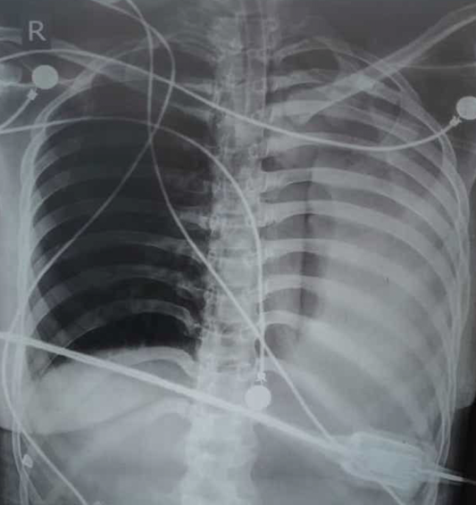
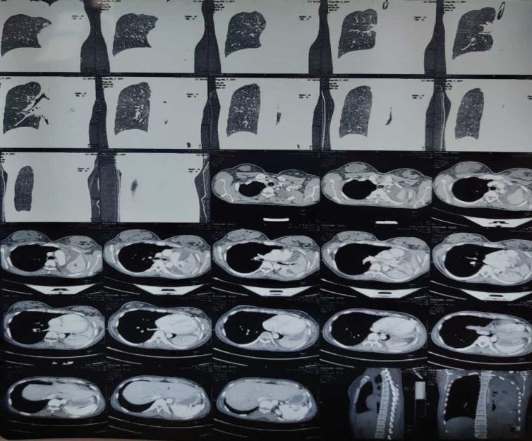
A 22-year-old female, status post-emergency laparotomy for cornual ectopic pregnancy with uterine rupture and hemoperitoneum, was referred to our hospital (throat swab reverse transcriptase polymerase chain reaction confirmed COVID-19). At presentation, she had an unstable hemodynamics with heart rate (HR) of 145/minute and blood pressure (BP) of 80/40 mmHg on infusion noradrenaline. She had peripheral oxygen saturation (SpO2) of 90% with fraction of inspired oxygen (fio2) of 1, on mechanical ventilation. Two days back she presented to nearest hospital with complain of bleeding per vagina. Her lower abdomen ultrasound showed cornual ectopic pregnancy, uni-cornuate uterus with left cornual rent and hemoperitoneum. She underwent emergency laparotomy. Intra-operatively, patient suffered hemorrhagic shock leading to two episodes of pulseless ventricular tachycardia. Later she was referred to our tertiary care center. Post resuscitation in our emergency department, she was shifted to intensive care unit (ICU) wherein her baseline vitals were: HR-140/minute, BP- 98/60 mmHg, SpO2- 90% on fio2 of 1. We put her on lung protective mechanical ventilation with tidal volume of 6 ml/kg and positive end expiratory pressure of 10 cmH2O. Mechanical thrombo-prophylaxis with pneumatic compression device was started. Proning was ruled out due to presence of laprostomy bag in view of burst abdomen. Her routine investigations showed hemoglobin of 9.8 gm/dl, international normalized ratio (INR) of 2.0, C - reactive protein (CRP) of 60 mg/dl, lactate dehydrogenase (LDH) of 563 IU/L, and total bilirubin of 2.6. Rest of the blood investigations was within normal limit. Bedside chest x-ray showed bilateral ground glass opacity congruent with COVID-19 diagnosis. Though her vitals continued to improve, her chest x-ray worsened from the time of admission with increased opacities in both lung fields. On the third day, the patient started showing a decreasing trend in SpO2 (fall from 95% to 85%). Bedside chest-x-ray revealed complete white out of the left lung, obliteration of left cardiac silhouette, suspected mediastinal shift and tracheal deviation, suggesting COVID-19 pneumonia with possible left lung collapse (Figure 1). Contrast-enhanced computed tomography chest confirmed complete collapse of left lung, with mediastinal shift and pleural effusion with pneumothorax (Figure 2). Intercostal drain (ICD) was inserted on left side and 200 ml serosanguinous fluid was drained. Subsequently, her oxygenation improved markedly (SpO2-90%, paO2- 88). Her pleural fluid examination showed: lymphocytes-30%, neutrophil-10%, red blood cell count- 68000, glucose-102mg/dl, LDH-783IU/L, microbiology negative. Prophylactic anticoagulation started after normalization of INR. By day six her routine bedside chest x-ray revealed remarkable improvement in bilateral lung fields with a little subcutaneous emphysema on left chest wall. On the 15th day patient was shifted out of ICU with stable vitals and later discharged for home isolation in good condition.
Discussion
The development of severe pneumonia in COVID-19 patients causes prolonged inflammatory damage to lung parenchyma leading to degenerative changes [7]. Direct invasion by SARS CoV2 virus, inflammation of visceral pleura and recruitment of inflammatory cytokines increase permeability of pleural surface. Pleural effusions likely present a more severe inflammatory, radiological evolution of COVID-19 pneumonia; their frequency increases during the hospital course with the concomitant progression of opacities from ground glass to consolidation on chest imaging [2]. Pleural effusion is usually identified after five to seven days of hospital admission and 11 days after the onset of COVID-19 symptoms [8].
Any comorbid pathology could contribute to pleural effusion; however when COVID-19 patients present with sepsis-like features (hemodynamic instability) often aggressive intravenous fluid administration leads to a clinical state of fluid overload. This can precipitate bilateral infiltrates and pleural effusion, attributable to depressed cardiac function, overwhelming systemic inflammatory response and third spacing of fluids.
An added ischemic insult to lung parenchyma, activation of fibroblasts, and lung fibrosis with inflammatory storm lead to exudates into alveoli and airway; this leads to check- valve obstruction and cyst formation in small airways. The progression of cyst formation from areas of consolidation has been radiologically corroborated by many case studies [9]. Mechanical ventilation appears to be a prominent risk factor for development of pneumothorax in COVID-19 patients. However pneumothorax has been reported in non- ventilated or spontaneously breathing COVID-19 patients [10]. Thus barotrauma alone cannot be an explanation for it. Spontaneous pneumothorax generally does not appear along with pleural effusion on X-ray as the increase in pleural pressure caused by pneumothorax inhibits transfer of interstitial fluid into pleural space. There are a limited number of reports, where spontaneous pneumothorax developed with pleural effusion [11].
Our patient presented to us in a critical state, already on mechanical ventilation and inotropes with unstable hemodynamics. She further went on to develop pleural effusion and pneumothorax on fifth day of her illness, which correlates with findings of Chong, et al. [8]. An important finding in our case was raised inflammatory markers, which is consistent with COVID-19-induced lung injury. Prophylactic anticoagulation was not given as per the hematology consultation due to raised INR at presentation. After seven days, INR returned to normal range and prophylactic anticoagulation was added. Aggressive resuscitative measures due to initial hemorrhagic shock along with severe inflammatory damage to lung parenchyma could be a reason for development of pleural effusion and pneumothorax in our patient.
In conclusion we would like to stress that although pleural effusion and pneumothorax are amongst rare complications of COVID-19 pneumonia, its implications on an already compromised cardio-respiratory status can be disastrous. These complications can be self-limiting or may require treatment. A timely diagnosis can be made with help of bedside chest-x-ray or a computed tomography scan. Mindfulness about these rare yet encountered complications can help us intervene and provide appropriate medical care to patient. More literature however, is required to clearly understand the pathophysiology of pleural effusion and pneumothorax occurring simultaneously in COVID-19 patient.
Author’s Contribution
- Sony Sony: This author helped in design of the work, substantial contributions to the conceptions, the acquisition of data, drafting the work and revising it critically for important intellectual content. All the authors approved the final version to be published and are accountable for all aspects of the work in ensuring that questions related to accuracy and integrity of any part of the work are appropriately investigated and resolved.
- Shivam Shekhar: This author helped in drafting of work, analysis, interpretation of data for the work and revising it critically for intellectual content.
- Pooja Ahuja: This author has substantial contribution to the conceptions and drafting of work and revising it critically for intellectual content.
- Mohaneesh Kumar Saiyam: This author has substantial contribution to the conceptions and drafting of work and revising it critically for intellectual content.
Conflicts of Interest and Sources of Funding
The authors have no conflicts of interest to declare.
Financial Support
None
Details of Previous Presentation
No work related to this article is published or presented.
References
-
Zheng Y, Wang L, Ben S (2021) Meta‐analysis of chest CT features of patients with COVID‐19 pneumonia. J Med Virol 93(1): 241-249.
-
Saha BK, Chong WH, Austin A, Kathuria R, Datar P, et al. (2021) Pleural abnormalities in COVID-19: a narrative review. J Thorac Dis 13(7): 4484-4499.
-
Chen KC, Chen PH, Chen JS (2020) New options for pneumothorax management. Expert Rev Respir Med 14(6): 587-591.
-
Martinelli AW, Ingle T, Newman J, Nadeem I, Jackson K, et al. (2020) COVID-19 and pneumothorax: a multicentre retrospective case series. Eur Respir J 56(5): 2002697.
-
Wakamatsu I, Yatomi M, Uno S, Oishi Y, Ikeuchi H, et al. (2021) A case of a patient with neurofibromatosis type I who developed pneumothorax and eosinophilic pleural effusion after suffering from COVID-19 pneumonia. Radiol Case Rep 16(11): 3504-3508.
-
Divisi D, Zaccagna G, Angeletti C, Cicerone E, De Vico A, et al. (2021) Pleural empyema associated with alveolar‐ pleural fistulas in severe acute respiratory syndrome coronavirus 2. Clin Case Rep 9(6): e04262.
-
Mallick T, Dinesh A, Engdahl R, Sabado M (2020) COVID-19 Complicated by Spontaneous Pneumothorax. Cureus 12(7): e9104.
-
Guan CS, Lv ZB, Yan S, Du YN, Chen H, et al. (2020) Imaging features of coronavirus disease 2019 (COVID-19): evaluation on thin-section CT. Academic radiology 27(5): 609-613.
-
Chong WH, Saha BK, Conuel E, Chopra A (2021) the incidence of pleural effusion in COVID-19 pneumonia: a state-of-the-art review. Heart & Lung 50(4): 481-490.
-
Zantah M, Castillo DE, Townsend R, Dikengil F, Criner GJ, et al. (2020) Pneumothorax in COVID-19 disease- incidence and clinical characteristics. Respir Res 21(1): 236.
-
Liu K, Zeng Y, Xie P, Ye X, Xu G, et al. (2020) COVID-19 with cystic features on computed tomography: A case report. Medicine Baltimore 99(18): e20175.
- Editorial on Multimodal Analgesia
- Surgical Incision Site Local Anaesthetic Infiltration and Superior Hypogastric Plexus Block in Total Abdominal Hysterectomy Under General Anaesthesia- A Placebo-Controlled, Randomized Clinical Trial
- Supraglottic Airway Insertion in Semi Fowler Position Due to Severe Thoracic Hyperkyphosis: A Case Report
- Anaesthetic Management of Cardiac Myxoma Patient with Systemic Involvement: A Case Report
- Current Problems in Pulmonary Respiratory Distress Syndrome (Literature Review)
- Evolution of Perioperative Hemodynamic Monitoring from the Hand on Pulse to Hypotension Prediction Index