Anaesthetic Management of Cardiac Myxoma Patient with Systemic Involvement: A Case Report
Although the metastatic involvement of heart is more common, the incidence of Primary Cardiac Tumour is very less (0.001- 0.03 percent of the population). Myxomas, the commonest Primary Cardiac Tumours, are usually benign. However, they can manifest symptoms of systemic involvement, depending upon their location. 75% of the myxomas are located in the left atria of the heart, although no chamber of the heart is immune. Commonly they have pedunculated morphology, with peduncle usually attached to the atrial inter-atrial septum. The symptoms of systemic involvement are common when the myxomas are located in left atria. Diagnosing these Cardiac myxomas can be challenging, especially when the presentation is diverse due to systemic involvement. Echocardiography is helpful in the diagnosis of these cases, although there are reports of various blood markers to be helpful. Once the diagnosis is made, tumour excision is to be undertaken in a timely manner. We present a case of patient left atrial myxoma that had signs of involvement of multiple organs- lungs, abdomen and nervous system. We will be discussing the manner in which we proceeded with the case, taking care of involved organs and optimization of their functions along with the literature review of such cases in this article.
Abbreviations
MCA: Middle Cerebral Artery; TTE: Through Transthoracic Echocardiography; TOE: Transesophageal Echocardiography.
Introduction
Myxomas are the commonest primary cardiac tumours. They are usually benign and generally asymptomatic but clinical features may vary according to their location, size and mobility [1]. Myxomas may be sporadic in origin or inherited as an autosomal dominant disorder called Carney’s complex [2]. Intracardiac myxomas, although benign, could cause serious consequences such as outflow obstruction, embolism, hemodynamic collapse, acute heart failure and also increases risk of neurological complications in 20-35% of patients [3]. Prompt diagnosis and management of these clinical manifestations is of prime importance. The current case study is representing the challenges during anaesthetic management of patients undergoing cardiac myxoma resection having residual neurological complication.
Case Report
A 27-year-old male, with no significant history of smoking or alcohol use, presented with complaints of cough, shortness of breath, and chest pain for one month. His medical history included a cerebrovascular accident (CVA) involving a left middle cerebral artery (MCA) infarct one year ago, treated conservatively. Over time, the patient developed progressive swelling in feet and the right forearm, eventually becoming bedridden two months prior to presentation due to weakness in his right arm and leg. He also had a history of pulmonary tuberculosis two years ago, for which he underwent incomplete treatment lasting only two months.
On examination, initially his vital signs were within normal limits. However, within a day, he experienced hypotension and reduced urine output. Physical examination revealed- presence of icterus, anemia, bilateral pitting pedal edema, and edema in the right forearm. On Auscultation decreased air entry present in the right lower lobe. Cardiovascular examination revealed a diastolic murmur at the mitral area and an irregular rhythm. The abdomen was tender and distended, Neurological evaluation indicated drowsiness, right-sided hemiparesis, and unequal limb power (4/5 on the right side, 5/5 on the left side). Laboratory tests revealed hemoglobin levels of 8.2 g/dL, raised serum bilirubin at 1.6 mg/dL. Serum creatinine- 1.3mg/dl, rest investigations were within normal limit.
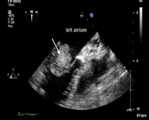
A non-contrast CT of the head showed evidence of an old left MCA infarct, but no acute abnormalities. Transthoracic echocardiogram (TTE) revealed a left atrial mass, measuring 3.6 x 5 cm (Figure 1) attached to the interatrial septum, filling the entire chamber and slightly protruding into the anterior mitral valve leaflet and left ventricle. Moderate mitral and tricuspid regurgitation were also noted. Abdominal ultrasound confirmed hepatomegaly and presence of gross ascites. A chest X-ray showed bilateral pleural effusion.
A diagnosis of left atrial myxoma was confirmed based on imaging studies, which also explained the patient’s residual neurological deficits. Due to unstable vitals, including low blood pressure, decreased urine output, and low spo2 levels, the patient was initially stabilized with oxygen therapy (4-6 L/min), dopamine infusion and diuretics (Dytor infusion). Strict monitoring of fluid intake/output and blood pressure was maintained. Once stabilized, he underwent left atrial myxoma resection with mitral valve repair.
Surgical intervention was performed under general anesthesia via median sternotomy and cardiopulmonary bypass. Intraoperative management included hemodynamic monitoring, maintenance of blood gases and electrolytes, and transfusion of one unit of packed red blood cells to manage anemia (Hb 8.4 g/dL). Following successful tumor resection, the patient was transferred to the ICU, where he was extubated the following day. The patient was discharged on postoperative day seven in stable condition.
Discussion
Primary cardiac tumors are exceedingly rare, with an incidence of less than 0.03% in the general population, and about 75% of these tumors are benign, with cardiac myxomas being the most common type [4]. Myxomas are typically located in the left atrium (75% of cases), but in 15- 20% of cases, they occur in the right atrium. Although benign, cardiac myxomas can have serious and even life-threatening complications due to embolization or obstruction of blood flow [5].
The symptoms of atrial myxomas are varied, often mimicking other cardiovascular and systemic conditions. In this case, the patient presented with a combination of neurological and cardiac symptoms, including right-sided hemiparesis, cough, shortness of breath, and chest pain. The neurological symptoms in particular, which included hemiparesis, likely stemmed from the embolic nature of the myxoma, with fragments of the tumor or thrombi being released into systemic circulation [6]. Embolic phenomena occur in 30-50% of patients with myxomas , most commonly affecting the cerebral arteries, which can lead to strokes. In fact, atrial myxomas account for a small but significant number of ischemic strokes (0.5% of all strokes) [7].
In this case, the patient had a prior cerebrovascular accident (CVA) involving the left middle cerebral artery, which may have been an early embolic event caused by the myxoma. Neurological symptoms are the presenting sign in 12-45% of patients with myxomas, depending on whether embolization occurs before the cardiac diagnosis is made [8, 9]. The progressive weakness and hemiparesis observed in our patient support the hypothesis that embolization had already occurred by the time of admission. Besides neurological symptoms, our patient had hepatic involvement also. In detail pre-anaesthetic checkup is very essential in these patients for proper optimisation of patient prior to surgery.
The diagnosis of atrial myxoma was confirmed through transthoracic echocardiography (TTE), which is the most sensitive diagnostic tool. In our case, TTE revealed a large mass measuring 3.6 × 5 cm in the left atrium, attached to the interatrial septum and slightly protruding into the mitral valve leaflet. This is a typical location and size for myxomas, which are often attached to the fossa ovalis of the atrial septum. Left atrial myxomas can cause obstruction of blood flow through the mitral valve, leading to symptoms resembling mitral stenosis (e.g.- dyspnea, orthopnea).
A critical component of managing patients with atrial myxomas is ensuring hemodynamic stability, especially prior to surgical resection. Large, mobile myxomas can cause obstructive symptoms, leading to acute hemodynamic decompensation. Our patient developed hypotension (BP 70/50 mmHg) and decreased urine output, which required immediate intervention. Hemodynamic support was initiated with dopamine infusion and diuretics, while strict monitoring of fluid input/output was maintained.
Inducing general anesthesia in patients with atrial myxomas poses significant risks. Intraoperative complications include the possibility of tumor embolization during manipulation or placement of invasive lines. The goal during anesthesia is to maintain stable systemic vascular resistance and ensure careful tumor handling to prevent embolization. Intraoperative transesophageal echocardiography (TOE) plays a crucial role in assessing the risk of embolization during surgery [10, 11].
The standard treatment for atrial myxomas is surgical resection. In our case, the patient underwent resection of the left atrial myxoma and mitral valve repair via median sternotomy, under cardiopulmonary bypass (CPB). Surgical excision is usually curative, with recurrence rates as low as 3% in sporadic cases . Given the tumor’s large size and its effect on the mitral valve, mitral valve repair was also necessary to restore proper valve function [12].
The surgical approach requires careful planning, particularly due to the risk of embolization during manipulation of the tumor. Intraoperative TOE helps surgeons monitor for signs of embolization or other complications. Additionally, anticoagulation is required during CPB to prevent clot formation, but this must be balanced with the risk of bleeding postoperatively [13, 14].
Postoperatively, the patient was transferred to the ICU for elective ventilation and was extubated the following day. The postoperative course was uneventful, and the patient was discharged on the seventh day. Although the prognosis after surgical resection is generally excellent, there are potential complications, including arrhythmias, atrial fibrillation, and thromboembolic events. Regular follow-up with echocardiography is essential to detect any recurrence of the myxoma, as well as to monitor for late complications such as valvular dysfunction.
Our patient had a previous history of a cerebrovascular event, which left him with residual right-sided hemiparesis. Postoperatively, the neurological symptoms showed no further progression. However, it is crucial to note that neurological recovery may vary depending on the extent of the embolic damage and the time elapsed before surgery. Studies have shown that early surgical intervention can help reduce the risk of further embolization and improve neurological outcomes, with improved prognosis [15].
Conclusion
This case highlights the complexity of managing atrial myxomas, particularly when systemic involvement present. Prompt diagnosis through echocardiography and surgical intervention is essential for preventing further complications such as embolization and heart failure. Although rare, atrial myxomas should be considered in the differential diagnosis of young patients presenting with stroke or other embolic phenomena. The prognosis after surgical resection is generally favourable, with low recurrence rates and good recovery of cardiac function.
References
-
Vivien HL, Heidi MC, Robert D, Brown Jr (2007) Central nervous system manifestation of cardiac myxoma. Arch Neurol 64: 1115-1120.
-
Anne A, Anandaswamy C, Suresh G, Gowshik R (2024) Anesthetic management of a patient with incidental left atrial myxoma for proximal femur nailing: A case report. Saudi J Anesthesia 18(2): 302-304.
-
Karabinis A, Samanidis G, Khoury M, Starvridis G, Perreas K (2018) Clinical presentation and treatment of cardiac myxoma in 153 patients. Med 97: 12397.
-
Saad EA, Mukherjee T, Gandour G, Fatayerji N, Rammal A, et al. (2024) Cardiac myxomas: causes, presentations, diagnosis, and management. Ir J Med Sci 193(2): 677- 688.
-
Kohno N, Kawakami Y, Hamada C, Toyoda G, Bokura H, et al. (2012) Cerebral embolism associated with left atrial myxoma that was treated with thrombolytic therapy. Case Rep Neurol 4(1): 38-42.
-
Oliveira R, Branco L, Galrinho A, Abreu A, Abreu J, et al. (2010) Cardiac myxoma: a 13-year experience in echocardiographic diagnosis. Rev Port Cardiol 29(7-8): 1087-1100.
-
Yuan SM, Humuruola G (2015) Stroke of a cardiac myxoma origin. Rev Bras Cir Cardiovasc 30(2): 225-234.
-
Wu Y, Fu XM, Liao XB, Zhou X (2018) Stroke and peripheral embolisms in a pediatric patient with giant atrial myxoma. Med 97(30): e11653.
-
Azhar AH, Ziyadi G, Zulkarnain H, Rahman MNG (2011) Atrial Myxoma Presenting As a Cerebellar Stroke. J Surg Acad 1(2): 36-40.
-
Melnyk V, Hackett PJ, Subramaniam K, Badhwar V, Esper SA (2018) Complex considerations and anesthetic management in patient with multiple intracardiac myxomas. J Cardiothorac Vasc Anesth 32(3): 1374-1376.
-
Wang Z, Chen S, Zhu M, Zhang W, Zhang H, et al. (2016) Risk prediction for emboli and recurrence of primary cardiac myxomas after resection. J Cardiothorac Surg 11: 22.
-
Fox J, Smith MM, Nuttall GA, Oliver WC (2018) Uncommon cardiac diseases. In: Kaplan JA, (Eds.), Kaplan’s essentials of cardiac anesthesia for cardiac surgery. 2nd (Edn.), Elsevier, pp: 426-434.
-
Zhang R, Tang Z, Qiao Q, Mahmood F, Feng Y (2020) Anesthesia management of atrial myxoma resection with multiple cerebral aneurysms: a case report and review of the literature. BMC Anesthesiol 20(1): 164.
-
Islam AKMM (2022) Cardiac myxomas: A narrative review. World J Cardiol 14(4): 206-219.
-
Guzman AG, Aseyev OI, Shah H, Sadreddini M (2022) Cardiac myxomas: clinical presentation, diagnosis and management. Heart 108: 827-833.
- Editorial on Multimodal Analgesia
- Surgical Incision Site Local Anaesthetic Infiltration and Superior Hypogastric Plexus Block in Total Abdominal Hysterectomy Under General Anaesthesia- A Placebo-Controlled, Randomized Clinical Trial
- Supraglottic Airway Insertion in Semi Fowler Position Due to Severe Thoracic Hyperkyphosis: A Case Report
- Current Problems in Pulmonary Respiratory Distress Syndrome (Literature Review)
- Evolution of Perioperative Hemodynamic Monitoring from the Hand on Pulse to Hypotension Prediction Index
- The Diverse Roles of Anaesthesiologists and Ambulance Services