Sled Dogs as a Model for Brown Fat Research
Brown adipose tissue (BAT) has the unique function of burning energy to create heat through the process of non-shivering thermogenesis. Activation of BAT in humans as a means of combating obesity is an active area of research, yet reliability of methods for recognizing and monitoring activation needs further research. This study assessed the feasibility of thermal imaging for monitoring BAT activation in an animal model, Alaskan sled dogs, which have been used extensively in coldweather metabolism and nutrition research. BAT activation was monitored monthly over four months in a cohort of five sled dog puppies. A change in temperature accompanies BAT activation. Therefore thermal images of the chest area were taken pre- and post-cold exposure. Concentrations of two plasma biomarkers associated with non-shivering thermogenesis, irisin and fibroblast growth factor 21 (FGF21) were also analyzed. No significant temperature change or concentration changes in irisin or FGF21 were observed between pre-and post-cold exposures. To our knowledge this is the first study in dogs, therefore the results provide methodology strategies for future study design. Given the potential benefits of BAT activation in humans, the validation of thermal imaging as a non-invasive monitoring technique and the use of an animal model could better inform the treatment of metabolic diseases.
Introduction
A National Health and Nutrition Examination Survey conducted in 2013-2014 estimated that the prevalence of obesity in US adults 20 years and older was 37.9% and the prevalence of extremely obese was at 7.7% [1]. This ongoing trend has spurred research efforts on new treatments that could help manage obesity and the associated health risks of metabolic syndrome. Brown adipose tissue, commonly referred to as BAT or brown fat, is a connective tissue in mammals that has the function of turning energy into heat through a process known as non-shivering thermogenesis. BAT cells are different from white adipose tissue in that instead of storing the energy in the form of a single lipid droplet, BAT cells have multiple smaller lipid droplets with abundant mitochondria that allow brown adipose cells to burn energy to create heat [2]. Through non-shivering thermogenesis, BAT provides an evolutionary advantage for mammals to survive in cold climates, especially young or small mammals that have a large surface-to-volume ratio [3]. Researchers have observed that as mammals age, the amount of BAT decreases; BAT was previously thought to be completely absent in adults. Several independent studies conducted in 2009 found that BAT is still present in most human adults [4, 5, 6, 7], and may be associated with outdoor activities [8]. The goals of this project were to use sled dog puppies as a model to 1) observe changes in brown fat through maturation and 2) activate BAT with a cold exposure protocol adapted from previous human studies [9].
The initial observations of BAT deposits in adult humans were found to be near the neck and supraclavicular region.
Most of the subsequent literature has been focused on ways to observe and monitor these BAT deposits. Currently, there have been no studies that observe BAT activation in dogs; therefore, the purpose of this pilot study was to design a protocol for monitoring BAT in dogs and to examine the feasibility of using a canine model for future studies. The use of an animal model in BAT activation research would provide additional knowledge about the role of BAT in the metabolic regulation and offer a model for testing of therapeutic treatments for obesity. The sled dog is a unique model for exploring the therapeutic potential of brown fat activation because sled dogs are well suited for cold and exercise treatments, both of which have shown to activate BAT [2, 10, 11].
The current gold standard for observing BAT in humans is positron emission tomography–computed tomography (PET-CT) technology to track the tracer fluorodeoxyglucose (F-FDG) through the body, but this method involves high levels of radiation, expensive equipment, and costly personnel [9]. The use of infrared thermography (IRT) to observe BAT is relatively new, but since heat is the final product of BAT activation, IRT can be used to measure the temperature of the skin that overlies BAT. One study used IRT technology to monitor non-shivering thermogenesis in voles and found that thermal imaging was useful in distinguishing the difference between skin surfaces where BAT was present and activated versus skin surfaces where BAT was not present or activated [12]. Therefore, for this study, a temperature increase in the supraclavicular region of the dogs was used to indicate the presence of active BAT. In a human study comparing PET-CT scans and IRT for monitoring BAT activation, the IRT image results correlated with the PET-CT scan results [9]. Several other studies also used IRT to detect BAT activation and demonstrated that IRT is an accurate, noninvasive method to observe and monitor BAT in real-time using either thermal images or videos [2, 10, 11].
The most studied activator of BAT is cold exposure, but other activators include hormone treatments [12] and ingestion of capsinoids [13]. Most human studies used a cold exposure treatment that involved placing the participant’s hands or feet in cold water as this method cools down the body without inducing shivering [2, 10, 11]. For this method, the length of the cold exposure needed to observe a significant cooling effect was 5 minutes. In comparison Jang, et al. [9] had participants in an air-conditioned room for two hours as a cold exposure. We employed a 15 to 20-minute cold exposure treatment in which the puppies were put outside in temperatures below 0°C.
In addition to thermal imaging, blood plasma concentrations of two biomarker associated with BAT activation was measured: irisin and fibroblast growth factor
21 (FGF21). Both biomarkers are known to be correlated with non-shivering thermogenesis in humans and to have increased concentrations in circulation with cold exposure [14, 15]. Irisin is a protein that assists in converting white adipose tissue into the energy-consuming BAT [16]. FGF21 is a stress response hormone that is involved with glucose and lipid metabolism. Increased FGF21 concentrations in the blood stream increase the amount of glucose that is taken up by the adipose tissue [17]. Coker, et al. hypothesized that irisin and FGF21 may foster changes in white and/or brown fat and promote elevations in the thermogenic profile [18].
We hypothesized that in a longitudinal study that followed five sled dogs puppies over a four-month time span, that there would be an increase in the skin temperature in the region overlying the chest BAT from the pre- to the post- cold exposure treatment using IRT. We also hypothesize that levels of irisin and FGF21 would increase from the pre- to post-cold exposure treatment with BAT activation.
Experimental Methods
Sample Collection
Five puppies starting from four months of age were used in this study and were sampled at approximately 1-month intervals (February 8th, March 3rd, April 3rd, and April 28th). A 12-hour postprandial blood collection was conducted pre- and post- cold exposure by collecting five milliliters of blood from the cephalic vein into EDTA anticoagulant vials. Blood samples were centrifuged at 3600 rpm for 15 minutes. One milliliter plasma aliquots were was collected into freezer vials and stored at -80°C.
Thermal Imaging
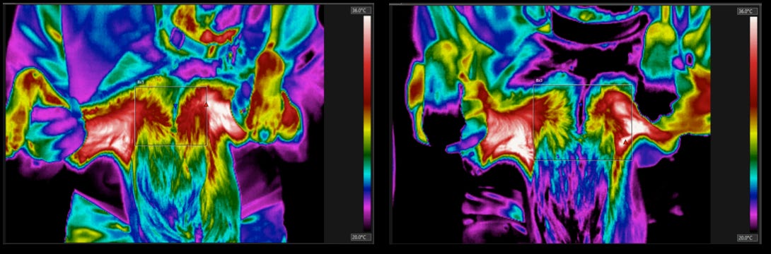
A FLIR T460 Thermal Imaging Camera was used to capture three to five images for each puppy, pre- and post- cold exposure. The dog was held in a seated position with the front legs positioned away from the body to allow for imaging of the chest region. The deposit was located along the sternum and in the supraclavicular regions. This region corresponds to where BAT deposits have been observed in humans using PET-CT scans [9].
The puppies were housed in outdoor kennels. The temperatures on the sampling days were -14°C, -30°C, 0.0°C, and -0.6°C, respectively. The coldest sample day was -30°C in the beginning of March and the warmest day was 0.0°C in the end of April. For the cold exposure treatment, the puppies were first allowed to acclimate at room temperature (20°C) for a minimum of two hours inside. Each puppy was then put outside in his or her kennel for 15 to 20 minutes. After the treatment, the puppies were brought inside again and immediately imaged and sampled.
The thermal images were analyzed using Forward Looking Infrared Radiometer (FLIR) Tools Software to measure the temperature differences between images from pre- to post-cold exposure. This was accomplished by creating a box that covered the dogs’ upper chest area in the sternal region, excluding the front legs. The images were analyzed and the software recorded the high temperature that occurred in the boxed region. One pre-treatment image and one post-treatment image for each dog was analyzed resulting in one high-temperature point for each dog for each time point (Figure 1).
Biochemical Analysis
Two different biomarkers were examined using the blood samples collected. All standards and samples were run in duplicate. Irisin concentration was measured using Irisin Enzyme Immunoassay Kit for use on human, rat, mouse, and canine (Phoenix Pharmaceuticals, Inc., Burlingame, CA, USA), which was completed according to the manufacturer’s directions.
This kit used a competitive enzyme-linked immunosorbent assay (ELISA) method in which the surfaces of the well-plates were pre-coated with a secondary antibody. A biotinylated primary antibody was added at same time as the plasma samples and standards. When the sample or standard was added, the peptide antibody competitively bound with the primary antibody. Then Streptavidin- horseradish peroxidase (SA-HRP) was added and interacted with the biotin on the primary antibody. SA-HRP catalyzed the substrate solution provided with kit and resulted in a color change. Hydrochloric acid was then added to turn the solution yellow; the intensity of the color was inversely proportional to the amount of peptide present. Absorbance was read at 450 nm a Synergy HT multi-mode microplate reader (Bio Tek, United States). A standard curve was established with known concentrations and the unknown sample concentrations were then interpolate from the standard curve. The plasma samples were not diluted for this kit because a study by Bell, et al. [19] used the same kit and found that the range of canine samples was 42 - 250 ng/mL, which was well within the range of the kit used. However, values in this study were concentrated on the higher side of the curve and therefore future studies may benefit from a 1:2 dilution so the values fall in the center of the curve.
The concentration of FGF21 was measured by Canine Fibroblast Growth Factor 21 (FGF21) ELISA Kit (MyBioSource Inc., San Diego, CA, USA) according to manufacturer’s instructions. This ELISA kit used the quantitative sandwich method in which the surface of the well plates was coated with capture antibodies specific for FGF21 proteins. When the sample plasma or standard was added, all FGF21 proteins present bound to the antibodies on the well-plate surface. Horseradish peroxidase (HRP)-conjugate reagent was then added and interacted with the protein to form a sandwich. When the chromogen substrate was added, it reacted with HRP- conjugate reagent to result in a color change in proportion to the concentration of FGF21 present. Absorbance was read at 450 Synergy HT multi-mode microplate readers (Bio Tek, United States). A standard curve was established with known concentrations and the unknown sample concentrations were interpolated from the standard curve.
Statistical Analysis
All statistical analysis was done using GraphPad Prism 5.0 statistical software. A one-way ANOVA followed by post hoc analysis with the Tukey-Kramer multiple comparison test was performed to compare the pre- and post-cold exposure from each of the samples.
Results
When analyzing the thermal images (Figure 2), there was not a significant difference in high chest temperatures between pre-and post-cold exposure groups for any of the sampling dates. No overall trend was observed between pre- and post-treatment groups on the same sample date. The outside temperature on the sampling day did not appear to correlate with the change in high chest temperatures, and high temperature also did not change over the 4-month sampling period.
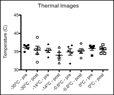
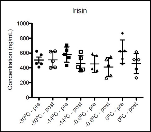
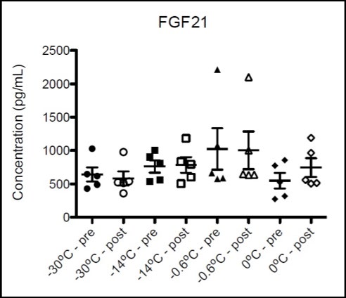
Qualitatively, there appeared to be a difference in the heat distribution between the pre- and post- images, but the varying positions of the dog in each image made accurate analysis difficult. The highest thermal signature was usually in the armpit area and it was difficult to avoid capturing that area in the thermal image without also eliminating a majority of the chest region. It appeared that there may be a small effect on the mean between pre and post when the temperature is below -14° C. There were no significant differences from pre- to post-cold exposure samples for either of the biomarkers tested on any of the sampling dates (Figures 3 &. 4).
Discussion
The lack of significant differences observed in this study could indicate that there was no BAT activation as a result of cold exposure in 4 to 8 month old puppies. Alternatively, and more convincingly, is that the area imaged and/or cold exposure used needs to be altered in future studies. Where and how to detect BAT and how to activate BAT are the central issues in this emerging field of BAT research. Both of these points will be discussed further.
The most difficult issue we faced was determining what body region to capture. In the literature, BAT monitoring in humans is focused on observing the supraclavicular BAT deposits from a frontal view [2, 9, 10, 11]. Our results indicate that this region is likely not ideal in dogs. To capture the supraclavicular BAT deposits, the position the puppies had to assume (sitting with front legs held extended from the body) was not an easy position for the handler to maintain, especially as the puppies grew in size and weight. As a result, the images were not replicable and this made data analysis difficult. Several studies have found that BAT deposits are also present in the interscapular region of human infants [20] and other mammals such as mice [21]. Subsequent necropsies, unrelated to this study, were conducted after the completion of our sample collection at the UAF veterinary school on a sled dog puppy and revealed a larger BAT deposit located between the scapulas and may be an easier and more profuse area to monitor. Future studies should consider capturing the interscapular region from a top view allowing for higher accuracy because this is a natural standing position for dogs. The disadvantage to this is that dogs’ hair is thicker on the back as opposed to the chest region, which could affect the precision of the temperature readings in the thermal images.
As mentioned previously, the difficulty in holding the dog in the position as well as the variation in size between the puppies as they grew from one month to the next, resulted in difficulty imaging the same area every time. Therefore, we analyzed the high temperature of the puppies’ entire chest regions. Another approach would be to measure the percentage of area that is above a certain temperature within the captured region in the image. Like other mammals, dogs are endothermic homeotherms and are able to maintain a constant internal temperature, so this new analysis approach would focus on the thermal signature of specific areas that BAT has shown to be present and may provide a more accurate analysis of the thermal images. This technique would be more suited for the intrascapular region compared to the supraclavicular region used in this study because of the relatively high underarm temperatures.
The use of a control would also improve the accuracy of the image data results, such as an internal temperature reading for each of the dogs or to include an object with a standard temperature in each of the images. One study stated that IRT was not able to detect BAT activation via cold exposure, but IRT could distinguish the difference between areas of skin where active BAT was and was not present [12]. Future studies that want to validate the use of IRT for BAT monitoring in animal models should consider using another BAT activation method such as hormone treatment or capsinoids.
Since there were no significant differences in the thermal images or the biomarker concentrations in this study, it is logical to assume that BAT was not activated. It is possible that the biomarkers tested are not fast acting, thus any concentration changes that may have occurred due to the cold exposure treatment were not seen in the short 15 to 20-minute time period tested. Literature is limited when looking at the effects of cold exposure on irisin and FGF21 concentrations in dogs. In humans, however, circulating concentrations of irisin and FGF21 have been observed to increase after a short cold exposure and are correlated with cold-induced thermogenesis, including both shivering and non-shivering thermogenesis [15].
Therefore a more plausible explanation for the lack of BAT activation is due to an ineffective cold exposure treatment. In theory, exposing an animal to below 0°C for a period of time would be a suitable cold exposure treatment. Sled dog puppies used in this study, however, lived at 64.8°N and were born in the autumn of the previous year and had been living outside in below 0°C temperatures for the majority of their lives. Since the puppies were all acclimatized to living in a cold environment prior to the start of the study, it is likely that a cold exposure within their normal temperature range would not elicit a cold stress response and therefore, not cause BAT activation. In fact, when all the puppies were brought indoors the morning of sampling, one of the study investigators noted that most of the puppies were panting before the start of sampling. This would suggest that bringing the puppies indoors might have been more of a thermally stressful situation than the cold exposure. Future studies should consider using a cold room for the cold exposure treatment to yield a consistent cold stress response and increase replicability. Puppies are usually born in late spring or early summer. This would make cold exposure treatments easier to achieve.
Another important point to consider with a cold exposure treatment is the animal’s thermoneutral zone. A thermoneutral zone is a range of temperatures in which an animal is able to maintain a stable body temperature without using energy to regulate heat loss or gain [22]. The National Research Council documented that the average domestic dog’s thermoneutral zone ranges from 20°C to 30°C [23] varying between breeds based on coat type and other physical traits. Sled dogs have adapted to live and thrive in Arctic conditions. One study assessed the thermoneutral zone in Inuit sled dogs living in the Arctic conditions of Greenland and found that the thermoneutral zone of these dogs ranged from 10°C in summer to -25°C in winter [24]. The sled dog puppies used in this study were short-haired, sprint type sled dogs and although they do not have thick fur coats like the long distance touring type sled dogs, the results and observations from this study suggest that their thermoneutral zone is more similar to that of the Inuit sled dogs than an average domestic dog. Future studies should consider looking into what temperature and length of cold exposure would be necessary to evoke a cold stress response in sled dogs.
A study testing BAT activation in human adult males via PET-CT scans compared a mild cold exposure (16°C) to a thermoneutral condition (22°C). Researchers observed BAT activation only with cold exposure but not with the thermoneutral condition [6]. This is further evidence that the lack of significant results in this pilot study was most likely due to an ineffective cold exposure treatment in a northern breed.
It is noteworthy to mention that even with the large temperature range over the course of the study (-30°C to 0°C), there was no correlation between the outside temperature and either the thermal images or the biomarker concentrations. A future study that followed the dogs over the course of a year would provide valuable insight into the effects of that Arctic seasonal weather changes have on BAT deposition and/or activation in sled dogs. Dogs are a well-established biomedical research model. Sled dogs in particular could provide valuable insight into cold exposure and BAT activation. The lack of literature in BAT activation in dogs makes these results particularly important in steering research design in future studies.
A study published in the Journal of American Preventative Medicine in 2012 used a model to forecast that the prevalence of obesity will increase to 42% and severe obesity prevalence will increase to 11%, with a projected increase in healthcare cost to $549.5 billion by the year 2030 if the current trends continue [25]. The relative new and promising field of BAT research is likely to continue to be an important focus in the medical field, as well as the need to develop suitable animal models.
Conclusion
Based on previous literature and research conducted so far, it is possible that in the future, treatments targeting BAT could have a role in assisting with the worldwide obesity epidemic by taking advantage of a naturally-occurring mechanism of metabolic regulation [26]. More research is needed in creating standardized protocols for observing BAT in both humans and dogs. The use of IRT in BAT monitoring provides a noninvasive technique that can have a widespread application. Using a canine model, specifically with sled dogs that have adaptive mechanisms for living in a cold climate, can help in advancing BAT therapeutic treatments for humans. Furthermore, future BAT activation studies with dogs should consider conducting a study that compares PET- CT scans to IRT, similar to the one performed by Jang et al. (2014) with adult humans.
Acknowledgments
Special thank you to the staff at Piledriver Kennel.
Financial Support
This work was supported by Nestlé Purina PetCare; the National Institutes of Health Common Fund, through the Office of Strategic Coordination, linked grant numbers, TL4GM118992, RL5GM118990, and UL1GM118991; and the Alaska Native-Serving and Native Hawaiian-Serving Institutions Education Competitive Grants Program (ANNH) grant no. 2018-38426-28900/ project accession no. 1017020 from the USDA National Institute of Food and Agriculture. The content is solely the responsibility of the authors and does not necessarily represent the official views of the National Institutes of Health. UA is an AA/EO employer and educational institution and prohibits illegal discrimination against any individual: www.alaska.edu/titleIXcompliance/ nondiscrimination.
Conflict Of Interest
None
Ethical Standards
The authors assert that all procedures contributing to this work comply with the ethical standards of the relevant national and institutional guides on the care and use of laboratory animals.
References
-
Fryar CD, Carrol MD, Ogden CL, Surveys DHNE (2016) Prevalence of overweight, obesity, and extreme obesity among adults aged 20 and over: United States, 1960- 1962 through 2013–2014. National Center for Health Statistics.
-
Ang QY, Goh HJ, Cao Y, Li Y, Chan SP, et al. (2017) A new method of infrared thermography for quantification of brown adipose tissue activation in healthy adults (TACTICAL): a randomized trial. J Physiol Sci 67(3): 395- 406.
-
Kajimura S, Saito M (2014) A new era in brown adipose tissue biology: molecular control of brown fat development and energy homeostasis. Annu Rev Physiol 76: 225-249.
-
Cypess AM, Lehman S, Williams G, Tal I, Rodman D, et al. (2009) Identification and importance of brown adipose tissue in adult humans. N Engl J Med 360(15): 1509- 1517.
-
Saito M, Okamatsu-Ogura Y, Matsushita M, Watanabe K, Yoneshiro T, et al. (2009) High incidence of metabolically active brown adipose tissue in healthy adult humans: effects of cold exposure and adiposity. Diabetes 58(7): 1526-1531.
-
van Marken Lichtenbelt WD, Vanhommerig JW, Smulders NM, Drossaerts JM, Kemerink GJ, et al. (2009) Cold- activated brown adipose tissue in healthy men. N Engl J Med 360(15): 1500-1508.
-
Virtanen KA, Lidell ME, Orava J, Heglind M, Westergren R, et al. (2009) Functional brown adipose tissue in healthy adults. N Engl J Med 360(15): 1518-1525.
-
Huttunen P, Hirvonen J, Kinnula V (1981) The occurrence of brown adipose tissue in outdoor workers. Eur J Appl Physiol Occup Physiol 46(4): 339-345.
-
Jang C, Jalapu S, Thuzar M, Law PW, Jeavons S, et al. (2014) Infrared thermography in the detection of brown adipose tissue in humans. Physiol Rep 2(11).
-
Robinson L, Ojha S, Symonds ME, Budge H (2014) Body mass index as a determinant of brown adipose tissue function in healthy children. J Pediatr 164(2): 318-322.
-
Symonds ME, Henderson K, Elvidge L, Bosman C, Sharkey D, et al. (2012) Thermal imaging to assess age-related changes of skin temperature within the supraclavicular region co-locating with brown adipose tissue in healthy children. J Pediatr 161(5): 892-898.
-
Jackson DM, Hambly C, Trayhurn P, Speakman JR (2001) Can non-shivering thermogenesis in brown adipose tissue following NA injection be quantified by changes in overlying surface temperatures using infrared thermography? J Therm Biol 26(2): 85-93.
-
Saito M, Yoneshiro T (2013) Capsinoids and related food ingredients activating brown fat thermogenesis and reducing body fat in humans. Curr Opin Lipidol 24(1): 71-77.
-
Lee P, Brychta RJ, Linderman J, Smith S, Chen KY, et al. (2013) Mild cold exposure modulates fibroblast growth factor 21 (FGF21) diurnal rhythm in humans: relationship between FGF21 levels, lipolysis, and cold- induced thermogenesis. J Clin Endocrinol Metab 98(1): E98-102.
-
Lee P, Linderman JD, Smith S, Brychta RJ, Wang J, et al. (2014) Irisin and FGF21 are cold-induced endocrine activators of brown fat function in humans. Cell Metab 19(2): 302-309.
-
Aydin S (2014) Three new players in energy regulation: preptin, adropin and irisin. Peptides 56: 94-110.
-
Gomez-Samano MA, Grajales-Gomez M, Zuarth-Vazquez JM, Navarro-Flores MF, Martinez-Saavedra M, et al. (2017) Fibroblast growth factor 21 and its novel association with oxidative stress. Redox Biol 11: 335-341.
-
Coker RH, Weaver AN, Coker MS, Murphy CJ, Gunga HC, et al. (2017) Metabolic Responses to the Yukon Arctic Ultra: Longest and Coldest in the World. Med Sci Sports Exerc 49(2): 357-362.
-
Bell MA, Levine CB, Downey RL, Griffitts C, Mann S, et al. (2016) Influence of endurance and sprinting exercise on plasma adiponectin, leptin and irisin concentrations in racing Greyhounds and sled dogs. Aust Vet J 94(5): 154- 159.
-
Rylander E, Pribylova H, Lind J (1972) A thermographic study of infants exposed to cold. Acta Paediatr Scand 61(1): 42-48.
-
Mo Q, Salley J, Roshan T, Baer LA, May FJ, et al. (2017) Identification and characterization of a supraclavicular brown adipose tissue in mice. JCI Insight 2(11).
-
Jordan M, Bauer AE, Stella JL, Croney C (2016) Temperature requirements for dogs. Are they tailored to promote dog welfare? Center for Animal Welfare Science.
-
NRC (2006) Nutrient requirements of dogs and cats. Washington, DC.
-
Gerth N, Redman P, Speakman J, Jackson S, Starck JM (2010) Energy metabolism of Inuit sled dogs. J Comp Physiol B 180(4): 577-589.
-
Finkelstein EA, Khavjou OA, Thompson H, Trogdon JG, Pan L, et al. (2012) Obesity and severe obesity forecasts through 2030. Am J Prev Med 42(6): 563-570.
-
Harms M, Seale P (2013) Brown and beige fat: development, function and therapeutic potential. Nat Med 19(10): 1252-1263.
- Pattern of Gonadal Hormones in Oral Testosterone-Supplimented Male Wistar Rats with Diabetes-Induced Hypogonadism
- Re-Evaluation of the Genotoxicity of Currently Used Food Dyes in Mouse Multiple Organs Via Continuous Administration by Drinking Using the Comet Assay
- Pharmacogenetics of Type 2 Diabetes Mellitus: Linking Genetic Variability to Drug Efficacy and its Cardiovascular Outcomes
- Exploratory Proteomic Profiling of SARS-CoV-2 Infected THP-1 Macrophages Reveals Alterations in Inflammatory Response and Cellular Metabolism
- Study of Genotoxicity of Hepatocarcinogens in Multiple Organs in Mice by Feeding and Drinking Using the Comet Assay
- Spirulina Polypeptides Inhibit the Growth of Human Lung Tumor (H460) Cells