COVID-19 and Long Covid as Complex, Multi-Organs Involvement and Multi-System Disease
Like any other infectious disease, the prognosis of COVID-19 is influenced by infecting agent, the SARS-CoV-2 virus load and the extent of organs affliction and damage. COVID-19 having a propensity for multiorgan involvement carries an adverse prognosis during the clinical course as well as later during the post-recovery period persisting as Long Covid.
Editorial
The Virus Infection, The Disease and Post- Disease
Like any other infectious disease, the prognosis of COVID-19 is influenced by infecting agent, the SARS-CoV-2 virus load and the extent of organs affliction and damage. COVID-19 having a propensity for multiorgan involvement carries an adverse prognosis during the clinical course as well as later during the post-recovery period persisting as Long Covid. The direct cytopathic effects of SARS-CoV-2 virus and the erratic and hyper-inflammatory response lead to tissue injury in various organs coupled with physiological dysfunctions and complications. In fact, the multi-system manifestations of COVID-19 are caused by a combination of specific host defence responses with associated inflammatory activity and vascular involvement with coagulopathy and a distinct propensity to develop thromboembolic complications. Simultaneously, comorbidities such as diabetes, hypertension and cardiovascular diseases influence the disease severity and mortality.
All age groups are susceptible to SARS-CoV-2 infection and advancing age and comorbidities are risk factors for infection, severe disease, and adverse prognosis. The routes of transmission of SARS-CoV-2 infection are through respiratory droplets released while talking, coughing, or sneezing; via aerosol as the virus is known to remain suspended in air specially in confined places; through mucosal membrane contact with fomites, and likely via faecal-oral transmission given the detection of viral RNA in stools in those with or without symptoms related to the gastrointestinal (GI) system as it has an abundant ACE2 expression. Following infection, viral shedding occurs in all those infected including asymptomatic individuals. There is a wide spectrum of clinical manifestations of COVID-19 ranging from asymptomatic or pauci-symptomatic forms to severe viral pneumonia with ARDS, multiorgan dysfunctions, septicaemia, and shock, and death.
The assessment of illness severity at admission through evaluation of the Sequential Organ Failure Assessment (SOFA) and Quick Sequential Organ Failure Assessment (qSOFA) scoring systems is helpful in identifying the COVID-19 patients with poor prognosis and a high risk for adverse outcome. Liu, et al. have documented that a SOFA score of ≥3 or a qSOFA score of ≥1 was associated with high mortality in severely ill COVID-19 patients [1]. As such, the SOFA score is prognostically superior to qSOFA in this setting, attributable to inclusion of higher number of clinical parameters and variables, which facilitates an accurate patient stratification. The parameters include mean arterial pressure, platelets count, bilirubin, creatinine, PaO2, FiO2, and D-dimer levels on admission [2]. The pathogenic mechanisms underlying alterations in these clinical correlates in COVID-19 patients are diverse.
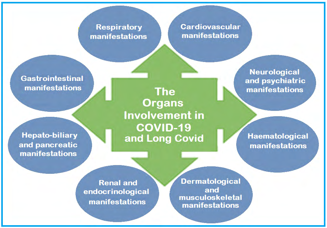
Organs Involvement in COVID-19 and Long COVID
Though primarily a respiratory viral disease, COVID-19 is a multi-organ and multi-system disease in a true sense. While it presents with common-cold-like symptoms in mild cases, the severe illness is characterised by multiorgan dysfunction and failure. The multi-organ involvement in COVID-19 can potentially lead to pneumonia and ARDS, acute liver dysfunction, acute kidney injury, cardiovascular disease, and a wide spectrum of gastro-intestinal (GI), neurological endocrinal, and haematological abnormalities (Figure 1).
The abundant expression of ACE2, the receptor for SARS- CoV2 in the respiratory, cardiovascular, GI, and endocrinal organs is partly responsible. The other factors include direct cytopathic injury, aberrant and over-active immune system manifesting as increased levels of inflammatory mediators and cytokine storm syndrome, and complications like endothelial dysfunction, coagulation disorders and thrombotic abnormalities.
The Respiratory System
The mild illness in COVID-19 resembles upper respiratory tract infection with nonspecific symptoms such as fever, headache, sore throat, cough (with or without sputum), anorexia, malaise, fatigue, muscle pain, and fatigue. Non-severe pneumonia may occur with moderate symptoms and without requirement for supplemental oxygen. Whereas severe pneumonia presents with fever, chest pain and respiratory discomfort with respiratory rate > 30 breaths/ min and reduced SpO2 on room air. The severe pneumonia may lead to acute respiratory distress syndrome (ARDS) of varying severity from mild to moderate to severe ARDS. The COVID-19 pneumonia has been categorized as type L with low lung elastance, low recruitability, and a poor response to positive end-expiratory pressure (PEEP) and type H as having high lung elastance, extensive consolidations on CT imaging, and significant response to higher PEEP [3]. In general, in about 80% of the COVID-19 patients, the respiratory involvement is mild and restricted to the upper airways, while the remaining 20% of the patients go on to develop pulmonary infiltrates as the virus invades the alveoli and infects the peripheral and subpleural units [4].
The Cardiovascular System
The patients with existing cardiovascular disease (CVD) are at a greater risk of suffering from severe COVID-19 and poor prognosis, whereas between 5% to 25% of the hospitalized COVID-19 patients show evidence of myocardial involvement in form of myocardial infarction, myocarditis, and cardiomyopathy without a prior diagnosis of hypertension or CVD [5]. There are multiple proposed aetiologies for adverse cardiovascular involvement and include the viral cytopathic effect via ACE2, a hyperinflammatory state, downregulation of ACE2 leading to pro-inflammatory and pro-fibrotic milieu, increased procoagulant activity, and increased cardiac physiologic demand. Given the hyperinflammatory and hypercoagulable state and increased metabolic demand COVID-19 patients may be at increased risk for acute myocardial ischaemia and infarction [6]. Elevated troponins have been found in COVID-19 patients denoting myocardial hypoxia and ischaemia or myocardial injury, whereas electrocardiogram (ECG) may show a range of findings mimicking ACS and echocardiography may show global dysfunction with myocarditis. The ECG and echocardiographic abnormalities are markers of severity in COVID-19 patients and correlated with adverse outcomes [7].
The Neurological System
Neurologic manifestations occur in more than one- third of patients hospitalized with COVID-19 [8]. Mao, et al. reported that in their cohort 36.4% of COVID-19 had neurologic manifestations [9]. The potential mechanisms of neurologic injury from COVID-19 include direct viral damage of nervous tissue, injury resulting from the excessive inflammatory response, erratic and hyper-immune response, and injury resulting from the effects of systemic illness. In fact, most COVID-related neurologic complications in critically ill patients fall into the latter category. A variety of neurological manifestations like headache, hyposmia, and hypogeusia are common, whereas the patients with severe COVID-19 are more likely to have neurological complications including delirium, encephalopathy, acute cerebrovascular disease, and other critical manifestations including skeletal muscle injury and myopathy [10]. On the serious side, meningoencephalitis, haemorrhagic encephalopathy, and acute necrotising encephalopathy may occur.
The Gastrointestinal System
The incidence of GI involvement ranges from 12–61% in patients with COVID-19 [11]. The commonly reported GI symptoms are diarrhoea, nausea, vomiting, and abdominal pain, and may be the sole presenting complaint in many cases. The hepatic injury occurs in 15% to 65% of COVID-19 patients ranging from mild and transient, the most common finding being abnormal transaminase levels, to severe damage in patients with severe COVID‐19 infection [12]. The virus, SARS‐CoV-2 has a tropism for the GI tract due to abundant presence of ACE2 in GI epithelial cells and the SARS‐CoV-2 RNA is readily detected in stool specimens, even when respiratory samples are negative [13]. The mechanisms of liver injury include viral cytopathic effect, immune‐ related injury, and drug hepatotoxicity. In patients with transaminitis, the medications with potential hepatotoxicity such as acetaminophen, statins, and hydroxychloroquine, should be used with caution. The patients with severe COVID-19 are at high risk of hepatobiliary, hypomotility, and ischemic GI complications due to small vessel thrombosis and viral enteroneuropathy [14].
The Renal System
The acute kidney injury (AKI), the abrupt loss of kidney function, is common in COVID-19 patients and the severity of AKI has been linked to adverse outcomes [15]. The alterations in renal function indicators such as blood urea, serum creatinine, proteinuria, and haematuria are predictors of AKI. The AKI occurs in 0.5–15% of hospitalized COVID-19 patients, and up to 23% of critically ill patients. The median onset of AKI from hospitalization ranges from 7 to 15 days, and it is an independent risk factor for morbidity and mortality [16]. The AKI is thought to occur through several proposed mechanisms, including intrinsic cellular injury by direct viral invasion of the renal tissue, acute tubular necrosis induced by septicaemia, hypoxia, hypovolemic state, and rhabdomyolysis. The management of AKI in COVID-19 patients must account for extent of renal damage. The renal replacement therapy (RRT) may be required in critically ill COVID-19 patients with AKI. The electrolyte abnormalities, such as hyperkalemia, hyponatremia, and hypernatremia, and metabolic acidosis may occur along with renal involvement and dysfunction.
The Endocrine System
While the effects of SARS-CoV-2 infection on the endocrine system remain largely unknown, given the expression of ACE2 in the majority of endocrine glands, dysfunction of various endocrine organs may occur in patients with severe COVID-19. In fact, a range of endocrine manifestations have been seen in COVID-19 patients without a pre-existing endocrine disease. The hospitalised COVID-19 patients commonly exhibit the abnormalities of glucose metabolism, such as worsened hyperglycemia, euglycemic ketosis, and diabetic ketoacidosis. Further, infection can cause new-onset diabetes [17]. Among exocrine pancreatic features, while pancreatitis is uncommon, elevated lipase or amylase have been documented in patients with COVID-19. In general, the diabetic patients are at higher risk for SARS- CoV-2 infection than the general population and more likely to have a severe disease due to compromised innate immunity and downregulated ACE2 levels [18]. Obesity is associated with severe COVID-19 due to ACE2 expression in adipose tissue and pro-inflammatory milieu. The SARS- CoV-2 infection affects the hypothalamus–pituitary axis either directly or via immune-mediated hypophysitis leading to central hypocortisolism and diabetes insipidus. The patients with severe COVID-19 are prone to develop critical illness-related corticosteroid insufficiency. Finally, COVID-19 may lead to subacute thyroiditis and other thyroid disorders.
The Musculoskeletal System
Myalgias are a common presenting symptom of COVID-19 and occur in more than one-third of patients and elevated creatinine kinase levels are prevalent in hospitalized patients and in patients with severe disease [19]. There is a possibility that myositis and rhabdomyolysis, the potential complication of COVID-19, are more common than reported due to muscle pains being a common symptom during the illness as well as creatine kinase and myoglobin levels are not being routinely tested. In a recent study, the incidence of rhabdomyolysis was 16.9% among all admitted COVID-19 patients [20]. In addition, the critically ill COVID-19 patients are at risk of developing myopathy and neuropathy due to prolonged immobility, systemic inflammation, corticosteroids, and the use of neuromuscular blocking agents. There has been recommended an early mobilization and initiation of physical therapy in these patients to ensure the best functional outcome [21].
The Dermatologic Manifestations
A variety of cutaneous manifestations such as erythematous rash, generalized urticaria, and chickenpox- like lesions have been described in COVID-19 and occur in up to 36% of the patients [22]. The skin manifestations may occur even before the onset of usual COVID-19 symptoms. There has not been established an association between cutaneous manifestations and COVID-19 severity, but dermatologic manifestations may reveal micro-thrombosis, hypercoagulability, and disseminated intravascular coagulation (DIC). The urticarial eruptions are typically consistent histologically with a viral exanthem. Livedo reticularis, a cutaneous manifestation is associated with DIC, whereas acrocyanosis and limb ischemia have been described in a cohort of critically ill patients with elevated D-dimer, fibrinogen, and fibrinogen degradation products [23]. The chilblains-like lesions, are erythematous areas on the feet, described as ‘Covid toes,’ may represent endotheliitis secondary to systemic COVID-19. A purpuric rash in patients with severe COVID-19 is probably caused localization of SARS-CoV-2 spike glycoprotein causing complement activation and thrombogenic vasculopathy.
The Haematological System
The COVID-19 patients present with several haematological abnormalities and thromboembolic complications. In general, leukocytosis (especially neutrophilia), lymphopenia, and thrombocytopenia are common and associated with adverse clinical course [24]. Simultaneously, the immune response to the virus infection is associated with increased circulating levels of pro- inflammatory cytokines, including tumor necrosis factor (TNF)-α, interleukins, granulocyte-colony stimulating factor, and chemokines. The hyper-inflammatory milieu combined with hypercoagulability carries the risk of venous thromboembolism and adverse outcomes [25]. Thus, the COVID-19 patients often present with both impaired haemostasis as well as thrombotic events [26]. Elevated fibrinogen and D-dimer levels are the most common coagulopathy seen in hospitalized COVID-19 patients. With the deranged coagulation cascade, DIC is another serious complication of COVID-19. The patients with COVID-19 are also at an increased risk of venous thromboembolic (VTE) disease and the use of anticoagulation with low molecular weight heparin (LMWH) or unfractionated heparin may improve clinical outcomes. Further, post-hospital discharge VTE prophylaxis may be considered in these patients depending on a case-by-case basis [27].
The Immunological System
The immune response is understandably the key determinant of the susceptibility to SARS-CoV-2 infection and severity of COVID-19 [28]. While weak immune system can increase the risk of acquiring SARS-CoV-2 infection and cause severe disease, the hyperinflammatory response to the infection is responsible for various complications by triggering organs involvement and damage [29]. The erratic immune response leads to the surge in inflammatory parameters like IL-2, IL-7, granulocyte-colony stimulating factor, interferon-γ inducible protein, monocyte chemoattractant protein 1, macrophage inflammatory protein 1-α, and tumor necrosis factor-α leading to an imbalanced and hyper-immune response responsible for cytokine storm. Throughout the disease course, there is required serial monitoring of lymphocyte count dynamics and inflammatory indices such as lactate dehydrogenase, C-reactive protein, and interleukin-6, for prognostic evaluation and measures for timely intervention.
The Clinical Manifestations, Course, and Fallouts
Given the world-wide viral pandemic with considerable morbidity and mortality, and post-recovery manifestations, the COVID-19 is an enigma. To simplify, the disease course can be divided into three distinct stages, beginning with acquiring the infection - Stage 1; which can sometimes progress to pulmonary involvement - Stage 2, with or without hypoxemia; and thereafter less frequently to systemic inflammation-Stage 3 [30]. The Stage 1 is the mild phase when the virus multiplies and establishes itself in the host tissues, predominantly in the respiratory tract. In Stage 2, there occurs viral multiplication and localized inflammation in the lungs, whereas the Stage 3 is marked by extra- pulmonary systemic hyper inflammation manifestations. The outcomes from the Stage 3 are often adverse. The Stage 3 is followed by recovery which may be afflicted by Long Covid manifestations for a variable period. There are certain comorbid conditions which worsen the outcome and include advanced age, hypertension, diabetes, coronary artery disease, chronic lung disease, and malignancies [31]. The studies have also documented variations in outcomes due to a dysregulated and hyperactive immune response.
Based on retrospective data from various studies, it seems that the progression of the disease can be predicted. From the therapeutic point, it appears that apart from exploring an effective anti-viral therapy, an important modality may be to down-regulate the inflammatory markers to limit the organs damage in COVID-19 [32]. In fact, the therapeutics of COVID-19 is still evolving, and recommendations may change, as apparent from the incredible volume and speed at which data is being published about the epidemiology, clinical manifestations, and treatment of COVID-19 [33].
References
-
Liu S, Yao N, Qiu Y, He C (2020) Predictive performance of SOFA and qSOFA for in-hospital mortality in severe novel coronavirus disease. Am J Emerg Med 38(10): 2074-2080.
-
Zaim S, Chong JH, Sankaranarayanan V, Harky A (2020) COVID-19 and Multiorgan Response. Curr Probl Cardiol 45(8): 100618.
-
Marini JJ, Gattinoni L (2020) Management of COVID-19 Respiratory Distress. JAMA 323(22): 2329-2330.
-
Wu Z, McGoogan JM (2020) Characteristics of and Important Lessons from the Coronavirus Disease 2019 (COVID-19) Outbreak in China: Summary of a Report of 72 314 Cases From the Chinese Center for Disease Control and Prevention. JAMA 323(13): 1239-1242.
-
Inciardi RM, Lupi L, Zaccone G (2020) Cardiac Involvement in a Patient with Coronavirus Disease 2019 (COVID-19). JAMA Cardiol 5(7): 819-824.
-
Ortega Paz L, Capodanno D, Montalescot G, Angiolillo DJ (2021) Coronavirus Disease 2019–Associated Thrombosis and Coagulopathy: Review of the Pathophysiological Characteristics and Implications for Antithrombotic Management. Journal of the American Heart Association 10(3): e019650.
-
Szekely Y, Lichter Y, Taieb P (2020) Spectrum of Cardiac Manifestations in COVID-19 - A Systematic Echocardiographic Study. Circulation 142(4): 342-353.
-
Liotta EM, Batra A, Clark JR (2020) Frequent neurologic manifestations and encephalopathy-associated morbidity in Covid-19 patients. Annals of Clinical Translational Neurology 7(11): 2221-2230.
-
Mao L, Jin H, Wang M (2020) Neurologic Manifestations of Hospitalized Patients With Coronavirus Disease 2019 in Wuhan, China. JAMA Neurol 77(6): 683-690.
-
Hosseini N, Nadjafi S, Ashtary B (2021) Overview of COVID-19 and neurological complications. Reviews in the Neurosciences 32(6): 671-691.
-
Cordie A, Gaber Y, AbdAllah M (2019) Gastrointestinal manifestations of human immunodeficiency virus and coronavirus disease 2019: Understanding the intersecting regions between the two epidemics. Arab J Gastroenterol 22(2): 75-87.
-
Marjot T, Webb GJ, Barritt AS (2021) COVID-19 and liver disease: mechanistic and clinical perspectives. Nat Rev Gastroenterol Hepatol 18(5): 348-364.
-
Van Doorn AS, Meijer B, Frampton CMA (2020) Systematic review with meta-analysis: SARS-CoV-2 stool testing and the potential for faecal-oral transmission. Aliment Pharmacol Ther 52(8): 1276-1288.
-
Kaafarani HMA, El Moheb M, Hwabejire JO (2020) Gastrointestinal Complications in Critically Ill Patients With COVID-19. Annals of Surgery 272(2): 61-62.
-
Arikan H, Ozturk S, Tokgoz B (2021) Characteristics and outcomes of acute kidney injury in hospitalized COVID-19 patients: A multicenter study by the Turkish society of nephrology. PLOS ONE 16(8): e0256023.
-
Hirsch JS, Ng JH, Ross DW (2020) Acute kidney injury in patients hospitalized with COVID-19. Kidney Int 98(1): 209-218.
-
Boddu SK, Aurangabadkar G, Kuchay MS (2020) New onset diabetes, type 1 diabetes and COVID-19. Diabetes Metab Syndr 14(6): 2211-2217.
-
Pal R, Bhansali A (2020) COVID-19, diabetes mellitus and ACE2: The conundrum. Diabetes Res Clin Pract 162: 108-132.
-
Chan KH, Farouji I, Abu Hanoud A, Slim J (2020) Weakness and elevated creatinine kinase as the initial presentation of coronavirus disease 2019 (COVID-19). Am J Emerg Med 38(7):1548.e1-1548.e3.
-
Haroun MW, Dieiev V, Kang J (2021) Rhabdomyolysis in COVID-19 Patients: A Retrospective Observational Study. Cureus 13(1): e12552.
-
Eggmann S, Kindler A, Andrea Perren A (2021) Early Physical Therapist Interventions for Patients With COVID-19 in the Acute Care Hospital: A Case Report Series. Physical Therapy 101(1): pzaa194.
-
Genovese G, Moltrasio C, Berti E, Marzano AV (2021) Skin Manifestations Associated with COVID-19: Current Knowledge and Future Perspectives. Dermatology 237(1): 1-12.
-
White Dzuro G, Gibson LE, Zazzeron L (2021) Multisystem effects of COVID-19: a concise review for practitioners. Postgrad Med 133(1): 20-27.
-
Tan L, Wang Q, Zhang D (2020) Lymphopenia predicts disease severity of COVID-19: a descriptive and predictive study. Signal Transduct Target Ther 5(1): 33.
-
Tang N, Li D, Wang X (2020) Abnormal coagulation parameters are associated with poor prognosis in patients with novel coronavirus pneumonia. J Thromb Haemost 18(4): 844-847.
-
Klok FA, Kruip MJHA, Van Der Meer NJM (2020) Incidence of thrombotic complications in critically ill ICU patients with COVID-19. Thromb Res 191: 145-147.
-
Tang N, Bai H, Chen X (2020) Anticoagulant treatment is associated with decreased mortality in severe coronavirus disease 2019 patients with coagulopathy. J Thromb Haemost 18(5): 1094-1099.
-
García LF (2020) Immune Response, Inflammation, and the Clinical Spectrum of COVID-19. Frontiers in Immunology 11: 1441.
-
Yazdanpanah F, Hamblin MR, Rezaei N (2020) The immune system and COVID-19: Friend or foe?. Life Sci 256: 117900.
-
Aguilar RB, Hardigan P, Mayi B (2020) Current Understanding of COVID-19 Clinical Course and Investigational Treatments. Frontiers in Medicine 7: 638.
-
Parker BS, Walker KH (2020) Clinical Course, Prognosis, and Epidemiology. Brigham and Women’s Hospital COVID-19 Clinical Guidelines.
-
Marik P (2020) EVMS Critical Care COVID-19 Management Protocol.
-
Massachusetts General Hospital (2020) Massachusetts General Hospital COVID-19 Treatment Guidance.
- Are the Vaccines the Only Solution to Prevent the COVID-19 Pandemic? Part Two
- Clinical Characteristics of Women in this New Global Immunodeficiency
- Cell Dynamics in HIV Pathogenesis: Insights and Implications
- Determination of the CDR (CDR1, CDR2) « Complementary- Determining Region Invertebrate Primitive Antibody from Sea Star »
- Prioritizing Care for High-Risk COVID-19 Patients in the EU: 10 Civic Recommendations to the Institutions
- Comprehensive Insights into ModRNA Vaccines: Persistent PP-Spike Recombinant Protein, Hyperimmune/Inflammatory Reactions, Thrombotic Vasculopathy, Chronic Organ Complications and Excess Deaths