Multifunctional Nanotherapeutics for Drug-Resistant Breast Cancer
Tumors are inherently resilient and always develop drug-resistance, leading to poor patient therapy effect. With the developing of sophisticated analytical tools, some novel strategy on improving targeting of anti-cancer delivery is developed to better thwart drug-resistance. This report demonstrates a multilayered nano-system to serve as a multifunctional platform for the treatment of drug-resistant breast cancers. This nano-system is composed of a poly (lactic-co-glycolic acid) core, a liposome second layer, and a hyaluronic acid outmost layer. The different types of drug, loaded in different layers, are released in a controlled and sequential manner upon internalization and localization. This recreated the time-staggering effect necessary for maximal efficacy
Short Communication
Breast cancer is one of the most devastating diseases known to mankind, and it unfortunately has no standard- of-care therapy. Currently, the most potent treatment, chemotherapy, still faces the problems of non-specificity, large doses of drug, and multidrug resistance effect. On one hand, the poor target and short-lived concentration of chemotherapeutics within tumor compel the use of high doses, increasing the risk of dose-related toxicities. On the other hand, suboptimal drug levels and single-agent based cancer treatment lead to the emergence of drug resistance and tumor relapse [1]. Some success has been achieved to date by implementing synergistic cancer genotoxic drugs using engineered delivery systems [2, 3, 4, 5, 6, 7, 8, 9, 10]. However, all current clinical treatment strategies primarily focus on the simultaneous release of drugs from a single delivery platform, and they pay no attention to how tumor survival/ drug resistant mechanisms and tumor cell-specific survival pathways may affect the treatment efficacy. New cancer treatment approaches must take into consideration of those mechanisms to improve efficacy.
Nanoparticle (NP) mediated drug delivery has shown tremendous promise for improved pharmacokinetics, dose- related toxicities, and chemical stability [11]. This strategy typically improves tumor specific delivery through either size dependent “passive” targeting [12] or “active” targeting that can be ligand directed or stimuli responsive [13, 14]. While active targeting often augments tumor cell killing, its efficacy is intrinsically limited by cellular heterogeneity that exists both within and among tumors [15, 16, 17]. Theoretically, tumor vessels are leaky, and tumor tissues lack well-defined lymphatic networks and effective lymphatic drainage, which favors diffusion of drug molecules to the center of the tumor through enhanced permeability and retention effect (EPR). However, selective accumulation of nanoparticles in tumor after systemic injection has been elusive so far. In addition, nanoparticles are easily subjected to opsonization and rapid clearance by the reticuloendothelial system (RES) [18]. To overcome rapid clearance, some polymers such as chitosan or polyethylene glycol are commonly applied to yield “stealth” particles that are invisible to the macrophage or provide a dynamic “cloud” on the surface to repel plasma protein resulting in prolonged half-life in blood and reduced allergic reaction and rejection by immune clearance [19, 20]. So far, single-drug therapies lack efficiency in cancer treatment due to the fact that cancer cells have been observed to develop mechanisms to survive single-agent chemotherapy [21]. At one point, traditional combinatorial chemotherapies provided a multi-pronged attack in hope of generating a long-lasting response through simultaneously using different drugs with varying modes of action. However, dual-drug treatment to overcome resistance is susceptible to failure since there is no intimate knowledge of the inner working of tumors. The novel combinatorial therapies exploit the molecular mechanisms that contribute to a tumor’s survival and attack the weakness of cancer. Two drugs can be used in conjunction such that one complements the other by preemptively cutting off alternative oncogenic signaling pathways [22]. For example, co-delivery of multiple anticancer drugs, which function via different mechanisms of hitting different targets and displaying different toxicity profiles, can improve the therapeutic index by achieving synergistic efficacy and increasing sensitivity [23, 24, 25]. Moreover, for free-drug formulations (absent of any carriers), the drug dose is often determined by the maximum amount a patient can tolerate, which is suboptimal. In contrast, nanoparticles can be engineered to co-encapsulate multiple drugs with precise control regardless of their physicochemical properties. Furthermore, nanoparticles can precisely control the timing of the drug release and how the drug is delivered upon reaching the target site [26, 27]. Although multi-drug delivery can be achieved by nanoparticles, some problems still need to be addressed. First, types of drugs that can be co-loaded are limited based on the specific nanoparticle delivery system. Second, the loading ratio of multiple drugs cannot be controlled precisely. Third, released drugs are easily extruded out of cancer cells by P-glycoprotein (P- gp), a plasma membrane ATP-binding cassette transporter overexpressed in tumor cells [28, 29, 30, 31]. P-gp is composed of four domains, two hydrophobic transmembrane domains and two cytosolic nucleotide-binding domains [32, 33] which are involved in ATP binding [34, 35]. The P-gp enhanced multidrug resistance (MDR) reduces the intracellular levels of cytotoxic drugs below lethal thresholds, making the drugs ineffective. This increased insight has led to the development of strategies that can weaken the P-gp mediated MDR effects. For example, flavonoids were shown to bind to nucleotide- binding domains with high affinity. Their binding site partially overlaps with both the ATP-binding site and the steroid-interacting region of P-gp, showing promise as a class of bifunctional modulators of P-gp [32, 35]. It is also found that special rewiring of signaling network for cancer cell could lead a new therapy strategy. Overexpression of epidermal growth factor (EGFR) on cancer cell could produce uncontrolled cell division, and EGFR inhibition dramatically sensitizes a subset of cancer cell to DNA damage if the genotoxic drugs are given sequentially [36].
The goal of this project is to develop multi-functional nanoparticles (MLNPs) Figure 1.
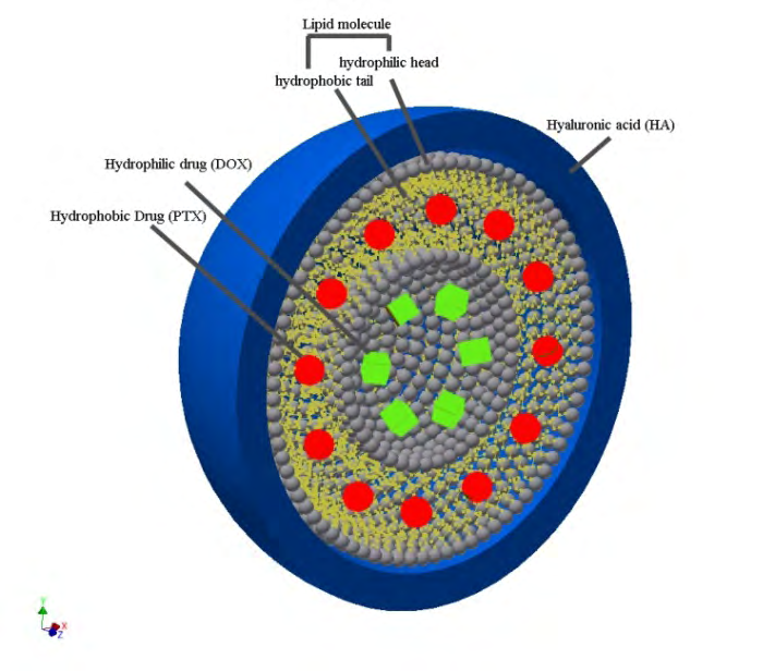
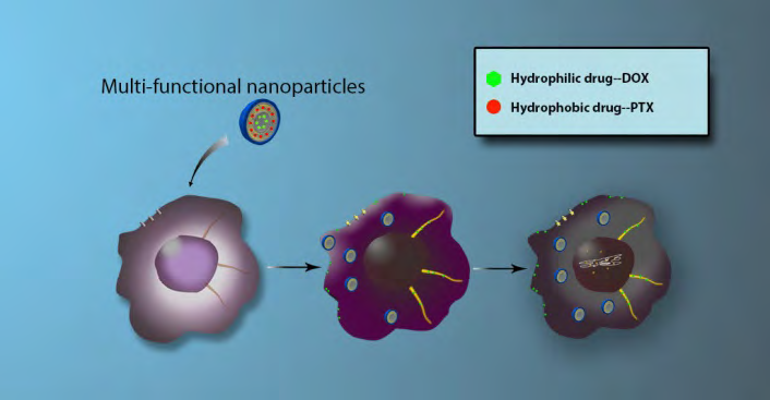
For effective MDR inhibition, effective EGFR inhibition, and controllable drug release for cancer therapy. MLNPs will be fabricated by depositing oppositely charged polymers on top of one another to build several highly stable films with high drug loading capacity. The layer-by-layer (L-B-L) fabrication method used here allows precise control at nanometer scale level. Liposome nanoparticles (around 75nm) was first prepared to enclose a liquid core. The liquid core was loaded with doxorubicin (DOX, a genotoxic drug targeting DNA to prevent cell replication), and the liposome layer was loaded with paclitaxel (PTX), which was proved to enhance the MDR inhibition systematically with Dox [37] Figure 2.
We hypothesis that drug molecules loaded in different layer of the multilayer nanoparticles will have a different temporal release profile once entered into cancer cells, and we further hypothesize that these drugs can work synergistically to effectively kill targeted cancer cells.
Utilizing an L-B-L fabrication method, this project generated multilayered multifunctional nanoparticles (NPs). Different from previously published reports [38, 39, 40, 41], these NPs will be made of materials that are dramatically different in their physicochemical properties, allowing the loading of drugs with significantly different physicochemical properties. Also, we target to inhibit a different drug-resistant mechanism of cancer cells first, and inhibit cancer cell division following. As shown in Figure 1, the multilayered NPs will be composed of an outside layer loaded with an MDR-enhanced inhibitor, and a liquid core loaded with cytotoxic drugs. This novel modular drug delivery platform can be tailored to achieve efficient intracellular delivery, extended serum half-life, and effective cancer cell killing. This multifunctional delivery system has superior capability for combinatory therapy by co-delivering drugs that targets P-gp, a drug efflux pump responsible for MDR effect of cancer cells, and doxorubicin, a genotoxic drugs to kill cancer. The NPs will be fabricated with a biopolymer, hyaluronic acid (HA), which is biodegradable and has low toxicity, and lipids, which are FDA-approved for chemotherapeutic treatment of several forms of cancer [42, 43]. HA has been shown to be biocompatible with living tissues since it does not cause allergic reaction and rejection [44, 45, 46]. Chitosan was also found to bind with CD44, which are overexpressed on surface of cancer cells [47].
In the experiment section, multi-layered nanoparticles were assembled by an L-B-L method. Briefly, 0.5 mg of DOX was encapsulated in 10 mg of poly (lactic-co-glycolic acid) (PLGA) through double-emulsion Figures 3A-3F [48, 49] and the DOX loaded PLGA was encapsulated in 4 mg of DOTAP (1,2-dioleoyl-3-trimethylammonium-propane), which was loaded with 1 mg of PTX through evaporation. The resulted two-layered particles (Figure 3D) were then coated with a hyaluronic acid (HA) layer. The three-layered NPs (HA-MLNPs, Figure 3E) were prepared in two different sizes and their structures were characterized by dynamic light scattering (Figure 3A), zeta potential (Figure 3B), transmission electron microscopy (TEM) (Figure 3C-E), and confocal laser scanning microscopy (Figure 3F). The mean diameter of the two particles is around 70 nm (HA-MLNPs-70) and 200 nm (HA-MLNPs-200) (Figure 3A). The size distribution can be significantly improved by purifying the particles through a size exclusion chromatography column [50]. The change of zeta potential (Figure 3B) shows different type of charge each layer carries. To visualize the three-layered structure, different materials used in the nanoparticle assembly were labeled with different fluorescent dye (Figure 3F), and the merged image (Figure 3F iv) shows the presence of each material in MLNPs. Figure 3 C-F shows unambiguously that the three-layered structure was indeed produced. HA- MLNPs-200 were used to test the drug release profile and intracellular uptake. The control group was fabricated to enclose both DOX and PTX in PLGA.
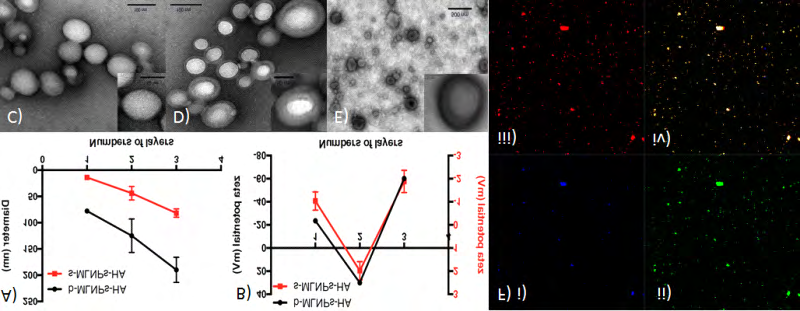
Figure 3: Assembly and characterization of multilayered nanoparticles. A)Hydrodynamic size, and B) Zeta potential. TEM images of C) the PLGA core, D) PLGA+liposome, and E) PLGA+liposome+HA. F) Confocal microscopic image of HA-MLNPs-200: i) HA was labelled with Alexa Fluor 350 (blue); ii) lipid with 1,2-diphytanoyl-sn-glycero-3-phosphoethanolamine-N-(7-nitro- 2-1,3-benzoxadiazol-4-yl) (ammonium salt) (NBD, green); iii) PLGA was labeled with Red Nile (red); iv) merged image.
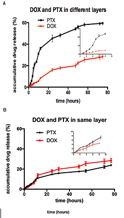
The drug release profiles were determined by a dialysis method [50] without the presence of serum. The results showed staggered release over 75 h with a fast release rate of PTX from the lipid film, compared to that of DOX from the PLGA core Figures 4A-4B In contrast, the release profiles of DOX and PTX from the control particles that had both drugs in the PLGA core are almost identical (Figure 4B). Compared with other reported drug release profiles from just PLGA without the extra layers [51, 52, 53, 54], the release of the drugs enclosed in PLGA (DOX in Figure 4A, and both DOX and PTX in Figure 4B) is delayed until around 10 h when the release rates increased significantly, showing the effect of the extra layers in the three-layered structure.
The assembled NPs in this project possess the following unique advantages: the structure of multilayered particles will enable loading of several different drugs, produce staggered drug release over time, and explore the treatment of drug-resistant cancers through inhibiting a unique cancer MDR mechanism, and EGFR signaling. The results indicated that the PTX was first be released from the outmost layer, block the P-gp pump, and rewire the cancer cell into a state more susceptible to the cytotoxic effects of genotoxic drugs (Figure 2). The release of PTX and the resulted MDR inhibition can potentially be tuned by the thickness of the outmost layer. In this subset, DNA of cancer cell could be damaged by subsequently released third drug, DOX, loaded in the hydrophilic aqueous compartment inside liposome [55]. The loaded drugs interfere with the cancer cells through different mechanisms, promoting synergistic pharmacological functions. Basically, the compartmentalization of the two drugs resulted in differential release. This recreated the time- staggering effect necessary for maximal efficacy. Because of the versatility of this novel multilayered nanoparticles, this project is undoubtedly inspiring the advancement of novel nanoparticles-based systems that are specifically designed based on the cancer cell biology for effective cancer treatment.
References
-
Vogelstein B, Papadopoulos N, Velculescu VE, Zhou SB, Diaz LA, et al. (2013) Cancer Genome Landscapes. Science 339(6127): 1546-1558.
-
Dai WB, Jin W, Zhang JL, Wang XQ, Wang JC, et al. (2012) Spatiotemporally Controlled Co-Delivery of Anti- vasculature Agent and Cytotoxic Drug by Octreotide- modified Stealth Liposomes. Pharm Res 29(10): 2902- 2911.
-
Sengupta S, Eavarone D, Capila I, Zhao GL, Watson N, et al. (2005) Temporal targeting of tumour cells and neovasculature with a nanoscale delivery system. Nature 436(7050): 568-572.
-
Aryal S, Hu CMJ, Zhang LF (2011) Polymeric Nanoparticles with Precise Ratiometric Control over Drug Loading for Combination Therapy. Molecular pharmaceutics 8(4): 1401-1407.
-
Fang RH, Aryal S, Hu CMJ, Zhang LF (2010) Quick Synthesis of Lipid-Polymer Hybrid Nanoparticles with Low Polydispersity Using a Single-Step Sonication Method. Langmuir 26(22): 16958-16962.
-
Hu CM, Aryal S, Zhang L (2010) Nanoparticle-assisted combination therapies for effective cancer treatment. Therapeutic delivery 1(2): 323-334.
-
Singh A, Dinawaz F, Mewar S, Sharma U, Jagannathan NR, et al. (2011) Composite Polymeric Magnetic Nanoparticles for Co-Delivery of Hydrophobic and Hydrophilic Anticancer Drugs and MRI Imaging for Cancer Therapy. Acs Appl Mater Inter 3 (3): 842-856.
-
Wong HL, Bendayan R, Rauth AM, Wu XY (2006) Simultaneous delivery of doxorubicin and GG918 (Elacridar) by new Polymer-Lipid Hybrid Nanoparticles (PLN) for enhanced treatment of multidrug-resistant breast cancer. J Controlled Release 116(3): 275-284.
-
Baeza A, Guisasola E, Ruiz-Hernandez E, Vallet-Regi M (2012) Magnetically Triggered Multidrug Release by Hybrid Mesoporous Silica Nanoparticles. Chem Mater 24(3): 517-524.
-
Patel NR, Rathi A, Mongayt D, Torchilin VP (2011) Reversal of multidrug resistance by co-delivery of tariquidar (XR9576) and paclitaxel using long-circulating liposomes. Int J Pharm 416(1): 296-299.
-
Liedtke C, Mazouni C, Hess KR, Andre F, Tordai A, et al. (2008) Response to neoadjuvant therapy and long-term survival in patients with triple-negative breast cancer. J Clin Oncol 26(8): 1275-1281.
-
Natarajan K, Xie Y, Baer MR, Ross DD (2012) Role of breast cancer resistance protein (BCRP/ABCG2) in cancer drug resistance. Biochem. Pharmacol 83(8): 1084-1103.
-
Whitehead KA, Langer R, Anderson DG (2009) Knocking down barriers: advances in siRNA delivery. Nature Reviews Drug Discovery 8(2): 129-138.
-
Gilleron J, Querbes W, Zeigerer A, Borodovsky A, Marsico G, et al. (2013) Image-based analysis of lipid nanoparticle-mediated siRNA delivery, intracellular trafficking and endosomal escape. Nat Biotechnol 31(7): 638-646.
-
Rehman ZU, Hoekstra D, Zuhorn IS (2013) Mechanism of Polyplex- and Lipoplex-Mediated Delivery of Nucleic Acids: Real-Time Visualization of Transient Membrane Destabilization without Endosomal Lysis. ACS nano 7(5): 3767-3777.
-
Bonner DK, Leung C, Chen-Liang J, Chingozha L, Langer R, et al. (2011) Intracellular Trafficking of Polyamidoamine- Poly(ethylene glycol) Block Copolymers in DNA Delivery. Bioconjug Chem 22(8): 1519-1525.
-
Sun TM, Du JZ, Yao YD, Mao CQ, Dou S, et al. (2011) Simultaneous Delivery of siRNA and Paclitaxel via a “Two-in-One” Micelleplex Promotes Synergistic Tumor Suppression. ACS nano 5(2): 1483-1494.
-
Nie SM (2010) Understanding and overcoming major barriers in cancer nanomedicine. Nanomedicine Uk 5(4): 523-528.
-
Behrens I, Pena AIV, Alonso MJ, Kissel T (2002) Comparative uptake studies of bioadhesive and non- bioadhesive nanoparticles in human intestinal cell lines and rats: The effect of mucus on particle adsorption and transport. Pharm Res 19(8): 1185-1193.
-
Kong M, Chen XG, Xing K, Park HJ (2010) Antimicrobial properties of chitosan and mode of action: A state of the art review. Int J Food Microbiol 144(1): 51-63.
-
Luo C, Sun J, Sun BJ, He ZG (2014) Prodrug-based nanoparticulate drug delivery strategies for cancer therapy. Trends Pharmacol Sci 35(11): 556-566.
-
Fang RH, Zhang LF (2014) Combinatorial Nanotherapeutics: Rewiring, Then Killing Cancer Cells. Sci Signal 7(325): 325-326.
-
Lv SX, Tang ZH, Li MQ, Lin J, Song WT, et al. (2014) Co-delivery of doxorubicin and paclitaxel by PEG- polypeptide nanovehicle for the treatment of non-small cell lung cancer. Biomaterials 35(23): 6118-6129.
-
Gao X, Wang BL, Wu QJ, Wei XW, Zheng FJ, et al. (2015) Combined Delivery and Anti-Cancer Activity of Paclitaxel and Curcumin Using Polymeric Micelles. J Biomed Nanotechnol 11(4): 578-589.
-
Zhao BY, Pritchard JR, Lauffenburger DA, Hemann MT (2014) Addressing Genetic Tumor Heterogeneity through Computationally Predictive Combination Therapy. Cancer Discov 4(2): 166-174.
-
Hu CMJ, Zhang LF (2012) Nanoparticle-based combination therapy toward overcoming drug resistance in cancer. Biochem Pharmacol 83(8): 1104-1111.
-
Hu CMJ, Zhang LF (2009) Therapeutic Nanoparticles to Combat Cancer Drug Resistance. Curr Drug Metab 10(8): 836-841.
-
Zhu X, Sui M, Fan W (2005) In vitro and in vivo characterizations of tetrandrine on the reversal of P-glycoprotein-mediated drug resistance to paclitaxel. Anticancer Res 25(3B): 1953-1962.
-
Katyal P, Batra N, Khajuria R (2014) Flavonoids and their therapeutic potential as anti-cancer agents: biosynthesis, metabolish and regulation. World Journal of Pharmacy and Pharmaceutical sciences 3: 2188-2216.
-
Modok S, Mellor HR, Callaghan R (2006) Modulation of multidrug resistance efflux pump activity to overcome chemoresistance in cancer. Curr Opin Pharmacol 6(4): 350-354.
-
Al-Lazikani B, Banerji U, Workman P (2012) combinatorial drug therapy for cancer in the post- genomic era. Nat Biotechnol 30(7): 679-692.
-
Singh DV, Godbol MM, Misra KA (2013) plausible explanation for enhanced bioavailability of P-gp substrates in presence of piperine: simulation for next generation of P-gp inhibitors. J Mol Mode 19(1): 227- 238.
-
Ren W, Qiao Z, Wang H, Zhu L, Zhang L (2003) Flavonoids: promising anticancer agents. Med Res Rev 23(4): 519- 534.
-
Di Pietro A, Dayan G, Conseil G, Steinfels E, Krell T, et al. (1999) P-glycoprotein-mediated resistance to chemotherapy in cancer cells: using recombinant cytosolic domains to establish structure function relationships. Braz J Med Biol Res 32(8): 925-939.
-
Cassidy CE, Setzer WN (2016) Cancer-relevant biochemical targets of cytotoxic Lonchocarpus flavonoids: a molecular docking analysis. J Mol Model 16(2): 311-326.
-
Lee M J, Ye AS, Gardino AK, Heijink AM, Sorger PK, et al. (2012) Sequential application of anticancer drugs enhances cell death by rewiring apoptotic signaling networks. Cell 149(4): 780-794.
-
Zang X, Wang G, Cai Q, Zheng, X, Zhang J, et al. (2018) A Promising Microtubule Inhibitor Deoxypodophyllotoxin Exhibits Better Efficacy to Multidrug-Resistant Breast Cancer than Paclitaxel via Avoiding Efflux Transport. Drug Metab Dispos 46(5): 542-551.
-
Cheng J, Teply BA, Sherifi I, Sung J, Luther G, et al. (2007) Formulation of functionalized PLGA-PEG nanoparticles for in vivo targeted drug delivery. Biomaterials 28(5): 869-876.
-
Guo JW, Gao XL, Su LN, Xia HM, Gu GZ, et al. (2011) Aptamer-functionalized PEG-PLGA nanoparticles for enhanced anti-glioma drug delivery. Biomaterials 32(31): 8010-8020.
-
Kumari A, Yadav SK, Yadav SC (2010) Biodegradable polymeric nanoparticles based drug delivery systems. Colloid Surface 75(1): 1-18.
-
Semete B, Booysen L, Lemmer Y, Kalombo L, Katata L, et al. (2010) In vivo evaluation of the biodistribution and safety of PLGA nanoparticles as drug delivery systems. Nanomed-Nanotechnol 6(5): 662-671.
-
Allen TM, Cullis PR (2013) Liposomal drug delivery systems: From concept to clinical applications. Adv Drug Del Rev 65(1): 36-48.
-
Barenholz Y, Peer D (2012) Liposomes and other assemblies as drugs and nano-drugs: From basic and translational research to the clinics Preface. J Controlled Release 160(2): 115-116.
-
Zhu LZ, Ma JW, Jia NQ, Zhao Y, Shen HB (2009) Chitosan-coated magnetic nanoparticles as carriers of 5-Fluorouracil: Preparation, characterization and cytotoxicity studies. Colloid Surface 68(1): 1-6.
-
Reverchon E, Antonacci A (2006) Chitosan microparticles production by supercritical fluid processing. Industrial & Engineering Chemistry Research 45(16): 5722-5728.
-
Akbuga J (1993) The Effect of the Physicochemical Properties of a Drug on Its Release from Chitosonium Malate Matrix Tablets. Int J Pharm 100(1-3): 257-261.
-
Rao W, Wang H, Han JF, Zhao ST, Dumbleton, et al. (2015) Chitosan-Decorated Doxorubicin-Encapsulated Nanoparticle Targets and Eliminates Tumor Reinitiating Cancer Stem-like Cells. ACS Nano 9(6): 5725-5740.
-
Zambaux MF, Bonneaux F, Gref R, Maincent P, Dellacherie E, et al. (1998) Influence of experimental parameters on the characteristics of poly(lactic acid) nanoparticles prepared by a double emulsion method. J Controlled Release 50(1-3): 31-40.
-
Yang YY, Chung TS, Ng NP (2001) Morphology, drug distribution, and in vitro release profiles of biodegradable polymeric microspheres containing protein fabricated by double-emulsion solvent extraction/evaporation method. Biomaterials 22(3): 231-241.
-
Huang W, Zhang CM (2011) Assembly and characterization of lipid-lipid binding protein particles. J Biotechnol 154(1): 60-67.
-
Avgoustakis K, Beletsi A, Panagi Z, Klepetsanis P, Karydas AG, et al. (2002) PLGA-mPEG nanoparticles of cisplatin: in vitro nanoparticle degradation, in vitro drug release and in Vivo drug residence in blood properties. Journal of Controlled Release 79(1-3): 123-135.
-
Govender T, Stolnik S, Garnett MC, Illum L, Davis SS (1999) PLGA nanoparticles prepared by nanoprecipitation: drug loading and release studies of a water soluble drug. Journal of Controlled Release 57(2): 171-185.
-
Fonseca C, Simoes S, Gaspar R (2002) Paclitaxel-loaded PLGA nanoparticles: preparation, physicochemical characterization and in vitro anti-tumoral activity. Journal of Controlled Release 83(2): 273-286.
-
Chan JM, Zhang LF, Yuet KP, Liao G, Rhee JW, et al. (2009) PLGA-lecithin-PEG core-shell nanoparticles for controlled drug delivery. Biomaterials 30(8): 1627-1634.
-
Morton SW, Lee MJ, Deng ZJ, Dreaden EC, Siouve E, et al. (2014) Nanoparticle-Based Combination Chemotherapy Delivery System for Enhanced Tumor Killing by Dynamic Rewiring of Signaling Pathways. Sci Signal 7(325): ra44.
- Effects of 5-HTP and Melatonin on the Sleep Cycle of Medical Students
- Adsorption of Bisphenol A on NH4OH- Modified Rice Husk and Sugar Cane Bagasse Biochar
- Comparative Assessment of the Reinforcement Efficiency of Palm Fruit Fibre and Coconut Fibre in High Density Polyethylene (HDPE) Matrix Composite
- Importance of Bio Compounds Naturally Present in Food with Functionality in Animal Metabolism
- Sub-Acute Study on the Cardiotoxic Effects of Monosodium Glutamate Ingestion in Albino Rat
- Weight Management and Its Natural Solutions: A Review