The Protective Effects of Different Types of Yemeni Honey on Hepatorenal Toxicity Induced by Gentamicin on Guinea Pigs
Aim: This study aimed to investigate the antioxidant properties of different types of Yemeni honey in reducing hepatotoxicity and nephrotoxicity caused by gentamicin in the female guinea pigs. Method and Results: The administration of Osaimi sider honey, Athel honey, Dam Al-Akhawain honey, Salam honey (5 mg/kg orally), Gentamicin (80 mg/kg i.p.), gentamicin+Osaimi sider honey, gentamicin+ Athel honey, gentamicin+ Dam Al-Akhawain honey, and gentamicin+ Salam honey for 7days caused a significant increase in the levels of AST and ALT, and also caused a significant decrease in the levels of total protein and albumin in gentamicin treated groups together with significant elevation in levels of urea and no significant increase in the level of creatinine. However, co-administration of gentamicin with different types of Yemeni honey used in this study ameliorated the harmful effects of gentamicin in most of the tested parameters. Different types of Yemeni honey used in this study have a protective effect on the histological changes of liver and kidney tissues induced either by gentamicin administration. Conclusion: The present study concluded that the use of antioxidants (Yemeni Honey) showed a highly significant protective effect on the functions and tissue of the liver and kidney when they were used as co-treatment. The effects of honey that were found in our experiment are due to the presence of many antioxidant compounds such as flavonoids, ascorbic acid, tocopherols, catalase, and phenolic compounds, that work together to provide a synergistic antioxidant effect, scavenging and eliminating free radicals. But the protective effect of the treatment with sider honey and Dam Al-Akhawain honey had a greater protective effect on the liver and the treatment with Dam Al-Akhawain honey and Athel honey had a greater protective effect on the kidney which was almost similar to the control.
Introduction
The process of drug discovery and development is challenging, rewarding, time consuming and complex. The attrition in drug development is not uncommon due to unpredicted toxicities and adverse effects in clinical studies. Hence, there is a need to identify, validate and evaluate various biomarkers that aid in decision making for the advancement of compound to next nonclinical or clinical stages of development. An emerging approach to achieve this objective is the use of biomarkers early in the lead optimization stage or Phase 1 clinical trial. Safety assessment of liver and kidney is mandatory component of preclinical and in clinical trials of new test substance. Drug- induced liver and kidney injury is one among the reasons for the compound attrition in drug development [1].
One of the most widely used classes of drugs is antibiotics. These drugs prevent many problems caused by infections. However, antibiotics have side effects and can damage various body organs including liver, kidney, brain, blood, skin, eyes, mouth, etc [2]. Aminoglycosides antibiotics, especially gentamicin is widely used to treat severe infections of Gram-negative bacteria [3]. Clinical use of gentamicin despite clinical benefits has been limited due to its side effects. The main side effects include liver damage which is one of the major factors of liver inefficiency in a significant number of people taking this medication. Therefore, taking these medications face limitations due to the fact that one of the major side effects of gentamicin is creating hepatotoxicity and nephrotoxicity [3, 4, 5]. It has been reported that gentamicin induces free radical generation which implicates a variety of pathological processes [6] and Reactive Oxygen Species (ROS). Increased production of ROS, which can be seen after the use of gentamicin in cells, is effective in inducing toxic impacts of this drug on the structure and function of tissues [7, 8].
Antioxidants protect key cell components from damage by neutralizing the free radicals [9]. Antioxidants that occur naturally in the body or are consumed through the diet may block damage to cells [10]. Therefore, supplementation of antioxidants can be considered as the alternative method to reduce such alterations. In fact, several studies demonstrated that cellular antioxidant activity is reinforced by the presence of dietary antioxidants [11]. Accordingly, interest has recently grown in the role of natural antioxidants used as a strategy to prevent oxidative damage as a factor in the pathophysiology of various health disorders [12]. Among antioxidants, the honey has the ability to counteract free radicals and protect the structure and function of proteins, DNA, and chromosomes against oxidation injury, and they are the most powerful in reducing storage and toxicity of reactive oxygen species [13, 14].
Numerous of biological and pharmacological properties of honey have been noted, including antibacterial, antifungal, anti-inflammatory, antioxidant, immunomodulatory, antiviral and anticarcinogenic properties [15, 16, 17]. The mentioned properties of the honey are mainly due to the presence of many chemical compositions such as polyphenolic composites including flavonoids, tannins, terpenoids and phenolic compounds, that are known to have a free-radical scavenging activity and reduce the levels of ROS [18, 19].
Pharmaceutical Antibiotics are very widely used, but their use is limited by their having harmful side effects. Honey has an antioxidant free from major side effects. Despite the foundation of many studies of the antibacterial, anti-inflammatory, and antioxidant activities in honey types with different botanical origins, it appears that there are no data for the antioxidant activities of Yemeni honey for the assessment of their quality and possible therapeutic potential, therefore, This study aimed to investigate the antioxidant properties of different types of Yemeni honey in reducing hepatotoxicity and nephrotoxicity caused by gentamicin in the female guinea pigs.
Materials and Methods
Animals
Female guinea pigs 3-4 months old, weighing 400 - 500 g was obtained from the zoo, Sana’a- Yemen. They were housed in stainless steel cages in a well-ventilated room at the medical laboratory-Faculty of Medical Sciences, Al-Razi University. The animals were kept under controlled environmental conditions with free access to a standard laboratory diet and water ad libitum during the entire period of the study. All animal experiments were carried out in accordance with the Guide for the Care and Use of Laboratory Animals published by the National Institute of Health (NIH, 1978). Animal handling and all related procedures were carried out by the procedures approved by the Al-Razi University Ethical Committee. Gentamicin.
Gentamycin was produced by Zhejian Ruixin Pharmaceutical Co., Ltd. Diagnostic kits for the aspartate aminotransferase (AST), alanine aminotransferase (ALT), total protein, albumin, urea and creatinine were obtained from Spinreact, S.A. Ctra. Santa Coloma, 7 E-17176 Sant Esteve DE Bas Spain. All of the other chemicals and reagents were of the greatest quality that could be found on the market.
Honey Samples
A total of 4 samples of Yemeni honey were directly obtained from the beekeepers during the period of 2019– 2020, living across different locations throughout Yemen.
Honey samples of different floral sources were obtained from the local beekeepers living in the different regions of Yemen as apparent in Table 1. All honey samples were kept at room temperature throughout the process of treatment.
| Parameters | Groups | |||
|---|---|---|---|---|
| AST(IU) Mean±S.D | ALT (IU) Mean±S.D | Total portions (mg/ dl) Mean±S.D | Albumin (mg/dl) Mean±S.D | |
| Control | 92±16.53 | 110.8±31.32 | 4.41±0.27 | 4±0.96 |
| Gentamycin 80mg/kg i.p | 184.8±31.32* | 138.4±34.24 | 3.66±0.82* | 2.46±0.51* |
| ↑101% | ↑25% | ↓17% | ↓39% | |
| Gentamycin+ sider honey(5 mg/kg) | 98.2±16.77# | 91.6±26.32# | 4.22±0.17 | 3.28±0.48 |
| ↑7% | ↓17% | ↓4% | ↓18% | |
| Gen+ Athel honey (5 mg/kg) | 144.8±20.38*/ | 119±16.50 | 4.13±0.21 | 3.42±0.65 |
| 57% | ↑7% | ↓6% | ↓15% | |
| Gen+ Dam Al-Akhawain honey (5 mg/kg) | 106.8±22.47# | 89.4±25.33# | 3.88±0.41 | 3.44±0.76 |
| ↑16% | ↓19% | ↓12% | ↓14% | |
| Gen+ Salam honey (5 mg/kg) | 138.6±21.63* | 110.4±29.42 | 4.05±0.47 | 2.93±0.33 |
| ↑50% | ↓0.36% | ↓ 8% | ↓27% |
Table 1: Biochemical parameters of liver after administration of gentamycin and different types of Yemeni honey on Geunia Pigs. V
Experimental Design
Animals were divided into six groups of five female guinea pig each as follow: Group 1: served as a negative control group with a single daily dose of saline solution (5 ml) as oral administration for 7 days. Group 2: Gentamicin (80 mg/kg i.p.) for 7 days Group 3: Osaimi sider honey (5 mg/kg orally) + gentamicin for 7 days. Group 4: Athel honey (5 mg/kg orally) + Gentamicin for 7 days. Group 5: Dam Al-Akhawain honey (5 mg/kg orally) + gentamicin for 7 days. Group 6: Salam honey (5 mg/kg orally) + gentamicin for 7 days.
Blood Sample Collection
At the end of the experiment, all animals were deprived of food for 12h. Blood samples were taken from the eye and collected into sterile tubes without anticoagulants and centrifuged at 3500 rpm for 20 min, and serum was separated for measurement of Liver functions tests and renal function tests and was measured by spectrophotometry in serum using Spinreact commercial kits.
Urine Collection
Urine samples were collected for about 15–16 h by keeping animals individually in Guinea pig metabolic cages. During the period of urine collection, access to food was restricted but the water was offered ad libitum. Urine was collected in chilled 25 ml conical polypropylene tubes kept in an ice container throughout the collection period. After low-speed centrifugation (400g) at 4 °C for 5 min, multiple aliquots of the supernatant urine were prepared and stored in polypropylene tubes at −80 °C until analysis. Urine samples were thawed completely, mixed well by vortexing, and centrifuged prior to use in the assay to remove particulates. No more than 2 freeze/thaw cycles were followed. Urine samples were analyzed for traditional markers.
Tissues Samples Collection
Animals were autopsied; small pieces of the liver and kidney of each guinea pig were removed, fixed in 10% formalin for 24 hours, and kept in 70% alcohol for histological preparation. The liver and kidney specimens of each guinea pig were dehydrated in a series of alcohol concentrations of
80%, 90%, and 100%, then cleared in xylene, embedded in paraffin wax at 58 °C. Blocks were cut at 4-5 µm thickness by using a rotary microtome (Leica, Germany) and stained with hematoxylin and eosin for histopathological examination under a light microscope.
Statistical Analysis
The mean ± S.E.M value of each parameter was computed considering data on five Guinea pigs in each group. The mean value of each parameter of the normal group and gentamicin group were compared using one-way analysis of variance (ANOVA) followed by Duncan’s new multiple range test fixing a minimum significance level of P < 0.05. The student’s t-test was used to compare mean values wherever there were only two groups.
Results
Biochemical Parameters of Liver
Results in the table (1) show that the administration of gentamicin alone (80 mg/kg b.w/day i.p) for 7 days in G2, caused a significant increase (P< 0.001) in the AST activity; the mean values increased by 101% in compared to that of the control group. Meanwhile, results show a non-significant increase in the activity of ALT by 25% and in comparison, to that of the control group. Meanwhile, results show a significant decrease in the level of total protein by 17% in comparison to that of the control group. In contrast, the level of albumin was significantly decreased by 39%, as compared to that of the control group. On the other hand, G3 that was treated with gentamicin concomitant with Sider honey (5 ml/kg b.w/day orally), showed a non-significant increase in the activity of AST by 7%, as compared to that of the control group. Meanwhile, results showed a non-significant decrease in serum levels of ALT, total portions, and albumin of the treated Guinea pig, which was 17%,4%, and 18% respectively in comparison to that of the control group. Table (1) in the G4 show that the treated guinea pig with gentamycin and Athel honey (5 ml/kg b.w/day orally), appeared a significant increase in the activity of AST which was 57%, as compared to that of the control group. Meanwhile, results show a non- significant increase in the activity of ALT by 7% in comparison to that of the control group. There was a non-significant decrease in the level of T. Portions by 6% and albumin by 15%, which were compared to that of the control group. Table (1) in the G5 shows that the combined administration of gentamicin and Dam Al-Akhawain honey (5 ml/kg b.w/ day orally) resulted in a non-significant increase in serum levels of AST by 16% when compared to that of the control group. Meanwhile, results showed a non-significant decrease in ALT by 19%, total portions by 12%, and albumin by 14%, as compared to the control group. Table (1) in the G6 show that the treated guinea pig with gentamicin and Salam honey (5 ml/kg b.w/day orally), appeared a significant increase in the activity of AST which was 50%, as compared to that of the control group. Meanwhile, results show a non-significant decrease in the activity of ALT by 0.36% and in comparison to that of the control group. There was a non-significant decrease in the level of T. Portions by 8% and albumin by 27%, which were compared to that of the control group. Interestingly, the treatment with sider honey (G3) and Dam Al-Akhawain honey (G5) had a greater protective effect on liver function tests which was almost similar to the control.
Biochemical Parameters of Kidney
Table 2 summarized the change in Biochemical parameters of the kidney of Guinea pigs treated with different types of Yemeni honey and gentamicin. In G2 treated with gentamicin (80 mg/kg b.w i.p), there were a significant increase in levels of urea by 26% and no significant increase in the level of creatinine by 50% when compared to the control group. The Guinea pig treated with sider honey (G3), caused no significant increase in the levels of urea by 10% when compared to the control, while showed no significant decrease in the level of urea in the Guinea pig that treated with Athel honey (G4) Dam Al-Akhawain honey (G5) and Salam honey (G6); the mean values decrease by 13%, 20%, and 1% respectively, as compared to that of the control group. The Guinea pigs treated with sider honey (G3), Athel honey (G4) Dam Al-Akhawain honey (G5), and Salam honey (G6), caused no significant decrease in the levels of creatinine; the mean values decrease by 23%, 32%, 25%, and 27% respectively, as compared to that of the control group. Interestingly, the treatment with Dam Al-Akhawain honey (G5) flowed Athel honey had a greater protective effect on kidney function testes which was almost similar to the control.
| Parameters | Groups | |
|---|---|---|
| Urea (mg/dl) Mean±S.D | Creatinine (mg/dl) Mean±S.D/% | |
| Control | 41.5±1.99 | 0.48±0.35 |
| Gentamycin 80mg/kg i.p | 52.4±2.42* | 0.72±0.31 |
| ↑26% | ↑50% | |
| Gentamycin+ sider honey(5 mg/kg) | 45.5±1.91 | 0.34±0.16 |
| ↑10% | ↓23% | |
| Gen+ Athel honey (5 mg/kg) | 36.2±5.81# | 0.44±0.15 |
| ↓13% | ↓32% | |
| Gen+ Dam Al-Akhawain honey (5 mg/kg) | 33.2±4.04# | 0.32±0.23 |
| ↓20% | ↓25% | |
| Gen+ Salam honey (5 mg/kg) | 41.1±6.41 | 0.36±0.23 |
| ↓1% | ↓27% |
Table 2: Urinary biomarkers after administration of gentamicin and different types of Yemeni honey for on Geunia Pigs. Values are
Urinary Biomarkers (Blood, Bilirubin, Urobilinogen, Ketone, Protein, Nitrate, Glucose PH and Specific Gravity)
Table 3 Shows that the administration of gentamicin alone (80 mg/kg b.w/day i.p) for 7 days in G2 no change in blood, bilirubin, urobilinogen, nitrate, glucose, and specific gravity when compared to the control group. Meanwhile, results show no significant increase in ketone by 0.5 and protein by +3 and show no significant decrease in PH by 8. The guinea pig treated with sider honey (G3), Athel honey (G4) Dam Al-Akhawain honey (G5), and Salam honey (G6) no change in the blood, bilirubin, urobilinogen, nitrate, glucose, PH (except G5), and specific gravity when compared to the control group. Meanwhile, results show a nonsignificant increase in ketone by 0.5 and protein by +3. Meanwhile, in Dam Al-Akhawain honey (G5) the results show no significant decrease in PH by 7.5.
| Parameters | Groups | |||||
|---|---|---|---|---|---|---|
| Control | Gentamycin 80mg/kg i.p | Gentamycin+ sider honey (5 mg/kg) | Gen+ Athel honey (5 mg/kg) | Gen+ Dam Akhawain honey (5 mg/kg) | Gen+ Salam honey (5 mg/kg) | |
| Blood | Negative | Negative | Negative | Negative | Negative | Negative |
| Bilirubin | Negative | Negative | Negative | Negative | Negative | Negative |
| Urobilinogen | 16 | 16 | 16 | 16 | 16 | 16 |
| ketone | 0 | 0.5 | 0.5 | 0.5 | 0.5 | 0.5 |
| Protein | 1 | 3 | 3 | 3 | 3 | 3 |
| Nitrite | Negative | Negative | Negative | Negative | Negative | Negative |
| Glucose | Negative | Negative | Negative | Negative | Negative | Negative |
| PH | 8.5 | 8 | 8.5 | 8.5 | 7.5 | 8.5 |
| Specific Gravity | 1 | 1.001 | 1.001 | 1.001 | 1.001 | 1.001 |
Table 3: Urinary biomarkers after administration of gentamicin and different types of Yemeni honey for on Geunia Pigs. Values are
Histological Observation of Lever
Light microscopic examination of liver in the control rats showed healthy looked cells; normal lobular structure, hepatocyte plates were arranged radiantly from a central vein, blood sinusoids separate the plates from each other’s, and Kupffer cells were clear (Figure 1a-h).
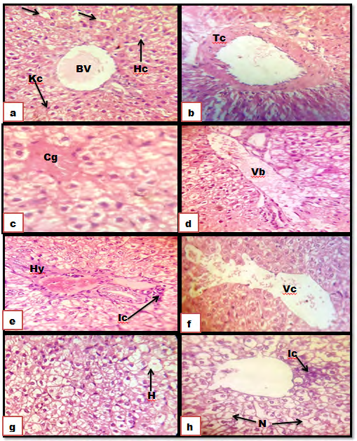
Figure 1: Photomicrographs of sections of the liver. (a): control group showing a normal architecture without pathological alterations. Hepatocyte (HP), Spherical nucleus (arrow), Sinusoids (S), Blood vessel (BV), and Kupffer cells (Kc). (b-h): IM group; showing obvious histopathological changes. Thickening in the central vein (Tc), Congestion of blood vessels (Cg), Vasodilatation in blood vessels (Vb), hyperplasia (Hy), Inflammatory cells infiltration (Ic), Vasodilatation in in the central vein (Vc), Hydropic changes (H), Necrosis (N). (HE) stain (X400).
Histological examination in lever after administration of gentamicin revealed thickening in the central vein, congestion of blood vessels and vasodilatation in blood vessels, as well as hyperplasia, Inflammatory cells infiltration, and Vasodilatation in the central vein, in addition to Hydropic changes and Necrosis (fig. 1b-g). Administration of gentamicin besides Osaimi sider honey in group 3, Athel honey in group 4, Dam Al-Akhawain honey in group 5, and Salam honey (Figures 2a-e) respectively, showed normal structure as in that of the control group.
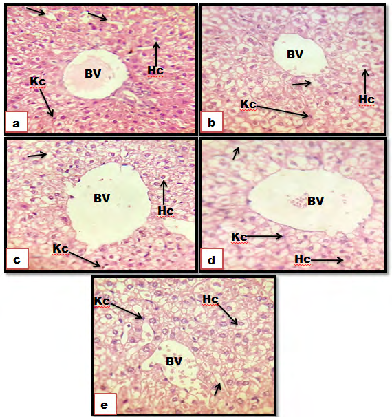
Figure 2: Photomicrograph of sections of the liver. (a): Control groups showing a normal architecture without pathological alterations. (b): Gen + Athel honey; (c): Gen + Dam Al-Akhawain honey; (d): Gen + Salam honey showing a normal liver structure as in control group. (e): Hepatocyte (HP), Spherical nucleus (arrow), Sinusoids (S), Blood vessel (BV), and Kupffer cells (Kc). (HE) stain (X400).
Histological Observation of Kidney
In the control group, kidney appeared renal corpuscles each one is formed of a glomerular tuft of blood capillaries surrounded by capsular space and Bowman’s capsule, the proximal convoluted tubules are more or less rounded in shape and formed of a single layer of cuboidal cells bearing a brush border in their luminal surfaces, and the distal convoluted tubules had large lumen than the proximal and their lumen has no brush border (Figures 3a-h).
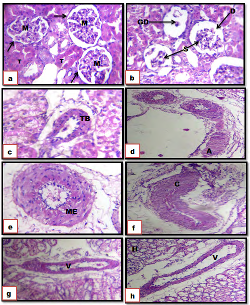
Figure 3: Photomicrograph of sections of the kidney. (a): Control groups showing normal histological structures of Malpighian corpuscles with its glomerulus (M) Bowman’s capsule (arrow), tubules (T). (b-h): Gen showing dilatation of Bowman’s capsule (D), glomerulus shrinkage (S), glomerular degeneration (GD), Thickened blood vessels (TB). Amyloid (A), Metaplasia (ME), Congestion and Compression of blood vessels (C), Vasodilatation of blood vessels (V), Hydropic change (H). (H&E X 100).
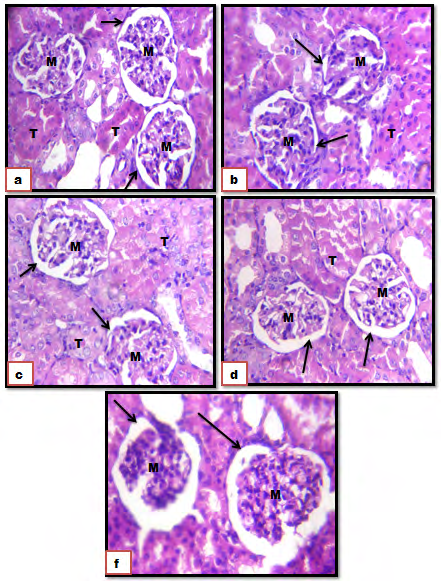
Figure 4: Photomicrograph of sections of the kidney. (a): Control groups showing a normal architecture without pathological alterations. (b): Gen+ Osaimi sider honey (c): Gen + Athel honey; (c): Gen + Dam Al-Akhawain honey; (d): Gen + Salam honey, showing a normal kidney structure as in control group. (f): Malpighian corpuscles with its glomerulus (M) Bowman’s capsule (arrow), tubules (T). (HE) stain (X400).
Histological examination of the cortex in the kidney after administration of gentamicin revealed severe shrinkage of glomeruli and glomerular degeneration, as well as thickened blood vessels, Amyloid and Metaplasia hydropic changes, in addition to congestion and Compression of blood vessels, Vasodilatation of blood vessels and Hydropic change (Fig. 3b- h). Administration of gentamicin besides Osaimi sider honey in group 3, Athel honey in group 4, Dam Al-Akhawain honey in group 5, and Salam honey (Figures 4a-f) respectively, resulted in that The cortex and medulla show nearly normal histological structures as in the control group.
Discussion
Biochemical parameters AST, ALT, GGT or Alkaline phosphatase and other parameters such as bilirubin and albumin levels and coagulating tests as prothrombin activity are commonly used as liver functions parameters and serve as the routine screening even in asymptomatic patients and the right evaluation of the results of the vital importance [20]. Liver transaminases (AST and ALT) are useful as biomarkers of liver injury in a patient with some degree of intact liver function. An increase in AST is the most specific marker of liver cell damage [21]. Results of this study confirmed that gentamicin at a dose of 80 mg/kg produces significant hepatotoxicity as evidenced by the increase in serum AST, ALT, and decrease in total portions, Albumin. In addition, gentamicin-induced severe hepatic damages as shown in the histopathological examination which coupled with markedly elevated levels of liver biochemical markers (AST, ALT, total portions, albumin. Our results agree with [22, 23, 24], whose results showed a significant increase in ALT and AST and a decrease in TP and albumin in serum of animals after administration of GM.
Gentamicin usually accumulates in the renal proximal tubules and enhances hydrogen peroxide generation by the mitochondria [1]. The hydrogen peroxide produced causes the release of iron from mitochondria membranes which complexes with gentamicin to evoke the generation of reactive oxygen species [6]. This antibiotic has been shown to dose-dependently induce nephrotoxicity when administered for more than 7-10 days [1]. In our study, serum creatinine and urea in gentamicin treated guinea pigs on Day 7 were comparable to control. However, an increase in creatinine at 80 mg/kg of gentamicin has been seen on Day 7 this result agrees with many studies [25, 26] reported significantly increased serum levels of blood urea nitrogen and creatinine in animals after administration of GM. This was associated with severe histopathological changes in kidneys indicating decline in kidney function (kidney damage and nepheotoxicity). Our result was gentamicin -induced glomerular congestion, per tubular and blood vessel congestion, epithelial desquamation, accumulation of inflammatory cells and necrosis of the kidney cells. This result agrees with many studies [27, 28].
Diminished glomeruli hypocellularity, moderately dilated tubules and mild loss of brush border, severe infiltration, extensive tubular degeneration, and presence of tubular cast. gentamicin can cause tissue injury such as nephrotoxicity, ototoxicity [29], and liver toxicity [30], possibly through the generation of free oxygen radicals. Nephrotoxicity of gentamicin arises due to its accumulation in renal cortical tubular epithelial cells. Qadir, et al. [31]. Although the pathogenesis of gentamicin-induced acute kidney injury (AKI) has been the focus of a large number of studies, the underlying mechanisms are not yet fully elucidated. Recent studies suggest that gentamicin nephrotoxicity is a complex and multifaceted process in which gentamicin triggers cellular responses involving multiple pathways that culminate in renal damage and necrosis [32].
In our study, we used different types of Yemeni honey as antioxidants. Honey is a natural antioxidant that may contain flavonoids, ascorbic acid, tocopherols, catalase, and phenolic compounds all of which work together to provide a synergistic antioxidant effect, scavenging and eliminating free radicals [18, 19].
In the present study, results revealed that the administration of different types of Yemeni honey ameliorates the changes that induce hepatotoxicity and nephrotoxicity. This result indicated different types of Yemeni honey prevented liver and kidney damage by its ability to eliminate free radicals generated by gentamicin. These results are in agreement with many studies which demonstrated that honey has a protective effect against gentamicin [15, 33], a similar observation was reported by [34, 35] against penicillin and streptomycin induce-hepatotoxicity and nephrotoxicity.
Conclusions
The present study concluded that the use of antioxidants (Yemeni Honey) showed a highly significant protective effect on the functions and tissue of the liver and kidney when they were used as co-treatment. The effects of honey that were found in our experiment are due to the presence of many antioxidant compounds such as flavonoids, ascorbic acid, tocopherols, catalase, and phenolic compounds, that work together to provide a synergistic antioxidant effect, scavenging and eliminating free radicals. but the protective effect of the treatment with sider honey and Dam Al- Akhawain honey had a greater protective effect on the liver and the treatment with Dam Al-Akhawain honey and Athel honey had a greater protective effect on the kidney which was almost similar to the control.
References
-
Noorani AA, Gupta K, Kale KM (2011) Protective Effect of Methanolic Leaf Extract of Caesaipinia Bonduc (L) On Gentamicin-Induced Hepatotoxicity and Nephrotoxicity in Rats. Iranian J Pharmacology Therapeutic 10(1): 21- 25.
-
Ayatollahi J (2005) Evaluation of knowledge and activities of medical students in the last two years of their education about chemoprophylaxis following contact with infectious diseases. IJCID 9(26): 54-59.
-
Stojiljkovic N, Stoiljkovic M (2006) Micromorphological characteristics of the liver and biochemical analyses in the blood of rats treated by gentamicin and verapamil. Acta medica Medianae 45(2): 5-9.
-
Masakazu K, Yoshiko E, Masashi E (2014) Acquired resistance of Listeria monocytogenes in and escaped from liver parenchymal cells to gentamicin is caused by being coated with their plasma membrane. Microbes and Infection 16(3): 237-243.
-
Venkatesha U, Prakash V (2019) Gentamicin induced acute renal damage and its evaluation using urinary biomarkers in rats. Toxicology Reports 6: 91-99.
-
Conlon BJ, Aran JM, Erre JP (1999) Attenuation of aminoglycoside induced cochlear damage with the metabolic antioxidant alphalipoic acid. Hear Res: 128(1- 2): 40-44.
-
Wojciec L, Vincent LP, Schacht J (2005) Ternary Complexes of Gentamicin with Iron and Lipid Catalyze Formation of Reactive Oxygen Species. Chemical Research in Toxicology 18(2): 357-364.
-
Erin E, Battin J (2009) Sulfur and selenium: A Review of reactiveOxygen species scavenging, glutathione peroxidase and metal-binding antioxidant mechanisms. Cell Biochem Biophys 55(1): 1-23.
-
Dekkers JC, Van Doornen LJ, Han CG, Kemper PJ (1996) The role of antioxidant vitamins and enzymes in the prevention of exercise-induced muscle damage. Sports Med 21(3): 213-238.
-
Cherubini A, Vigna GB, Zuliani G, Ruggiero C, Senin U, et al. (2005) Role of antioxidants in atherosclerosis: epidemiogical and clinical update. Curr Pharm Des 11(16): 2017-2032.
-
Prior RL, Cao G (2000) Antioxidant phytochemicals in fruits and vegetables; diet and health implications. Hortic j Sci 35(4): 588-592.
-
Shireen KF, Pace RD, Mahboob M, Khan AT (2008) Effects of dietary vitamin E, C and soybean oil supplementation on antioxidant enzyme activities in liver and muscles of rats. Food Chem Toxicol 46(10): 3290-3294.
-
Al-Yahya M, Ramzi M, Al-Said M, Al-Dosari M, Al-Musayeib N, et al. (2013) Attenuation of CCl4-Induced Oxidative Stress and Hepatonephrotoxicity by Saudi Sidr Honey in Rats. Evidence Based Comple Altern Med 10: 1-10.
-
Abdel-El Denshary ES, Al-Gahazali MA, Mannaa FA, Salem HA, Hassan NS, et al. (2012) Dietary honey and ginseng protect against carbon tetrachloride-induced hepatonephrotoxicity in rats. Exp Toxicol Patho 64(7-8): 753-760.
-
Abd El-Ghany MA, Ramadan AM, Ghozy SF (2012) Nutraceutical Effects of Curcuma, Ginger, Celery, Yeast and Honey on Side Effects of Gentamicin Induced Nephrotoxicity in Rats. World Appl Sci J 16 (5): 646-655.
-
Ramos AF, Miranda JL (2007) Honey: a review of its anti- inflammatory and healing actions. J Venom Anim Toxins incl Trop Dis 13(4): 679-710.
-
Al-Awar MS, Al-Qalah TA, Ghaleb AD, Rashed MM (2020) Anti-inflammatory Activities of Different Types of Yemeni honey on Rats. North American Academic Research 3(11): 170-189.
-
Gheldof N, Wang XH, Engeseth NJ (2002) Identification and quantification of antioxidant components of honeys from various floral sources. J Agric Food Chem 50(21): 5870-5877.
-
Chandane RD, Jugalkishor B, Jaju Manik S, Ghadlinge R, Bhosale A, et al. (2013) Effect of honey on hepatotoxicity induced by antitubercular drugs in albino rats. Int J Basic Clin Pharmacol 2(2): 177-181.
-
Borque AM, Moreno LG, Jiménez JM, Buey LG, Otero RM (2007) Utility of analytical parameters in the diagnosis of liver disease. An Med Interna 24(1): 38-46.
-
Chatterjea MN, Shinde R (2005) Text Book of Medical Biochemistry. 6th(Edn.), Jaypee Broth, New-Delhi, pp: 644.
-
Ansah C, Moomin A, Boadu KM (2016) Terminalia ivorensis A. Chev. Ethanolic Stem Bark Extract Protects Against Gentamicin-Induced Renal and Hepatic Damage in Rats. J App Pharmacol Sci 6(4): 175-182.
-
Elgebaly HA, Mosa NM, Germoush MO, Brahim AC (2016) The Nephro-Protective Effects of Olive Oil and Bee Honey against Gentamicin-Induced Nephrotoxicity in Rabbits. AUMJ 1-7.
-
Govindappa PK, Gautam V, Tripathi SM, Sahnib YP, Raghavendra HLS (2019) Effect of Withania somnifera on gentamicin induced renal lesions in rats. Revista Brasileira de Farmacognosia 29(2): 234-240.
-
Rathod NS, Halagali KS, Nidavani RB, Shalavadi MH, Biradar BS, et al. (2016) Protective Effect of Punica Granatum L. On Gentamicin Induced Acute Renal Failure In Adult Rats. J Clin Exp Pharmacol 6(3): 2161-1459.
-
Udupa V, Prakash V (2019) Gentamicin induced acute renal damage and its evaluation using urinary biomarkers in rats. J Tox Rep 6: 91-99.
-
Randjelović P, Slavimir V, Stojiljković N, Sokolović D, Ilić I (2017) Gentamicin Nephrotoxicity in Animals: Current Knowledge and Future Perspectives. EXCLI Journal 16: 388-399.
-
Hafez OL, A Ali FA, El-Ghoneimy AI, Abdel-Aziz M (2019) Nephro-Protective Effect of Wheat Germ Oil on Gentamicin-Induced Acute Nephrotoxicity in Wistar Albino Rat. J Inte Vete Sci 2(1): 51-67.
-
Alarifi S, Al-Doaiss A, Alkahtani S, Al-Farraj S, Al-Eissa M, et al. (2012) Blood chemical changes and renal histological alterations induced by gentamicin in rats. Saudi J Biol Sci 19(1): 103-110.
-
Aubrecht J, Goad M, Simpson E (1997) Expression of hygr intransgenic mice causes resistance to toxic effects of hygromycin B in vivo. J Pharmacol Exp Ther 281(2): 992-997.
-
Qadir M, Tahir M, Lone K, Munir B, Sam W (2011) Protective role ofginseng against gentamicin induced changes in kidney ofalbino mice. J Ayub Med Coll Abbottabad 23(4): 53-57.
-
Gonzalez A, Jimenez A, Vazquez D (1978) Studies on the mode of action of hygromycin B, an inhibitor of translocation in eukaryotes. Biochem Biophys Acta 521(2): 459-469.
-
Chilwant KS, Kothekar MA, Muglikar AG, Jaju JB, Mateenuddin MD (2004) Effect of honey on gentamicin induced nephrotoxicity in rats. Indian J Pharmacol 36 (2): 112-126.
-
Al-Arami AMJ, Al-Awar MSA (2013) Hepatoprotective and hepatocurative effect of Sider honey in penicillin induce-hepatotoxicity. JPS 2(5): 34-40.
-
Al-Awar MSA, AL-Shaibani EAS, Salih EMA, Al-Eryania MAY (2013) The Protective Effect of Sider honey Against Pathological Effects of Penicillin and Streptomycin on Histological Structure and Functions of Guinea pigs Liver. J App Pharma Sci 3(4): 1-6.
- Effects of 5-HTP and Melatonin on the Sleep Cycle of Medical Students
- Adsorption of Bisphenol A on NH4OH- Modified Rice Husk and Sugar Cane Bagasse Biochar
- Comparative Assessment of the Reinforcement Efficiency of Palm Fruit Fibre and Coconut Fibre in High Density Polyethylene (HDPE) Matrix Composite
- Importance of Bio Compounds Naturally Present in Food with Functionality in Animal Metabolism
- Sub-Acute Study on the Cardiotoxic Effects of Monosodium Glutamate Ingestion in Albino Rat
- Weight Management and Its Natural Solutions: A Review