Association of Elevated Levels of Some Intra-Thyroidal Trace Elements with Goiter and Cancer Risk of Female Thyroid
Introduction: Advancing age is known to influence the formation of adenomatous goiter and thyroid cancer. An excess or deficiency of specific TEs contents in thyroid can play an important role in goitro- and carcinogenesis of this gland. Objective: This study aimed to assess the variation with age of fifty trace element (TE) contents in normal female thyroid. Methods: Samples of the human thyroid were obtained from randomly selected autopsy specimens of 33 females (EuropeanCaucasian) aged 3.5 to 87 years after a sudden death mainly from trauma. The mass fractions of TEs in normal female thyroids were investigated using neutron activation analysis and inductively coupled plasma mass spectrometry. Tissue samples were divided into two portions. One was used for morphological study while the other was intended for TEs analysis. Results: This work revealed that there is a statistically significant increase in Co, Cs, Fe, La, Pb, Rb, Sb, Se, Sn, Tl, and Zn mass fraction in the normal thyroid of female during a lifespan. Contents of such carcinogenic or potentially carcinogenic TEs as Co, La, Pb, Sb, Sn, and Tl in thyroid of seniors are 5-10 times higher than those in thyroid of girls or young women. Conclusions: From results of our study, a goitrogenic and carcinogenic effect of elevated Co, Cs, Fe, La, Pb, Rb, Sb, Se, Sn, Tl, and Zn levels in the thyroid of elderly females is shown to be a very likely consequence.
Introduction
The prevalence of benign and malignant nodules is increased in the elderly, reaching a frequency of nearly 50% by the age of 65 [1, 2, 3, 4]. Both prevalence and aggressiveness of thyroid malignancy increase with age [1]. Women are affected by thyroid benign and malignant nodules two to five times more often than men [1, 3, 4, 5]. Aging is characterized by the accumulation of molecular damage in DNA, proteins and lipids, and also by an increase in intracellular reactive oxygen species (ROS) [6, 7]. Oxidative damage to cellular macromolecules which induce age-related diseases, including cancer, can also arise through overproduction of ROS and faulty antioxidant and/or DNA repair mechanisms [8]. Overproduction of ROS is associated with many factors, including overload of certain trace elements (TE), or deficiency of other TE with antioxidant properties [9, 10, 11, 12, 13, 14, 15]. The imbalance in the composition of TE in cells, tissues and organs may cause different types of pathology. The importance of appropriate levels of many TE is indisputable, due to their beneficial roles when present in specific concentration ranges, while on the other hand they can cause harmful effects with excessively high or low concentrations [12].
In our previous studies Zaichick V [16, 17, 18, 19, 20, 21, 22, 23, 24] the high mass fraction of iodine and some other TE were observed in the intact human thyroid gland when compared with their levels in non-thyroid soft tissues of the human body. However, the age-dependence of TE mass fraction in thyroid of adults and, particularly, in elderly females still remain unanswered. One valuable way to elucidate the situation is to compare the mass fractions of TE in young adults (the control group) with those in older adult and geriatric thyroids. The other way is to calculate a correlation between age and TE content in the thyroid. Findings of the excess or deficiency of TE contents in thyroid may indicate their roles in the higher prevalence of thyroid benign and malignant nodules in the elderly.
Reliable data on TE mass fractions in normal geriatric thyroids is apparently extremely limited. There are a few studies reporting TE content in human thyroids, using various chemical techniques and instrumental methods. However majority of the analytical methods currently used and validated for the determination of TE in thyroid are based on techniques requiring sample digestion. The most frequently used digestion procedures are the traditional dry ashing and high-pressure wet digestion that cause destruction of organic matter of the sample. Sample digestion is a critical step in elemental analysis and due to the risk of contamination and analytes loss, a digestion step contributes to the systematic uncontrolled analysis errors [25, 26, 27]. Moreover, only a few of the previous studies employed quality control using certified/standard reference materials (CRM/SRM) for determination of the TE mass fractions. In previously published papers there is no data on the age effects on TE content of normal female thyroids. Therefore, nondestructive technique such as instrumental neutron activation analysis with high resolution spectrometry of long-lived radionuclides (INAA- LLR) combined with subsequent, destructive inductively coupled plasma mass spectrometry (ICP-MS) provides a good method for multi TE determination in samples of thyroid parenchyma. This combination of methods provides a possibility to ensure data quality assurance using a comparison of results obtained for some TE by both methods. The primary purpose of the study was to determine reliable values for TE mass fractions in the normal (intact) thyroid of subjects ranging from children to elderly females using INAA-LLR and ICP-MS. The second aim was to compare the TE mass fractions determined in thyroid gland of age group 2 (adults and elderly persons aged 40 to 87 years), with those of group 1 (from 3.5 to 40 years). The final aim was to find the correlations between age and TE contents.
Materials and Methods
Samples of the human thyroid were obtained from randomly selected autopsy specimens of 33 females (European-Caucasian) aged 3.5 to 87 years within 48 hours after a sudden death. All the deceased were citizens of Obninsk and had undergone routine autopsy at the Forensic Medicine Department of City Hospital, Obninsk. Obninsk is a small city of a non-industrial region 105 km south-west from Moscow. Age ranges for subjects were divided into two age groups, with group 1, 3.5-40 years (30.9±3.1 years, M±SEM, n=11) and group 2, 41–87 years (66.3±2.7 years, M±SEM, n=22). These groups were selected to reflect the condition of thyroid tissue in the children, teenagers, young adults and first period of adult life (group 1) and in both the second period of adult life and also in the old age (group 2). The available clinical data were reviewed for each subject. None of the subjects had a history of an intersex condition, endocrine disorder, or other chronic disease that could affect the normal development of the thyroid. None of the subjects were receiving medications or used any supplements known to affect thyroid trace element contents. None of the subjects had a professional contact with trace elements, radionuclides and radiation. The typical causes of sudden death of most of these subjects included trauma or suicide and also untreated acute illness (cardiac insufficiency, stroke, pulmonary artery embolism and alcohol poisoning).
All studies were approved by the Ethical Committees of MRRC. All the procedures performed in studies involving human participants were in accordance with the ethical standards of the institutional and/or national research committee and with the 1964 Helsinki declaration and its later amendments, or with comparable ethical standards.
All right lobes of thyroid glands were divided into two portions using a titanium scalpel [28]. One tissue portion was reviewed by an anatomical pathologist while the other was used for the TE content determination. A histological examination was used to control the age norm conformity as well as the absence of any microadenomatosis and latent cancer.
After the samples for TE analysis were weighed, they were transferred to an environment at temperature -20°C and stored until the day of transportation to the Medical Radiological Research Center, Obninsk, where all samples were freeze-dried and homogenized [29]. The samples, each weighing about 50 mg, were used for TE measurement by INAA-LLR. Biological synthetic standards (BSS) were used as standards [30]. In addition to BSS, aliquots of commercially available pure compounds were also used. A vertical channel of nuclear reactor with a neutron flux of 1.3×10¹³ n×cm- 2×s-1 was applied to determine the content of Ag, Co, Cr, Fe, Hg, Rb, Sb, Sc, Se, and Zn in thyroid samples by INAA-LLR.
After determination TE by INAA-LLR the thyroid samples were used for ICP-MS. Before ICP-MS analysis the samples were decomposed in autoclaves. After autoclaving sample aliquots were used to determine the content of Ag, Al, As, Au, B, Be, Bi, Cd, Ce, Co, Cr, Cs, Dy, Er, Eu, Ga, Gd, Hg, Ho, Ir, La, Li, Lu, Mn, Mo, Nb, Nd, Ni, Pb, Pd, Pr, Pt, Rb, Sb, Se, Sm, Sn, Tb, Te, Th, Ti, Tl, Tm, U, Y, Yb, Zn, and Zr by ICP- MS using an ICP-MS Thermo-Fisher “X-7” Spectrometer (Thermo Electron, USA). The TE concentrations in aqueous solutions were determined by the quantitative method using multi elemental calibration solutions ICP-MS-68A and ICP- AM-6-A produced by High-Purity Standards (Charleston, SC 29423, USA). Indium was used as an internal standard in all measurements. Information detailing with the NAA-LLR and ICP-MS methods used and other details of the analysis was presented in our previous publication [31, 32, 33, 34, 35, 36, 37].
For quality control, ten subsamples of the certified reference materials (CRM) IAEA H-4 Animal Muscle from the International Atomic Energy Agency (IAEA), and also five sub- samples INCT-SBF-4 Soya Bean Flour, INCT-TL-1 Tea Leaves and INCT-MPH-2 Mixed Polish Herbs from the Institute of Nuclear Chemistry and Technology (INCT, Warszawa, Poland) were analyzed simultaneously with the investigated thyroid tissue samples. All samples of CRMs were treated in the same way as the thyroid tissue samples. Detailed results of this quality assurance program were presented in earlier publications [31, 32, 33, 34, 35, 36, 37].
A dedicated computer program for INAA-LLR mode optimization was used [38]. All thyroid samples were prepared in duplicate, and mean values of TE contents were used in final calculation. Using Microsoft Office Excel, a summary of the statistics, including, arithmetic mean, and standard deviation, standard error of mean, minimum and maximum values, median, percentiles with 0.025 and 0.975 levels was calculated for TE contents. The difference in the results between the two age groups was evaluated by the parametric Student’s t-test and non-parametric Wilcoxon- Mann-Whitney U-test. For the construction of the “age vs TE mass fraction” diagrams (including trend lines with age) and the estimation of the Pearson correlation coefficient between age and TE mass fraction the Microsoft Office Excel programs were also used. To identify the trend of the age dependency of TE contents, we applied approximation methods using exponential, linear, polynomial, logarithmic and power function. The maximum of corresponding values of R2 parameter, reflecting the accuracy of approximation, was used for the selection of function.
Results
The comparison of our results for the Ag, Co, Cr, Fe, Hg, Rb, Sb, Se, and Zn mass fractions (mg/kg, dry mass basis) in the normal thyroid of female obtained by both NAA-LLR and ICP-MS methods is shown in Table 1.
| Element | NAA-LLR M1 | ICP-MS M2 | ∆, % |
|---|---|---|---|
| Ag | 0.0140±0.0020 | 0.0129±0.0041 | 7.9 |
| Co | 0.0505±0.0064 | 0.0479±0.0069 | 5.1 |
| Cr | 0.573±0.049 | 0.496±0.057 | 13.4 |
| Fe | 232±22 | 217±19 | 6.5 |
| Hg | 0.0389±0055 | 0.0471±0.0087 | -21.1 |
| Rb | 6.16±0.48 | 6.38±0.53 | -3.6 |
| Sb | 0.116±0.012 | 0.098±0.014 | 15.5 |
| Se | 2.22±0.23 | 2.16±0.23 | 0.5 |
| Zn | 85.7±7.4 | 83.2±8.1 | 2.9 |
Table 1: Comparison of the mean values (M±SEM) of the chemical element mass fractions (mg/kg, on dry-mass basis) in the normal th
Table 2 represents certain statistical parameters (arithmetic mean, standard deviation, standard error of mean, minimal and maximal values, median, percentiles with 0.025 and 0.975 levels) of the Ag, Al, As, Au, B, Be, Bi, Cd, Ce, Co, Cr, Cs, Dy, Er, Eu, Fe, Ga, Gd, Hg, Ho, Ir, La, Li, Lu, Mn, Mo, Nb, Nd, Ni, Pb, Pd, Pr, Pt, Rb, Sb, Sc, Se, Sm, Sn, Tb, Te, Th, Ti, Tl, Tm, U, Y, Yb, Zn, and Zr mass fractions in intact (normal) thyroid of females. The As, Au, Eu, Ho, Ir, Lu, Pd, Pt, Te, Th, Tm, Yb, and Zr mass fractions were determined in a few samples.
The possible upper limit of the mean (≤M) for these TE was calculated as the average mass fraction, using the value of the detection limit (DL) instead of the individual value when the latter was found to be below the DL The upper limits of mass fraction of these TE were: As ≤ 0.0045, Au ≤ 0.0039, Eu ≤ 0.00041, Ho ≤ 0.00036, Ir ≤ 0.00027, Lu ≤ 0.00018, Pd ≤ 0.023, Pt ≤ 0.0014, Te ≤ 0.0050, Th ≤ 0.0031, Tm ≤ 0.00015, Yb ≤ 0.00093, and Zr ≤ 0.097.
| Element | M | SD | SEM | Min | Max | Median | P 0.025 | P 0.975 |
|---|---|---|---|---|---|---|---|---|
| Ag | 0.0132 | 0.0092 | 0.002 | 0.0016 | 0.0331 | 0.0121 | 0.00225 | 0.0321 |
| Al | 7.43 | 4.49 | 1.24 | 2.5 | 17.2 | 5.5 | 2.77 | 16.6 |
| As | ≤0.0045 | - | - | <0.003 | 0.01 | - | - | - |
| Au | ≤0.0039 | - | - | <0.002 | 0.0101 | - | - | - |
| B | 0.418 | 0.257 | 0.074 | 0.2 | 1 | 0.315 | 0.2 | 0.89 |
| Be | 0.00067 | 0.00089 | 0.0003 | 0.0001 | 0.0031 | 0.00035 | 0.0001 | 0.00261 |
| Bi | 0.018 | 0.03 | 0.0084 | 0.001 | 0.1 | 0.007 | 0.00106 | 0.0902 |
| Cd | 1.63 | 1.73 | 0.48 | 0.011 | 5.84 | 1.18 | 0.0443 | 5.21 |
| Ce | 0.00897 | 0.00785 | 0.0023 | 0.0014 | 0.0253 | 0.0059 | 0.00195 | 0.0251 |
| Co | 0.0493 | 0.0332 | 0.0066 | 0.016 | 0.14 | 0.038 | 0.0166 | 0.13 |
| Cr | 0.535 | 0.254 | 0.051 | 0.233 | 1.22 | 0.456 | 0.27 | 1.11 |
| Cs | 0.0185 | 0.0105 | 0.0029 | 0.0022 | 0.0368 | 0.0182 | 0.00289 | 0.036 |
| Dy | 0.00173 | 0.00328 | 0.001 | 0.0003 | 0.0121 | 0.00084 | 0.0003 | 0.00913 |
| Er | 0.0005 | 0.00057 | 0.0002 | 0.0001 | 0.0022 | 0.00032 | 0.00013 | 0.00181 |
| Eu | ≤0.00041 | - | - | <0.0002 | 0.0019 | - | - | - |
| Fe | 225 | 98 | 20 | 52 | 435 | 199 | 64.2 | 391 |
| Ga | 0.0309 | 0.0209 | 0.006 | 0.01 | 0.081 | 0.0285 | 0.01 | 0.0725 |
| Gd | 0.00147 | 0.00174 | 0.0005 | 0.0004 | 0.0065 | 0.0009 | 0.0004 | 0.0056 |
| Hg | 0.04 | 0.0274 | 0.0057 | 0.007 | 0.1 | 0.031 | 0.0125 | 0.1 |
| Ho | ≤0.00036 | - | - | <0.0001 | 0.0012 | - | - | - |
| Ir | ≤0.00027 | - | - | <0.00004 | 0.0002 | - | - | - |
| La | 0.0055 | 0.00368 | 0.0011 | 0.001 | 0.0118 | 0.0045 | 0.00128 | 0.0117 |
| Li | 0.0153 | 0.0078 | 0.0024 | 0.0015 | 0.0251 | 0.0152 | 0.003 | 0.025 |
| Lu | ≤0.00018 | - | - | <0.00001 | 0.0005 | - | - | - |
| Mn | 1.32 | 0.84 | 0.22 | 0.55 | 4.04 | 1.1 | 0.603 | 3.28 |
| Mo | 0.0755 | 0.0608 | 0.0169 | 0.0104 | 0.252 | 0.0567 | 0.015 | 0.217 |
| Nb | 0.641 | 0.722 | 0.2 | 0.013 | 2.21 | 0.336 | 0.013 | 2 |
| Nd | 0.0046 | 0.0042 | 0.0012 | 0.00054 | 0.0165 | 0.0035 | 0.00086 | 0.0139 |
| Ni | 0.385 | 0.192 | 0.056 | 0.2 | 0.89 | 0.31 | 0.211 | 0.81 |
| Pb | 0.2 | 0.11 | 0.032 | 0.023 | 0.45 | 0.185 | 0.0442 | 0.406 |
| Pd | ≤0.023 | - | - | <0.014 | 0.051 | - | - | - |
| Pr | 0.00123 | 0.00102 | 0.0003 | 0.0002 | 0.0039 | 0.0009 | 0.00029 | 0.00345 |
| Pt | ≤0.0014 | - | - | <0.0002 | 0.014 | - | - | - |
| Rb | 6.27 | 2.43 | 0.48 | 1.21 | 12.8 | 6.3 | 2.5 | 30.8 |
| Sb | 0.107 | 0.066 | 0.013 | 0.0115 | 0.248 | 0.09 | 0.0183 | 0.247 |
| Sc | 0.0086 | 0.0158 | 0.0046 | 0.0002 | 0.057 | 0.0034 | 0.00034 | 0.0453 |
| Se | 2.19 | 1.17 | 0.23 | 0.32 | 5.32 | 2.07 | 0.745 | 4.85 |
| Sm | 0.00057 | 0.00046 | 0.0001 | 0.0001 | 0.0014 | 0.00043 | 0.00012 | 0.0014 |
| Sn | 0.112 | 0.093 | 0.026 | 0.009 | 0.263 | 0.0621 | 0.0165 | 0.26 |
| Tb | 0.0002 | 0.00015 | 4E-05 | 0.00008 | 0.0006 | 0.00018 | 0.00009 | 0.00052 |
| Te | ≤0.0050 | - | - | <0.003 | 0.013 | - | - | - |
| Th | ≤0.0031 | - | - | <0.002 | 0.008 | - | - | - |
| Ti* | 3.74 | 4.2 | 1.16 | 0.44 | 13.6 | 2.3 | 0.521 | 12.8 |
| Tl | 0.0007 | 0.00036 | 0.0001 | 0.0001 | 0.0013 | 0.00072 | 0.00016 | 0.00125 |
| Tm | ≤0.00015 | - | - | <0.0001 | 0.0003 | - | - | - |
| U | 0.0006 | 0.00069 | 0.0002 | 0.0001 | 0.0026 | 0.00033 | 0.00012 | 0.00219 |
| Y | 0.00304 | 0.00282 | 0.0008 | 0.001 | 0.011 | 0.0019 | 0.001 | 0.0093 |
| Yb | ≤0.00093 | - | - | <0.0003 | 0.0057 | - | - | - |
| Zn | 84.5 | 38.9 | 7.6 | 7.3 | 166 | 83.5 | 23 | 156 |
| Zr | ≤0.097 | - | - | <0.03 | 0.45 | - | - | - |
Table 2: Some statistical parameters of 51 trace element mass fraction (mg/kg, dry mass basis) in the normal thyroid of female. M
Table 2: Some statistical parameters of 51 trace element mass fraction (mg/kg, dry mass basis) in the normal thyroid of female. M – arithmetic mean, SD – standard deviation, SEM – standard error of mean, Min – minimum value, Max – maximum value, P 0.025 – percentile with 0.025 level, P 0.975 – percentile with 0.975 level. To estimate the effect of age on the Ag, Al, B, Be, Bi, Cd, Ce, Co, Cr, Cs, Dy, Er, Fe, Ga, Gd, Hg, La, Li, Mn, Mo, Nb, Nd, Ni, Pb, Pr, Rb, Sb, Sc, Se, Sm, Sn, Tb, Ti, Tl, U, Y, and Zn contents we examined two age groups, described above Table 3.
| Element | Female Thyroid Tissue | Ratio | |||
|---|---|---|---|---|---|
| AG1 3.5-40 years n=11 | AG2 41-87 years n=22 | T-test p£ | U-test p | AG2 to AG1 | |
| Ag | 0.0125±0.0032 | 0.0138±0.0027 | 0.757 | >0.05 | 1.02 |
| Al | 5.95±0.97 | 9.8±2.7 | 0.233 | >0.05 | 1.65 |
| B | 0.361±0.070 | 0.533±0.177 | 0.419 | >0.05 | 1.48 |
| Be | 0.00088±0.00046 | 0.00037±0.00013 | 0.333 | >0.05 | 0.42 |
| Bi | 0.018±0.012 | 0.017±0.012 | 0.961 | >0.05 | 0.94 |
| Cd | 1.34±0.53 | 2.10±0.95 | 0.51 | >0.05 | 1.57 |
| Ce | 0.0070±0.0030 | 0.0118±0.0034 | 0.323 | >0.05 | 1.69 |
| Co | 0.0311±0.0046 | 0.0635±0.0099 | 0.0078 | ≤0.01 | 2.04 |
| Cr | 0.495±0.066 | 0.566±0.076 | 0.483 | >0.05 | 1.14 |
| Cs | 0.0160±0.0032 | 0.0225±0.0056 | 0.342 | >0.05 | 1.41 |
| Dy | 0.0021±0.0014 | 0.00095±0.00014 | 0.44 | >0.05 | 0.45 |
| Er | 0.00059±0.00025 | 0.00032±0.00005 | 0.31 | >0.05 | 0.54 |
| Fe | 174±25 | 264±25 | 0.018 | ≤0.01 | 1.52 |
| Ga | 0.0285±0.0086 | 0.0358±0.0069 | 0.524 | >0.05 | 1.26 |
| Gd | 0.00141±0.00074 | 0.00156±0.00055 | 0.875 | >0.05 | 1.11 |
| Hg | 0.0402±0.0078 | 0.0391±0.0084 | 0.923 | >0.05 | 0.97 |
| La | 0.00423±0.00120 | 0.00728±0.00176 | 0.191 | >0.05 | 1.72 |
| Li | 0.0131±0.0033 | 0.0178±0.0032 | 0.342 | >0.05 | 1.36 |
| Mn | 1.21±0.14 | 1.54±0.45 | 0.622 | >0.05 | 1.27 |
| Mo | 0.072±0.027 | 0.081±0.014 | 0.768 | >0.05 | 1.13 |
| Nb | 0.349±0.155 | 1.11±0.40 | 0.131 | >0.05 | 3.18 |
| Nd | 0.0046±0.0019 | 0.0046±0.0008 | 0.996 | >0.05 | 1 |
| Ni | 0.357±0.051 | 0.424±0.120 | 0.626 | >0.05 | 1.19 |
| Pb | 0.140±0.026 | 0.284±0.047 | 0.034 | ≤0.01 | 2.03 |
| Pr | 0.00118±0.00044 | 0.00132±0.00028 | 0.793 | >0.05 | 1.12 |
| Rb | 4.93±0.64 | 7.26±0.57 | 0.013 | ≤0.01 | 1.47 |
| Sb | 0.0762±0.00084 | 0.1296±0.0198 | 0.023 | ≤0.01 | 1.7 |
| Sc | 0.0290±0.0281 | 0.0045±0.0043 | 0.543 | >0.05 | 0.16 |
| Se | 1.80±0.24 | 2.47±0.35 | 0.128 | >0.05 | 1.37 |
| Sm | 0.00039±0.00017 | 0.00083±0.00017 | 0.104 | >0.05 | 2.13 |
| Sn | 0.076±0.023 | 0.168±0.048 | 0.139 | >0.05 | 2.21 |
| Tb | 0.000213±0.000060 | 0.000183±0.000048 | 0.704 | >0.05 | 0.86 |
| Ti* | 3.39±1.30 | 4.30±2.39 | 0.748 | >0.05 | 1.27 |
| Tl | 0.00060±0.00016 | 0.00083±0.00010 | 0.25 | >0.05 | 1.38 |
| U | 0.00063±0.00029 | 0.00052±0.00020 | 0.757 | >0.05 | 0.83 |
| Y | 0.0032±0.0012 | 0.0027±0.0006 | 0.713 | >0.05 | 0.84 |
| Zn | 58.3±9.3 | 103.7±8.5 | 0.0016 | ≤0.01 | 1.78 |
Table 3: Differences between mean values (M±SEM) of trace element mass fractions (mg/kg, dry mass basis) in the normal female thy
In addition, the Pearson correlation coefficient between age and TE mass fraction was calculated Table 4.
| Element r | Ag 0.15 | Al 0.46 | B 0.26 | Be -0.25 | Bi 0.26 | Cd 0.26 | Ce 0.44 | Co 0.56b | Cr 0.31 | Cs 0.50a |
|---|---|---|---|---|---|---|---|---|---|---|
| Element r | Dy 0.06 | Er 0.07 | Fe 0.26 | Ga 0.38 | Gd 0.19 | Hg -0.08 | La 0.59a | Li 0.45 | Mn 0.16 | Mo 0.39 |
| Element r | Nb 0.32 | Nd 0.29 | Ni 0.05 | Pb 0.74b | Pr 0.34 | Rb 0.60c | Sb 0.51b | Sc -0.39 | Se 0.50b | Sm 0.47 |
| Element r | Sn 0.55a | Tb 0.18 | Ti 0.23 | Tl 0.62a | U 0.14 | Y 0.15 | Zn 0.65c |
Table 4: Correlations between age (years) and trace element mass fractions (mg/kg, dry mass basis) in the normal thyroid of femal
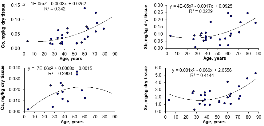
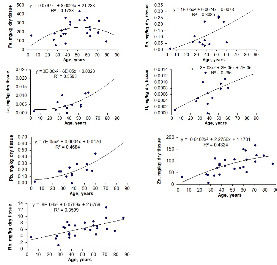
Figure 1 shows the individual data sets for the Co, Cs, Fe, La, Pb, Rb, Sb, Se, Sn, Tl, and Zn mass fraction in all samples of thyroid, and also lines of trend with age. Since the age dependency of these TE contents was best described by a polynomial function, this approximation was reflected in Figure 1.
Discussion
A good agreement of our results for the Ag, Al, As, Au, B, Be, Bi, Cd, Ce, Co, Cr, Cs, Dy, Er, Eu, Fe, Ga, Gd, Hg, Ho, Ir, La, Li, Lu, Mn, Mo, Nb, Nd, Ni, Pb, Pd, Pr, Pt, Rb, Sb, Sc, Se, Sm, Sn, Tb, Te, Th, Ti, Tl, Tm, U, Y, Yb, Zn, and Zr mass fractions with the certified values of CRM IAEA H-4 Animal Muscle, INCT-SBF-4 Soya Bean Flour, INCT-TL-1 Tea Leaves, and INCT-MPH-2 Mixed Polish Herbs [31, 32, 33, 34, 35, 36], as well as the similarity of the means of the the Ag, Co, Cr, Fe, Hg, Rb, Sb, Se, and Zn mass fractions in the normal thyroid of male determined by both NAA-LLR and ICP-MS methods (Table 1) demonstrates an acceptable precision and accuracy of the results obtained in the study and presented in Tables 2–4 and Figure 1.
A statistically significant age-related increase in Co, Fe, Pb, Rb, Sb, and Zn mass fraction was observed in thyroids of females when the two age groups were compared (Table 3). In the second group of females with mean age 66.3 years the mean of Co, Fe, Pb, Rb, Sb, and Zn mass fraction in thyroids were 1.5-2 times higher than in thyroids of the first age group (mean age 30.9 years) of females. A statistically significant increase in Co, Pb, Rb, Sb, and Zn mass fraction with age was confirmed by the Pearson’s coefficient of correlation between age and mass fractions of these TE (Table 4).
Moreover, a statistically significant increase in Cs, La, Se, Sn, and Tl content was shown by the Pearson’s coefficient of correlation between age and mass fractions of these TE (Table 4, Figure 1).
Thus, only positive (increase with age) correlations were observed for TE contents in the normal female thyroid. The increase of Cs and Fe was fast during the first four-five decades, after which the changes in mass fractions of these TE were rather limited. In contrast, contents of Co, La, Sb, and Se remain fairly stable until the fifth decade, after which a strong but variable accumulation takes place. The Pb, Rb, Sn, Tl, and Zn mass fraction showed a progressive accumulation during lifespan, herein, content of Pb, Sn, and Tl increases approximately 10 times (Fig. 1). Co, La, and Sb mass fractions are about quintuple from age 30 until age 70-80 (Fig. 1). According to the authors’ currently available information, no published data referring to age-related changes of Ag, Al, B, Be, Bi, Cd, Ce, Co, Cr, Cs, Dy, Er, Fe, Ga, Gd, Hg, La, Li, Mn, Mo, Nb, Nd, Ni, Pb, Pr, Rb, Sb, Sc, Se, Sm, Sn, Tb, Ti, Tl, U, Y, and Zn mass fractions in female thyroid is available. An age-related increase and excess in TE mass fractions in thyroid tissue may contribute to harmful effects on the gland. There are good reasons for such speculations since many reviews and numerous papers raise the concern about toxicity and tumorigenesis of TE. Co is an essential TE since it is the component of vitamin B12. However, on the one hand, Co is genotoxic and carcinogenic, mainly caused by oxidative DNA damage by reactive oxygen species, perhaps combined with inhibition of DNA repair [39]. In humans blood cesium concentrations were inversely associated with fT3 and T3 [40]. It means that impaired thyroid function is a common effect of Cs exposure, for example, through drinking water. However, Cs was not identified as carcinogenic, because there are no studies regarding stable cesium and cancer. Fe is an essential element for humans, but the beneficial Fe concentration window is narrow. Many recent studies have highlighted adverse effects of Fe overload. Indeed, Fe overload may be involved in oxidative stress, dwindling thyroid function, and increasing risk of goiter or cancer [41]. Rare earth elements, including La and lanthanides, are not described as essential for humans, because no biochemical function has been directly connected to it. At this stage of our knowledge, no doubt that La and lanthanides overload negatively impact human health [42]. Pb is highly cytotoxic. It affects hormonal secretion and hormonal-induced cell responses. The epidemiological evidence for an association between Pb exposures and human cancer risk has been strengthened by many studies Silbergeld EK [43]. Rb has no known biological role. No negative environmental effects have been reported. Rb is only slightly toxic on an acute toxicologic basis and would pose an acute health hazard only when ingested in large quantities [44]. Animal carcinogenicity data were concluded sufficient for Sb. Possible mechanisms of action includes potential to produce active oxygen species and to interfere with DNA repair system [45]. The high level of Se content found just in the thyroid gland of old females cannot be regarded as pure chance. The seleno-protein characterized as Se-dependent glutathione peroxidase (Se- GSH-Px) is involved in protecting cells from peroxidative damage. This enzyme may reduce tissue concentration of free radicals and hydroperoxides. It is particular important for the thyroid gland, because thyroidal functions involve oxidation of iodide, which is incorporated into thyreoglobulin, the precursor of the thyroid hormones. For oxidation of iodide thyroidal cells produce a specific thyroid peroxidase using of physiologically generated hydrogen-peroxide (H2O2) as a cofactor [46]. It follows that the thyroid parenchyma must be continuously exposed to a physiological generation of H2O2 and in normal conditions must be a balance between levels of Se (as Se-GSH-Px) and H2O2. Thus, it might be assumed that the elevated level of Se in thyroid of old females reflects an increase in concentration of free radicals and hydroperoxides in gland parenchyma. Some studies suggest that Sn is an essential TE for humans. However, organotin compounds have been proven to be of toxicological relevance and to initiate the multistage process of carcinogenesis by influencing steroid hormonal metabolism [47]. Tl is a ubiquitous natural metal considered as the most toxic among TE. Moreover, Tl is a suspected human carcinogen [48]. Zn is active in more than 300 proteins and over 100 DNA-binding proteins, including the tumor suppressor protein p53, a Zn- binding transcription factor acting as a key regulator of cell growth and survival upon various forms of cellular stress. p53 is mutated in half of human tumors and its activity is tightly regulated by metals and redox mechanisms. On the one hand, Zn ions are cofactors of the superoxide dismutase enzymes, which prevent the onset and progression of tumors through cell protection against substances that cause the formation of free radicals and ROS. The role of Zn is to act as a membrane stabilizer and to participate in antioxidative protection and oxidative stress inhibition. Therefore, the elevated Zn level in thyroid of elderly females may reflect an increase in inflammation of female thyroid parenchyma at age about 60 years and above. On the other hand, excessive intracellular Zn concentrations may be harmful to normal metabolism of cells. By now much data has been obtained related both to the direct and indirect action of intracellular Zn on the DNA polymeric organization, replication and lesions, and to its vital role for cell division [49]. Moreover, it is known that Zn is an inhibitor of the Ca-dependent apoptotic endonuclease, which takes part in the internucleosomal fragmentation of DNA. Consequently there is a reduction of cell apoptosis. Other actions of Zn have been also described [50]. All these facts imply that age-related overload Zn content in female thyroid is probably one of the factors in etiology of goiter and carcinoma.
Thus, if we accept the levels of TE mass fractions in thyroid glands of females in the age range up to 40 years as a norm, we must conclude that after age 40 years the Co, Cs, Fe, La, Pb, Rb, Sb, Se, Sn, Tl, and Zn contents are significantly higher normal levels. Elevated levels in thyroid parenchyma all these TE may contribute to harmful effects on the gland, including goitrogenic and carcinogenic.
This study has several limitations. Firstly, the sample size of our study was relatively small. Nonetheless, our data showed the significant increase in Co, Cs, Fe, La, Pb, Rb, Sb, Se, Sn, Tl, and Zn mass fraction in the normal thyroid of female during a lifespan. This tendency was found by two ways of statistical data manipulation. Age-dependence of Fe, Cs, La, Se, Sn, and Tl was confirmed by only one from two ways of statistical data manipulation used in the study. Therefore, large studies are needed in the future to confirm the effect of age on Fe, Cs, La, Se, Sn, and Tl contents in normal thyroid of females and also to investigate age-related changes of inter- correlations of these TE with others. Secondly, generalization of our results may be limited to Russian women. Moreover, TE contents in thyroid can depend on a place of residence inside Russia. Thirdly, TE contents only in female thyroid were investigated in our study, because women are affected by thyroid benign and malignant nodules two to five times more often than men [1, 3, 4, 5]. Therefore, additional study is needed in the future on TE contents in male thyroid and their role in etiology of thyroid goiter and cancer. In spite of these limitations, this is the first study that evaluated the relationship between age and fifty TE levels in normal thyroid of females.
Conclusion
Goiter and thyroid cancer are multietiological and multifactorial complex diseases. The complete understanding of the role of inadequate levels of some TE in thyroid parenchyma in the etiology of goiter and carcinoma requires a global vision of their different mechanisms of action, which is not yet possible with the present state of knowledge. However, our data elucidate that there is a statistically significant increase in in Co, Cs, Fe, La, Pb, Rb, Sb, Se, Sn, Tl, and Zn mass fraction in the normal thyroid of female during a lifespan. Contents of such carcinogenic or potentially carcinogenic TE as Co, La, Pb, Sb, Sn, and Tl in thyroid of seniors are 5-10 times higher than those in thyroid of girls or young women. Thus, from results of our study, a goitrogenic and carcinogenic effect of elevated Co, Cs, Fe, La, Pb, Rb, Sb, Se, Sn, Tl, and Zn levels in the thyroid of elderly females is shown to be a very likely consequence.
Acknowledgements
We are grateful to Dr. Yu. Choporov, Head of the Forensic Medicine Department of City Hospital, Obninsk, for supplying thyroid samples. We are also grateful to Dr. Karandaschev V., Dr. Nosenko S., and Moskvina I., Institute of Microelectronics Technology and High Purity Materials, Chernogolovka, Russia, for their help in ICP-MS analysis.
References
-
Mitrou P, Raptis SA, Dimitriadis G (2011) Thyroid disease in older people. Maturitas 70(1): 5-9.
-
Gesing A (2015) The thyroid gland and the process of aging. Thyroid Research 8(Suppl 1): A8.
-
Kwong N, Medici M, Angell TE, Liu X, Marqusee E, et al. (2015) The influence of patient age on thyroid nodule formation, multinodularity, and thyroid cancer risk. J Clin Endocrinol Metab 100(12): 434-440.
-
Mazzaferri E (1993) Management of a solitary thyroid nodule. NEJM 328: 553-559.
-
Smailyte G, Miseikyte-Kaubriene E, Kurtinaitis J (2006) Increasing thyroid cancer incidence in Lithuania in 1978-2003. BMC Cancer 6: 284.
-
Olinski R, Siomek A, Rozalski R, Gackowski D, Foksinski M, et al. (2007) Oxidative damage to DNA and antioxidant status in aging and age-related diseases. Acta Biochim Pol 54(1): 11-26.
-
Minelli A, Bellezza I, Conte C, Culig Z (2009) Oxidative stress-related aging: A role for prostate cancer? Biochim Biophys Acta 1795(2): 83-91.
-
Klaunig JE, Kamendulis LM, Hocevar BA (2010) Oxidative stress and oxidative damage in carcinogenesis. Toxicol Pathol 38(1): 96-109.
-
Zaichick V, Zaichick S (1999) Role of zinc in prostate cancerogenesis In: Anke M, et al. (Eds.), Mengen und Spurenelemente, 19 Arbeitstagung. Jena: Friedrich- Schiller-Universität, pp: 104-115.
-
Järup L (2003) Hazards of heavy metal contamination. Br Med Bull 68: 167-182.
-
Zaichick V (2004) INAA and EDXRF applications in the age dynamics assessment of Zn content and distribution in the normal human prostate. J Radioanal Nucl Chem 262(1): 229-234.
-
Zaichick V (2006) Medical elementology as a new scientific discipline. J Radioanal Nucl Chem 269(2): 303- 309.
-
Toyokuni S (2008) Molecular mechanisms of oxidative stress-induced carcinogenesis: from epidemiology to oxygenomics. IUBMB Life 60(7): 441-447.
-
Gupte A, Mumper RJ (2009) Elevated copper and oxidative stress in cancer cells as a target for cancer treatment. Cancer Treat Rev 35(1): 32-46.
-
Lee JD, Wu SM, Lu LY, Yang YT, Jeng SY (2009) Cadmium concentration and metallothionein expression in prostate cancer and benign prostatic hyperplasia of humans. Taiwan yi zhi 108(7): 554-559.
-
Zaichick V, Tsyb A, Vtyurin BM (1995) Trace elements and thyroid cancer. Analyst 120(3): 817-821.
-
Zaichick V, Choporov Yu (1996) Determination of the natural level of human intra-thyroid iodine by instrumental neutron activation analysis. J Radioanal Nucl Chem 207(1): 153-161.
-
Zaichick V, Zaichick S (1997) Normal human intrathyroidal iodine. Sci Total Environ 206(1): 39-56.
-
Zaichick V (1998) Iodine excess and thyroid cancer. J Trace Elem Exp Med 11: 508-509.
-
Zaichick V (1998) In vivo and in vitro application of energy-dispersive XRF in clinical investigations: experience and the future. J Trace Elem Exp Med 11: 509-510.
-
Zaichick V, Iljina T (1998) Dietary iodine supplementation effect on the rat thyroid 131I blastomogenic action. In: Anke M, et al. (Eds.), Mengen und Spurenelemente, 18 Arbeitstagung. Jena: Friedrich-Schiller-Universität; pp: 294-306.
-
Zaichick V, Zaichick S (1999) Energy-dispersive X-ray fluorescence of iodine in thyroid puncture biopsy specimens. J Trace Microprobe Tech 17(2): 219-232.
-
Zaichick V (1999) Human intrathyroidal iodine in health and non-thyroidal disease. In: Abdulla M, et al. (Eds.), New aspects of trace element research. London and Tokyo: Smith-Gordon and Nishimura, pp: 114-119.
-
Zaichick V (2000) Relevance of and potentiality for in vivo intrathyroidal iodine determination. Ann N Y Acad Sci 904: 630-631.
-
Zaichick V (1997) Sampling, sample storage and preparation of biomaterials for INAA in clinical medicine, occupational and environmental health. In: Harmonization of Health-Related Environmental Measurements Using Nuclear and Isotopic Techniques. Vienna: IAEA, pp: 123-133.
-
Zaichick V (2004) Losses of chemical elements in biological samples under the dry ashing process. Trace Elements in Medicine 5:17-22.
-
Zaichick V, Zaichick S (2000) INAA applied to halogen (Br and I) stability in long-term storage of lyophilized biological materials. J Radioanal Nucl Chem 244(2): 279- 281.
-
Zaichick V, Zaichick S (1996) Instrumental effect on the contamination of biomedical samples in the course of sampling. The Journal of Analytical Chemistry 51: 1200- 1205.
-
Zaichick V, Zaichick S (1997) A search for losses of chemical elements during freeze-drying of biological materials. J Radioanal Nucl Chem 218: 249-253.
-
Zaichick V (1995) Applications of synthetic reference materials in the medical Radiological Research Centre. Fresenius J Anal Chem 352: 219-223.
-
Zaichick S, Zaichick V (2011) The effect of age on Ag, Co, Cr, Fe, Hg, Sb, Sc, Se, and Zn contents in intact human prostate investigated by neutron activation analysis. J Appl Radiat Isot 69(6): 827-833.
-
Zaichick V, Zaichick S (2013) INAA application in the assessment of Ag, Co, Cr, Fe, Hg, Rb, Sb, Sc, Se, and Zn mass fraction in pediatric and young adult prostate glands. J Radioanal Nucl Chem 298: 1559-1566.
-
Zaichick V, Zaichick S (2014) Relations of the Al, B, Ba, Br, Ca, Cl, Cu, Fe, K, Li, Mg, Mn, Na, P, S, Si, Sr, and Zn mass fractions to morphometric parameters in pediatric and nonhyperplastic young adult prostate glands. BioMetals 27(2): 333-348.
-
Zaichick S, Zaichick V, Nosenko S, Moskvina I (2012) Mass fractions of 52 trace elements and zinc trace element content ratios in intact human prostates investigated by inductively coupled plasma mass spectrometry. Biol Trace Elem Res 149(2): 171-183.
-
Zaichick V, Zaichick S (2013) Use of neutron activation analysis and inductively coupled plasma mass spectrometry for the determination of trace elements in pediatric and young adult prostate. AJAC 4(12): 696-706.
-
Zaichick V, Zaichick S (2014) Use of INAA and ICP-MS for the assessment of trace element mass fractions in adult and geriatric prostate. J Radioanal Nucl Chem 301: 383- 397.
-
Zaichick S, Zaichick V (2010) The effect of age and gender on 37 chemical element contents in scalp hair of healthy humans. Biol Trace Elem Res 134(1): 41-54.
-
Korelo AM, Zaichick V (1993) Software to optimize the multielement INAA of medical and environmental samples. In: Activation Analysis in Environment Protection. Joint Institute for Nuclear Research, Dubna, Russia, pp: 326-332.
-
Simonsen LO, Harbak H, Bennekou P (2012) Cobalt metabolism and toxicology--a brief update. Sci Total Environ 432: 210-215.
-
Harari F, Bottai M, Casimiro E, Palm B, Vahter M (2015) Exposure to lithium and cesium through drinking water and thyroid function during pregnancy: A Prospective Cohort Study. Thyroid 25(11): 1199-1208.
-
Toyokuni S (2009) Role of iron in carcinogenesis: cancer as a ferrotoxic disease. Cancer Sci 100(1): 9-16.
-
Zaichick S, Zaichick V, Karandashev V, Nosenko S (2011) Accumulation of rare earth elements in human bone within the lifespan. Metallomics 3(2): 186-194.
-
Silbergeld EK (2003) Facilitative mechanisms of lead as a carcinogen. Mutation Research/Fundamental and Molecular Mechanisms of Mutagenesis 533(1-2): 121- 133.
-
Johnson GT, Lewis TR, Wagner WD (1975) Acute toxicity of cesium and rubidium compounds. Toxicol Appl Pharmacol 32(2): 239-245.
-
De Boeck M, Kirsch-Volders M, Lison D (2003) Cobalt and antimony: genotoxicity and carcinogenicity. Mutat Res 533(1-2): 135-152.
-
Aaseth J, Frey H, Glattre E, Norheim G, Ringstad J, et al. (1990) Selenium concentrations in the human thyroid gland. Biol Trace Elem Res 24(2): 147-152.
-
Manente S, Iero A, Bragadin M (2012) Organotins as mitochondrial toxins. In: Pagliarani A, Trombetti F, Ventrella V, et al. (Eds.), Biochemical and Biological Effects of Organotins. Bentham EBooks.
-
Emsley J, Thallium (2005) The element of murder: A history of poison. Oxford University Press Inc, New York.
-
Bozym RA, Chimienti F, Giblin LJ, Gross, GW, Korichneva I, et al. (2010) Free zinc ions outside a narrow concentration range are toxic to a variety of cells in vitro. Exp Biol Med (Maywood) 235(6): 741-750.
-
Zaichick V, Zaichick S, Wynchank S (2016) Intracellular zinc excess as one of the main factors in the etiology of prostate cancer. J Anal Oncol 5: 124-131.
- Effects of 5-HTP and Melatonin on the Sleep Cycle of Medical Students
- Adsorption of Bisphenol A on NH4OH- Modified Rice Husk and Sugar Cane Bagasse Biochar
- Comparative Assessment of the Reinforcement Efficiency of Palm Fruit Fibre and Coconut Fibre in High Density Polyethylene (HDPE) Matrix Composite
- Importance of Bio Compounds Naturally Present in Food with Functionality in Animal Metabolism
- Sub-Acute Study on the Cardiotoxic Effects of Monosodium Glutamate Ingestion in Albino Rat
- Weight Management and Its Natural Solutions: A Review