Serological Survey of Epizootic Fish Viral Diseases in Some Rearing Rainbow Trout Farms by Enzyme-Linked Immunoabsorbent Assay (Elisa) in Iran
Immunological methods (e.g. FAT, IHC, ELISA) enable the rapid specific detection of fish pathogens. Enzyme-Linked Immunosorbent Assay (ELISA) method can also be used in serology (detection of pathogen-specific antibodies in the host fishes) as a means of disease surveillance programs in aquaculture. IHN, IPN and VHS are serious viral diseases in rainbow trout farms. In the present study, ELISA as a serologic method was carried out for the detection of the causative pathogens and confirmation of virology examination. Fifty-three serums of rainbow trout broodstocks (29 female and 24 male) were examined for detection of antibodies against IHNV, IPNV and VHSV that were collected from 13 hatchery and rearing farms in three provinces in Iran (March 2002 until November 2003). Also in order to confirm the results of virology examination two samples that had revealed CPE in cell lines were chosen. Finally, 44 serum specimens plus 24 negative control samples (8 for each suspect virus: IHNV, IPNV and VHSV) were selected for ELISA examination. Blood samples were taken from a caudal vein and Serum was separated after centrifuging, nine samples were omitted for some technical problems such as hemolysis, finally, serum specimens were transferred in sturdy, leak-proof plastic vials and stored at -20ºC freezers. Then was used of ELISA examination and all Optical Density (O.D) were obtained. All (O.D)s more than Cut-off Point were considered as positive samples. So regarding ELISA findings and disease incidence percentage (Number of all positive case standing above Cut-off point divided to all examined suspected samples), IHNV had more percentage of disease in ELISA test with 23.25% in comparison with other relevant viral diseases i.e. VHSV with 14.29% and IPNV with 7.31 %. These findings and disease incidence percentage show that IHN could be one of the most important viral diseases in Iran. In fact, serology findings in ELISA could be supported strongly from virology conclusion and confirmed Rhabdovirus-like particles (that was observed previously in virology examination) as the main causative agent in the occurrence of IHN in the hatchery and rearing Rainbow trout farms in Iran. Rapid and sensitive diagnosis methods of fish viral infectious diseases are very critical if dissemination of the viruses causing these diseases is to be controlled because no effective vaccines or treatment currently exist for their control and prevention in the country.
Bejo3, Mehdi Soltani4 and Roozbeh Fallahi5
Iran
Agricultural Research Education and Extension Organization (AREEO), Iran
Email: zorrieh@yahoo.com
percentage (Number of all positive case standing above Cut-off point divided to all examined suspected samples), IHNV had more percentage of disease in ELISA test with 23.25% in comparison with other relevant viral diseases i.e. VHSV with
14.29% and IPNV with 7.31 %. These findings and disease incidence percentage show that IHN could be one of the most important viral diseases in Iran. In fact, serology findings in ELISA could be supported strongly from virology conclusion and confirmed Rhabdovirus-like particles (that was observed previously in virology examination) as the main causative agent in the occurrence of IHN in the hatchery and rearing Rainbow trout farms in Iran. Rapid and sensitive diagnosis methods of fish viral infectious diseases are very critical if dissemination of the viruses causing these diseases is to be controlled because no effective vaccines or treatment currently exist for their control and prevention in the country.
Keywords: Enzyme-Linked Immunoabsorbent Assay (ELISA); IHN; VHS; IPN; Rainbow trout; Optical Density (O.D);
Cut-off Point; Iran
Abbreviations: ELISA: Enzyme-Linked Immunoabsorbent Assay; IPN: Infectious Pancreatic Necrosis; IHN: Infectious Haematopoietic Necrosis; VHS: Viral Haemorrhagic Septicaemia; IVO: Iranian Veterinary Organization; IHC: Immunohistochemistry.
Introduction
Viral diseases such as Infectious pancreatic necrosis (IPN), infectious haematopoietic necrosis (IHN) and viral haemorrhagic septicaemia (VHS) are hazarding diseases that causing high mortality and economic loss in rainbow trout farms.
With reference to fish age, water temperature, clinical signs and characteristics of mortality in cold water farms most probable disease to be considered would be IHN, VHS and IPN as the possible important causative agent of FMS in Iran. Recently, some investigations have been carried out on coldwater fish viral diseases in Iran. Some serological studies of suspected viral diseases, IHN and IPN, in cultured rainbow trout (Oncorhynchus mykiss) were carried out in Fars province [1]. Study of fry trout mortality syndrome in some farmed rainbow trout was done in Tehran and Lorestan provinces [2]. An IHN-like virus was identified in cold water farms in Haraz region and the finding was confirmed by ELISA and FAT examinations [3]. When some suspected rainbow trout tissues were collected from farms in Markazi and Khorasan provinces and examined by immunohistochemistry (IHC), histopathology and RT-PCR, an IHN-like virus was identified [4]. According to an Mohammad Jalil Zorriehzahra, et al. Serological Survey of Epizootic Fish Viral Diseases in Some Rearing Rainbow Trout Farms by Enzyme-Linked Immunoabsorbent Assay (ELISA) in Iran. Cell Cellular Lif Sci J 2019, 4(1): 000139.
official report of Iranian Veterinary Organization (IVO), between April-June 2005 suspected cases of IHN, VHS and IPN were reported from a few provinces in Iran. The outbreaks occurred in a limited area in Saveh (Markazi province), Nayshabour, Bojnourd and Ortekand (Khorasan-e-Razavi), and Meshkinshahr (Ardabil province). The mortality rate was reported to be between 20% -30% and disease was detected by only PCR examination and its serotype was unknown (Quarterly Diseases Report, Asia and Pacific Region, OIE, 2005).
Enzyme-Linked Immunosorbent Assay (ELISA) can be used in serology (detection of pathogen-specific antibodies in the host animal) as a means of disease surveillance, although such methods are presently underused in aquaculture. ELISA is designed for detecting and quantitating substances such as peptides, proteins, antibodies and hormones [5].
In ELISA, an antigen must be immobilized to a solid surface. The antigen is then complexed with an antibody that is linked to an enzyme. Detection is accomplished by incubating this enzyme-complex with a substrate that produces a detectable product. The most crucial element of the detection strategy is a highly specific antibody- antigen interaction [6].
Two methods of ELISA are available for detection such as Sandwich ELISA and Competitive ELISA.
The development of Rapid Kits and multiplex tests has recently added a new dimension to fish immunodiagnostics. The former is based on lateral flow Copyright© Mohammad Jalil Zorriehzahra, et al.
technology and enable rapid sensitive detection of pathogens without the need for specialized equipment. Multiplex assays, using Luminex technology, are also under development for the simultaneous detection and identification of a variety of fish pathogens [7].
Materials & Methods
Blood Samples Preparation
Fifty-three serums of rainbow trout broodstocks (29 female and 24 male) were taken for detection of antibodies against IHNV, IPNV and VHSV that were collected during March till November 2003 from 13 hatcheries and rearing farms in three provinces in Iran Blood samples were taken from caudal vein of suspected broodstocks and it was clotted at room temperature. The clotted blood was centrifuged at 2,000 to 3,000 rpm for approximately 15 minutes. Then separated serum was transferred to Eppendorf tubes. Also in order to confirm virology examination two samples that had revealed CPE were chosen. Nine samples were omitted for some technical problems such as hemolysis. Finally, 44 serum specimens were transferred in sturdy, leak-proof plastic vials and stored at -20ºC freezer.
The Procedure of ELISA Examination
In the current study, Sandwich ELISA examination was selected for detection of antibody against of IHN, VHS and IPN in elected rainbow trout farms.
ELISA examination for antibody detection against IHNV in broodstock serum consists of the preparation of 1/180 dilution for inactivated IHNV antigen for the coating operation. According to manufactory protocol, 100µl of viral antigen was used for coating. Therefore 70 wells consist of 44 suspected samples; negative control and blank well were selected from an ELISA 96 plate for the coating operation. Required viral agents were calculated according to formula as follows: Request IHNV antigen = 70 (sample) × 100 µl (antigen) = 7000µl Recommended dilution = 1/180 Total necessary inactivated viral antigen = 7000 ÷ 180 ≈ 40 µl In fact, 40 µl inactivated viral antigen were needed so for preparation of request solution with 1/180 dilution it should be solved in 7ml (7000 µl) of carbonate buffer [8].
Mohammad Jalil Zorriehzahra, et al. Serological Survey of Epizootic Fish Viral Diseases in Some Rearing Rainbow Trout Farms by Enzyme-Linked Immunoabsorbent Assay (ELISA) in Iran. Cell Cellular Lif Sci J 2019, 4(1): 000139.
Well Selection for the Viral Antigen Coating Operation
In order to examine primary stages and initial set up, four binary rows were selected. So eight rows were chosen for viral antigen coating and altogether 64 wells were coated (16 × 4 = 64). Then 100 µl of diluted viral antigens was added to each well of ELISA plate with 1/180 dilution. Consequently, sample plates were covered with parafilm to prevent evaporation and then refrigerated (4ºC) for one day in favour of completion of coating procedure. After that ELISA plate was removed from the refrigerator on next day and discharged well contents completely. Then wells were washed with 250µl carbonate buffer (Coating buffer) for three times and discharged wells contents completely (Dilution of buffer was 0.25%). Then 250 µl of Bovine Serum Albumin (BSA) 3% were added to each well. Again sample plates were covered with parafilm and then stored at room temperature (22ºC) for one hour. Also, wells were washed with 250µl PBS Tween solution four times and discharged wells contents completely. In this stage wells, content discharge was done after 1 -2 minutes. {PBS tween = 1000 ml PBS+ 0.5 ml tween20}
Dilution Preparation of Suspected Serum Samples
The dilution of the whole 44 suspected serum samples were 1:2 and it was made as follows:
Test sample = (100μl serum sample + 100μl PBS Tween). Then 100 µl of diluted serum samples with 1:2 dilution was added to each well of ELISA plate and then were covered with parafilm and incubated at 25ºC for 1.5 hours. Eight serums were collected from good health condition farms in the same provinces and selected it as negative control sera. These serums were examined in virological examination and IFAT versus IHNV and no CPE was observed and findings were negative.
Then ELISA plate was removed from the incubator and after discharge of its contents, it was washed with PBS Tween four times. Consequently, Anti Rainbow trout IgM MAbs removed from -70ºC freezer and kept in room temperature. Next 100 µl was added to each well after MAbs melting. Then it was covered with parafilm and incubated at 25ºC for 1.5 hours. Then the plate was removed from the incubator and it was washed with PBS Tween. Next 100 µl of Anti-mouse IgG HRP conjugate which was diluted 1/3000 were added to each well and stored at room temperature at least for one hour. After Copyright© Mohammad Jalil Zorriehzahra, et al.
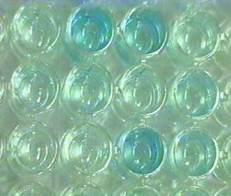
that 100 µl substrate solutions were added to each well. It was used pure and no need to dilute. After ten minutes the special reaction was occurred and some color changes in positive cases were observed. These changes were in the range of light blue till dark blue and mentioned color changes were observed in some wells and were recorded (Figure 1).
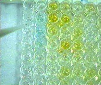
Consequently, 50 µl stop solution was added after ten minutes to each well. This solution was changed blue color to yellow color in positive cases. The rate of change of color was the same in the last stage and depended to blue color in each well. In negative cases that blue color was not observed so yellow color was absent automatically (Figure 2).
In the final stage, the ELISA plate arrived in ELISA reader machine and all Optical Density (O.D) was Mohammad Jalil Zorriehzahra, et al. Serological Survey of Epizootic Fish Viral Diseases in Some Rearing Rainbow Trout Farms by Enzyme-Linked Immunoabsorbent Assay (ELISA) in Iran. Cell Cellular Lif Sci J 2019, 4(1): 000139.
obtained. Then the result could be printed. For easy analysis, it was recommended that change in colors should be marked by a circle around of good number.
The first stage of ELISA optimization for antibody detection against IPNV and VHSV in brood stock serum consists of Protein measurement of IPNV and VHSV antigens. First of all, protein measurement of IPNV and VHSV antigens should be estimated that it as measured by Bio Photometer (Eppendorf®, 739 models) and its procedure carried out as follows:
Carbonate buffer (pH=9.6) was used as a BLANK in the test. For the preparation of the first dilution (1/60) 10 µl inactivated IPNV was solved in 590 µl Carbonate buffer. Therefore:
(10+590=600), 10/600 = 1/60 dilution.
The rate of requested viral antigen protein was 0.020 and its protein was measured by Photometer and it was 0.067 (about three times of requested rate = 0.020) so it was diluted three times (So the ratio of 0.060 reached to 0.020). Then 5 µl of viral antigen was solved in 895 µl carbonate buffer and 1/180 dilution was obtained. The same method was performed for VHSV and 1/180 dilution produced for both of them.
Sixteen serums were collected from good health condition farms in the same provinces and selected as negative control sera. Eight sera for IPNV and eight sera for VHS were specified. These serums were examined in virological examination and IFAT versus IPNV and VHSV and no CPE was observed and findings were negative.
For coating operation, 100 µl of viral antigens diluted (IPNV & IHNV) (with 1/180 dilution) were added to each well (Rows: A-H). Then a plate of ELISA was covered by parafilm and stored at 4ºC refrigerator for one day in favor of high completion of coating procedure. Buffers washing (with high and low salt) were carried out and all wells washed according to last protocol. The ELISA plate was removed from the refrigerator on the next day and discharged well contents completely. Again wells were washed with 250 µl low salt solution for three times and discharged wells contents completely. Then 50 µl of Bovine Serum Albumin (BSA) 3% were added to each well. The sample plates were covered with parafilm and then stored at room temperature (22ºC) for one hour. Again wells were washed with 250 µl low salt solution four times and discharged wells contents completely.
Copyright© Mohammad Jalil Zorriehzahra, et al.
Sample Selection Stage
Suspected serum samples were diluted with 1:1 dilution with PBS Tween. (100 µl serum sample + 100 µl PBS Tween). Serum samples from 1-7 for IPNV and 38-44 for VHSV were selected and 100 µl suspected serum was added to each well of ELISA plate and then were covered with parafilm and incubated at 25ºC for 1.5 hours.
The ELISA plate was removed from the incubator and after discharge of its contents; it was washed with high salt solution five times. Next 100 µl Anti Rainbow trout IgM MAbs were added to A, B, C, D, E, F, and H wells. G well-kept empty for positive control.
Positive Control Stage
A row of G well considered as a positive control in premature procedure with using of a polyclonal antibody that had produced before through inactivated viral antigens injection to rabbit but it was not ready so using of the positive control was decided for all samples.
Negative Control Stage
The negative control group is essential for comparison and analysis of findings in ELISA examination. Therefore 24 sera samples were collected from five same farms in mentioned provinces with high-quality health status. All fish have been satisfied health and no clinical signs were observed in the selected population.
All sera were examined in virology and IFAT assay and results were divided into three categories separately for three mentioned viral diseases. Then collected sera distributed in small vial aliquots and kept in -20ºC freezer. After that ELISA plate was covered with para film and stored at room temperature for one hour. All wells were washed with a high salt solution for five times. Anti- mouse IgG HRP conjugate (100 µl) with 1/3000 dilution was added to each well and incubated at 25ºC for 45-60 minutes. Again all wells were washed with high salt solution same last stage. Then 100 µl substrate solutions were added to each well. Primary results revealed that blue color changes were not observed for IPNV after 10 minutes. In wells of 40 and 42 reaction of blue color changes were observed for VHSV after 1-2 minutes. Then 100 µl stop solutions were added to each well and yellow color again was observed in wells of 40 and 42. In the final stage, ELISA plate arrived in ELISA reader machine with 450 wavelength nm and all O.D was obtained and results were printed.
Mohammad Jalil Zorriehzahra, et al. Serological Survey of Epizootic Fish Viral Diseases in Some Rearing Rainbow Trout Farms by Enzyme-Linked Immunoabsorbent Assay (ELISA) in Iran. Cell Cellular Lif Sci J 2019, 4(1): 000139.
Main ELISA Examination for Antibody Detection against IPNV and VHSV in Broodstock Serum Coating Stage for Viral Diluted Antigens
In purpose of good coating, 30 µl of IPN viral antigen were solved in 5.5 ml carbonate buffer and obtained 1/180 dilution for coating. Similar to the above stage, again 30 µl of VHS viral antigen were solved in 5.5 ml carbonate buffer and obtained 1/180 dilution for coating. Then 100 µl viral diluted antigens (IPNV and VHSV) with 1/180 dilution were added to each well and stored at 4ºC for coating operation overnight. After that suspected serum samples were distributed in ELISA plate but some serum samples such as numbers 5, 6, 13, 14, 15, 18, 19, 26 and 26 were removed for several reasons (serum hemolysis and serum low amount). Samples No = 40 and No= 42 were divided to more dilutions ¼, ⅛, 1/16, 1/32, 1/64 and 1/128 (because they had shown before some blue color changes in the optimization stage).
The ELISA plate was covered by parafilm and stored at 4ºC for overnight in favor of high completion of coating procedure. The ELISA plate was removed from the refrigerator on the next day and discharged well contents completely. After that wells were washed with 250 µl low salt solution for three times and discharged wells contents completely. Then 250 µl of Bovine Serum Albumin (BSA) 3% were added to each well and stored at temperature room for one hour. Again wells were washed with 250 µl low salt solution four times and wells contents discharged completely.
Sample Adding Stage
First of all 100µl suspected serum samples were added to each well which had been diluted with 50/50 dilution with PBS tween (100 µl serum sample + 100 µl PBS tween). Then ELISA plate was covered with parafilm and incubated at 25ºC for 1.5 hours. Next, ELISA plate was removed from incubator and wells were washed with 250 µl high salt solution five times and discharged wells contents completely.
Anti-Rainbow Trout IgM MAbs Adding Stage
First of all, 100 µl Anti Rainbow trout IgM MAbs were added to all wells and also blank well. Then 100 µl PAbs were added to special well (Rabbit Hyper Immune Serum) it had produced before through injection to the rabbit. (This antibody were diluted 1/20 with PBS tween). Then ELISA plate was covered with parafilm and stored at room temperature for one hour. Next, all wells were Copyright© Mohammad Jalil Zorriehzahra, et al.
washed with 250 µl high salt solution five times and discharged wells contents completely (last time was extended 5 minutes). After that 100 µl of Anti-Mouse IgG HRP conjugate with 1/2000 dilution were added to each well (It had before diluted in PBS tween). Consequently, 100 µl of Anti-Rabbit IgG HRP conjugate with 1/20000 dilution was added to a well of Rabbit Hyper Immune Serum (this conjugate had been diluted in PBS tween). Then ELISA plates stored at room temperature for 1 hour. Next, all wells were washed with 250 µl high salt solution five times and discharged wells contents completely (last time was 5 minutes). Next 100 µl substrate solution were added to all wells and waited for 15 minutes. Some blue color changes reaction were observed in some wells that it considered as positive samples. Then 50 µl stop solution were added to all wells and yellow color changes were observed in blue wells. In the final stage, ELISA plates arrived in ELISA reader machine with 450 nm wavelengths and all O.D findings were printed.
When 100 µl substrate solutions were added to wells of Rabbit Hyper Immune Serum after a few seconds a blue color reaction was observed immediately that it could be revealed this ELISA examination was desirable and optimization was done in good condition.
Results
The ELISA method for the detection of antibodies against IHNV, IPNV and VHSV was performed essentially according to the standard protocol. ELISA plates were read on a Dynatech® MR4000 plate reader at an absorbance of 450nm-630 nm. The optical density of mentioned sera was obtained and categorized separately in three sections for three mentioned viral diseases IHNV, IPNV and VHSV as potential hazard factors in the hatchery and rearing rainbow trout farms in Iran.
In the current study, the Cut-off value was calculated according to the average of OD in this control group plus three times the standard deviation. In fact, Cut-off value was determined as follows:
Cut-off point = [M+ (3 × SD)]
M= the mean of OD values of all Negative Control samples SD= Standard deviation of Negative Control samples Ref.: Medina et al, (2000), Carnevale et al, (2001) [9]. All findings summarized in Table 1.
| IHN Virus | IPN Virus | VHS Virus | |
|---|---|---|---|
| Negative Control Samples | |||
| Statistical analysis for Cut-off Point calculation | |||
| NC1 | 0.032 | 0.013 | 0.042 |
| NC2 | 0.045 | 0.015 | 0.031 |
| NC3 | 0.251 | 0.019 | 0.022 |
| NC4 | 0.061 | 0.021 | 0.013 |
| NC5 | 0.121 | 0.017 | 0.061 |
| NC6 | 0.21 | 0.029 | 0.017 |
| NC7 | 0.11 | 0.096 | 0.078 |
| NC8 | 0.052 | 0.019 | 0.017 |
| Mean | 0.11 | 0.029 | 0.035 |
| SD | 0.081 | 0.027 | 0.023 |
| C.V | 74% | 97% | 67% |
| Alpha | 0.05 | 0.05 | 0.05 |
| Sample Size | 8 | 8 | 8 |
| Confidence level | 0.056 | 0.019 | 0.016 |
| Mean + 3*SD | 0.354 | 0.112 | 0.106 |
| Mean + 3.08*SD | 0.36 | 0.114 | 0.108 |
| Mean + Confidence | 0.166 | 0.048 | 0.051 |
| Cut-off Point | 0.354 | 0.112 | 0.106 |
Table 1: Summary of optical density of negative control samples in ELISA examination and statistical analysis for Cut-off point m
The OD cases that standing above Cut-off point was considered as positive cases.
The Cut-off value, Positive Samples of Samples size and Percentage of detection for IHN, VHS and IPN showed in Table 2.
| Characterization | ||||||||||||||
|---|---|---|---|---|---|---|---|---|---|---|---|---|---|---|
| Cut-off Point | Samples size | Positive Samples | Percentage of detection | |||||||||||
| Disease name | ||||||||||||||
| IHN | 0.354 | 43 | 10 | 23.25% | ||||||||||
| VHS | 0.106 | 35 | 5 | 14.29% | ||||||||||
| IPN | 0.112 | 41 | 3 | 7.31% |
Table 2: Summary of ELISA final result for detection of probably important viral agents in fry rainbow trout Table 2: Summary of
Mohammad Jalil Zorriehzahra, et al. Serological Survey of Epizootic Fish Viral Diseases in Some Rearing Rainbow Trout Farms by Enzyme-Linked Immunoabsorbent Assay (ELISA) in Iran. Cell Cellular Lif Sci J 2019, 4(1): 000139.
Copyright© Mohammad Jalil Zorriehzahra, et al.
Discussion
Rapid and sensitive diagnosis methods of fish viral infectious diseases are very critical if dissemination of the viruses causing these diseases is to be controlled because no effective vaccines currently exist for their prevention. Rapid diagnosis test is desirable during the acute phase of the disease, whereas highly sensitive diagnosis is necessary to detect viruses during the carrier or chronic phase of the disease. Methods for diagnosis of viral diseases can be categorized into two groups, those measuring the presence of the virus and those detecting the specific immune response of the host. To detect the virus in fish it is necessary first to sample the fish population correctly, second to amplify the content of the sample, and third to identify the virus. To identify the virus cytopathic effect (CPE) on cell culture, proteins, nucleic acids and/or morphology could be measured. For most of these methods reagents such as polyclonal or monoclonal antibodies (Abs) and DNA probes are needed, all of which should be complementary to structural components of the virus to be identified. To detect the reaction of the host we can measure either their humoral or their cellular immunological responses [10].
Medina, et al. (1992) was reported his achievements about “Diagnosis of infectious hematopoietic necrosis virus in Atlantic salmon (Salmo salar) by enzyme-linked immunosorbent assay” that several techniques including fluorescent antibody detection in cell culture, immunoblot assay, detection of viral nucleic acids by means of polymerase chain reaction and the use of a biotinylated DNA probe have been used for IHNV detection [11, 12, 13, 14]. Although these techniques are sensitive, they are time- consuming and technically intensive. An alternate method, the enzyme-linked immunosorbent assay (ELISA), is generally less expensive and more rapid.
Rainbow trout fry mortality syndrome is a serious disease in farmed salmonid fish. In the present study, Serology method was carried out for the detection of the causative pathogens and confirmation of virology examination. The method was an Enzyme Linked Immunoabsorbent Assay (ELISA).
Serology examination was selected in order to confirm previous virology findings and applied on collected serums. Fifty-three serums of rainbow trout broodstocks (29 female and 24 male) were examined for detection of antibodies against IHNV, IPNV and VHSV that were collected from 13 hatchery and rearing farms in three Mohammad Jalil Zorriehzahra, et al. Serological Survey of Epizootic Fish Viral Diseases in Some Rearing Rainbow Trout Farms by Enzyme-Linked Immunoabsorbent Assay (ELISA) in Iran. Cell Cellular Lif Sci J 2019, 4(1): 000139.
provinces in Iran (March 2002 until November 2003). Also in order to confirm the results of virology examination two samples that had revealed CPE in cell lines were chosen. Finally, 44 suspected serum specimens plus 24 negative control samples (8 for each suspect virus: IHNV, IPNV and VHSV) were selected for ELISA examination.
Regarding ELISA findings and disease percentage (Number of all positive case standing above Cut-off point divided to all examined suspected samples), IHNV had more percentage of disease in ELISA test with 23.25% in comparison with other relevant viral diseases i.e. IPNV with 7.31% and VHSV with 14.29 %.
Finally, regarding these findings and percentage of detection in the current study, it could be concluded that the IHN virus could be one of the most important etiologies of fry mortality syndrome (FMS) in Iran. In fact, serology findings in IFAT and ELISA could be supported strongly from virology conclusion and confirmed Rhabdovirus-like particles as the main causative agent in the occurrence of rainbow trout fry mortality syndrome in the hatchery and rearing Coldwater farms in Iran [15].
Acknowledgement
The authors thank Dr. Kim Thompson (Moredun Research Institute, UK) for her kindly efforts in the technical protocol. The authors also are grateful to colleagues in the Iranian Fisheries Science Research Institute (IFSRI), Razi Vaccine and Serum Research Institute (RVSRI) and Faculty of Veterinary Medicine of Tehran University for providing necessary facilities for this study.
References
-
Akhlaghi M (2000) Immunological study of suspected viral diseases, infectious haematopoietic and pancreatic necrosis in cultured rainbow trout (Oncorhynchus mykiss). Iranian Journal of Veterinary Research 1(2): 85-95.
-
Soltani M, Rostami M, Ebrahimzadeh Mousavi HA, Mirzargar SS, Fallahi R (2002) Study of fry trout mortality syndrome in some farmed rainbow trout in Tehran and Lorestan Provinces. Pajouhesh-va- Sazandegi 14(4): 2-5. Copyright© Mohammad Jalil Zorriehzahra, et al.
-
Fallahi R, Soltani M, Kargar R, Zorriehzahra MEJ, Shchelkunov I, et al. (2003) Isolation and identification of the infectious haematopoietic necrosis virus (IHNV)-like agent from farmed rainbow trout (Oncorhynchus mykiss) from Iran. Archives of Razi Institute 56: 37-45.
-
Haghighi Khiabanian Asl A, Soltani M, Kazemi B, Sohrabi Haghdoust I, Sharifpour I (2007) Use of Immunohistochemical and PCR Methods in Diagnosis of Infectious Haematopoietic Necrosis Disease in Some Rainbow Trout Hatcheries in Iran. Pakistan Journal of Biological Sciences 10(2): 230-234.
-
Zorriehzahra MEJ, Rezvani S (2005) Preliminary study of infectious agents (Viral and Bacterial) of Rainbow trout (Oncorhynchus mykiss) Fry Mortality Syndrome in Iran. Research Final Report, pp: 250.
-
Zorriehzahra MEJ (2008) Aetiologic Agents of Fry Mortality Syndrome in the Coldwater fish farms in Iran, Ph.D thesis, pp: 280.
-
Adams A, Thompson KD (2006) Biotechnology offers revolution to fish health management. Trends Biotechnol 24(5): 201-205.
-
Charriaut-Marlangue C, Aggoun-Zouaoui D, Represa A, Ben-Ari Y (1996) Apoptotic features of selective neuronal death in ischemia, epilepsy and gpI20 toxicity. Trends in neurosciences 19(3): 109-114.
-
Medina E, Paglia P, Rohde M, Colombo MP, Guzman CA (2000) Modulation of host immune responses stimulated by Salmonella vaccine carrier strains by Mohammad Jalil Zorriehzahra, et al. Serological Survey of Epizootic Fish Viral Diseases in Some Rearing Rainbow Trout Farms by Enzyme-Linked Immunoabsorbent Assay (ELISA) in Iran. Cell Cellular Lif Sci J 2019, 4(1): 000139. using different promoters to drive the expression of the recombinant antigen. European Journal of Immunology 30(3): 768-777.
-
Sanz F, Coll J (1992) Techniques for diagnosing viral disease of salmonid fish. Diseases Aquatic Organisms 13: 211-223.
-
LaPatra SE (1996) The use of serological techniques for virus surveillance and certification of finfish. Annual Review of Fish Diseases 6: 15-28.
-
McAllister PE, Schill WB (1986) Immunoblot assay: a rapid and sensitive method for the identification of salmonid fish viruses. Journal of Wildlife Diseases 22(4): 468-474.
-
Arakawa CK, Deering RE, Higman KH, Oshima KH, O'Hara PJ, et al. (1990) Polymerase chain reaction (PCR) amplification of a nucleoprotein gene sequence of infectious hematopoietic necrosis virus. Diseases of Aquatic Organisms 8: 165-170.
-
Deering RE, Arakawa CK, Oshima KH, O'Hara PJ, Landolt ML, et al. (1991) Development of a biotinylated DNA probe for detection and identification of infectious hematopoietic necrosis virus. Diseases of aquatic organisms 11(1): 57-65.
-
Office International des Epizooties, OIE (2004) Aquatic Animals Health Code, Part 2, Section 2.1, Chapter 2.1.2, OIE, Paris. Copyright© Mohammad Jalil Zorriehzahra, et al.
- The Muculent Bleb-Mucinous Cystic Neoplasm-Hepatobiliary Region
- Insulin Sensitizers as Anti-Aging Agents: Unveiling Synergies with Albumin, GLP-1RA, Klotho Protein, and Metformin in the Quest to Combat Aging
- Reprogramming of GLP-1 Response at Prediabetes for the Prevention of Type 2 Diabetes: The Role of Albumin and GLP-1 Receptor Agonists
- The Mingled Allies-Combined Hepatocellular Carcinoma and Cholangiocarcinoma
- Compilation and Embodiment-Leydig Cell Tumour Testis
- Glucolipotoxicity: A Novel Different Perspective on the Causes of Cancer