Ovarian Low-Grade Papillary Serous Cystadenocarcinoma with Osseous Metaplasia- Case Report and Review of Literature
Introduction: The stromal osseous metaplasia is extremely rare finding in serous cystadenocarcinoma of the ovary. Case report: A 45-year old female presented with pain in lower abdomen of about 2 months, breathlessness of 10days. On physical examination per abdomen, a large pelvic mass identified. She had pleural and pericardial effusion. Pericardiocentesis was done. Abdomen and pelvis ultrasound revealed a large heterogeneous right adnexal ovarian mass measuring 13.5 × 13.1 × 10.8cm, volume about 916 ml. It was well defined, oval, cystic, partially solid mass, the impression of ovarian neoplasm was given. There was no significant lymphadenopathy. The left ovary, uterus, cervix, fallopian tubes and rest of abdominal organs were normal. Serum CA 125 was 75.5 U/ml. She underwent total abdominal hysterectomy. On histopathological findings reported as right ovarian low-grade papillary serous cystadenocarcinoma with osseous metaplasia. Conclusion: The osseous metaplasia in ovarian serous cystadenocarcinoma is extremely rare.
Introduction
Ovarian surface epithelial serous tumors are common neoplasms [1]. However osseous metaplasia in non- teratomatous ovaries is extremely rare [2]. The pathogenesis of osseous metaplasia in epithelial tumors of the ovary is unclear. The various mechanisms are given in which a metaplastic process involving multipotential stromal stem cells results in bone formation is postulated. In most of these cases osseous metaplasia is secondary to inflammation, tissue damage, or substances such as bone morphogenetic proteins released from neoplastic cells. Osseous metaplasia most likely occurs by osteoblasts differentiating from fibroblasts. We report a case of right ovarian low-grade papillary serous cystadenocarcinoma with osseous metaplasia. To the best of our knowledge, this is a sixth rare case of low-grade papillary serous cystadenocarcinoma with osseous metaplasia.
A 45-year old female presented with pain in lower abdomen of 2 months, breathlessness of 10 days. On physical examination, per abdomen a large pelvic mass was detected. She had pleural and pericardial effusion. Pericardiocentesis was done. On cytology fluid was negative for malignat cells. Her obstetric history was P2, L2, A0. There was no significant contributary family or past history .Abdomen and pelvis ultrasound revealed a large heterogeneous right adenexal ovarian mass measuring 13..5 × 13.1 × 10.8cm, volume about 916 ml. It was well defined, oval, cystic, partially solid mass with anechoic and Hypoechoic lesion with focal calcifications .the impression of ovarian neoplasm was given. Left ovary was normal measuring 3.2x2, 2x2. cm. There was no significant lymphadenopathy. The uterus, cervix and fallopian tube was normal. Rest of abdominal organs were normal. Serum CA 125 was 75.5 U/ml. She underwent total abdominal hysterectomy.
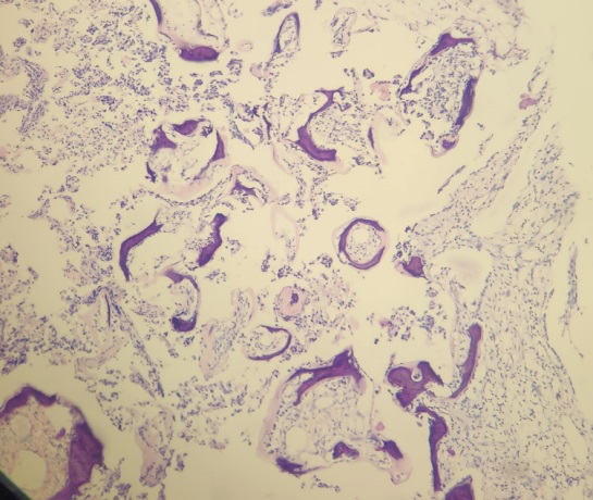
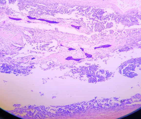
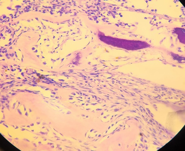
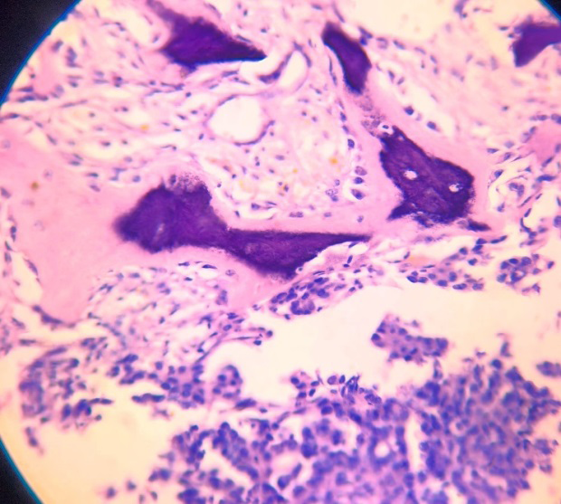
We received panhysterectomy specimen. The right ovarian mass measured 16x14x8.2 cm, weighing 800 gm. External surface was smooth, congested. On cut open showed a solid cystic mass measuring 5x3.5x1 cm. The soild area showed papillary growth, cut section of it was grey white, firm to hard (Figures 1 & 2). The rest of organs were normal. Microscopy showed a tumor composed of neoplastic cells arranged in complex papillae with broad fibrovascular cores, in glandular cystic, and solid sheets. The neoplastic cells were round ,uniform having moderate nuclear atypia with inconspicuous nucleolus and scant cytoplasm. The low mitotic index of < 10 mitotic figures per 10 high power fields were noted. The desmoplastic stroma showed tumor invasion with areas of bone matrix and psammoma bodies. There were no necrosis. Tumor was limited to right ovary. On histopathological findings reported as right ovarian low- grade papillary serous cystadenocarcinoma with osseous metaplasia, areas of psammomatous calcifications (Figures 3-6). The left ovary, endometrium, myometrium, bilateral fallopian tubes were normal.


Discussion
Serous ovarian tumors are common cystic neoplasms and constitute about one fourth of all ovarian tumors [2]. The ovarian carcinomas have five common types are high- grade serous (70%), endometrioid (10%), clear cell (10%), mucinous (3%), and low-grade serous carcinomas (<5%) [3]. Low-grade serous carcinomas are relatively uncommon, accounting for less than 5% of ovarian carcinomas [4].
Osseous metaplasia in non-teratomatous ovaries is extremely rare. This phenomenon may occur in various non-neoplastic or neoplastic ovarian diseases. The most common type of ovarian tumor containing osseous elements is a teratoma [5]. The other condition may also be associated with bone formation is heterologous mixed mesodermal tumors of the ovary [6]. It is observed that exceptionally osseous metaplasia develops in ovarian stroma-rich tumors. In histopathological examination Psammoma bodies are very common findings in ovarian papillary serous tumors, but evidence of bone formation is extremely rare. Miliaras et al, study showed that the presence of extensive heterologous bone formation, or osseous metaplasia is rare. While the presence of Psammomatous calcifications is relatively common. The osseous metaplasia in non-teratomatous ovarian tumors have only been reported in 10 other cases, including a benign serous cystadenoma and two cases of papillary serous cystadenocarcinomas [7].
A first case by Barua R, et al. [8] observed occurrence of bone in serous cystadenocarcinoma of the ovary. Other case report presented by Bosscher J, et al. [9] as the finding of osseous metaplasia within an ovarian papillary serous cystadenocarcinoma. Alghamdi DA reported the fourth case of serous cystadenocarcinoma with ossification and bony metaplasia in a 33-year old female [10]. Xavier Catteau et al, reported the case of low-grade serous carcinoma with osseous metaplasia and a BRAF mutation in ovary [11].
Fifth case by Ribeiro RR, et al. [12] of low-grade serous carcinoma with extensive osseous metaplasia arising from ovarian serous cystadenofibroma was reported. To the best of our knowledge, our is a sixth rare case of low-grade papillary serous cystadenocarcinoma with osseous metaplasia.
Osseous metaplasia in non-teratomatous ovaries is extremely rare [13]. The pathogenesis of osseous metaplasia remains unclear. One mechanism described as ,the pathogenesis for bone formation in an ovarian papillary serous cystadenocarcinoma is a metaplastic process of the multipotential stromal cell. The other physiopathology for osseous metaplasia could be due to the release of tumor necrosis factor and bone morphogenic protein-7, growth differentiation factor-5were given. Teot LA, et al. [14] noted that the transforming growth factor-ß produce by the tumor cells has been implicated as osteoinductive in osteogenesis within these neoplasms. Toyran S, et al. [15] observed that the growth differentiation factor-5 (GDF-5), bone morphogenic protein-7 (BMP-7), and transforming growth factor beta-1 (TGF β1) are multifunctional cytokines that have important roles in bone formation.
The ovarian low grade serous adenocarcinoma shows excellent prognosis with surgical excision alone if confined to the ovary. It requires to study that a osseous metaplasia of the ovary have any prognostic or pathological significance in patient management.
Conclusion
The osseous metaplasia in ovarian serous cystadenocarcinoma is extremely rare. This is unique case of ovarian low grade serous papillary cystadenocarcinoma with osseous metaplasia presented for its histopathological findings.
References
-
Ovary RJ (2004) Ackerman’s Surgical Pathology. 9th (Edn.), Mosby St Louis, 2: 1649-1736.
-
Godbole P, Outram A, Sebire N (2005) Osseous metaplasia in a benign ovarian cyst in association with cloacal anomaly. J Clin Pathol 58(3): 334-335.
-
Gilks CB, Prat J (2009) Ovarian carcinoma pathology and genetics: recent advances. Hum Pathol 40(9): 1213- 1223.
-
Gershenson DM, Sun CC, Lu KH, Coleman RL, Sood AK, et al. (2006) Clinical behavior of stage II–IV low-grade serous carcinoma of the ovary. Obstet Gynecol 108(2): 361-368.
-
Kurman RJ, Ellenson LH, Ronnett BM (2019) Blaustein’s Pathology of the Female Genital Tract. 17th (Edn.), Springer International Publishing, New York.
-
Jagtap SV, Jagtap SS, Gudur R, Billawaria S (2022) Primary ovarian malignant mixed Müllerian tumor: a rare case report. Ther Adv Rare Dis 3: 1-6.
-
Miliaras D, Ketikidou M, Pervana S (2007) Osseous metaplasia in ovarian tumors: a case with serous cystadenoma. J Clin Pathol 60(5): 582-583.
-
Barua R, Cox LW (1982) Occurrence of bone in serous cystadenocarcinoma of the ovary. Aust N Z J Obstet Gynaecol 22(3): 183-186.
-
Bosscher J, Barnhill D, O’Connor D, Doering D, Nash J, et al. (1990) Osseous metaplasia in ovarian papillary serous cystadenocarcinoma. Gynecol Oncol 39(2): 228- 231.
-
Alghamdi D, Abdullah L, Mousa AA, Shinawi SA (2016) Stromal osseous metaplasia in ovarian serous cystadenocarcinoma. JKAU Med Sci 23(1): 41-45.
-
Catteau X, Preat F, D’haene N, Jean-Christophe N (2021) Osseous Metaplasia in Low-grade Ovarian Serous Carcinoma With a BRAF Mutation: A Case Report International Journal of Gynecological Pathology 40(5): 448-451.
-
Ribeiro RR , Valenzuela A, Beffa L, Sung CJ, Quddus MR (2021) Low-grade serous carcinoma with extensive osseous metaplasia arising from ovarian serous cystadenofibroma. Gynecologic Oncology Reports 36: 10070.
-
Jagtap SV, Kshirsagar NS, Jagtap SS, Boral S, Nasre N (2019) Ovarian teratomas: clinicopathological study at tertiary care center. Int J Reprod Contracept Obstet Gynecol 8(8): 3318-3322.
-
Teot LA, O’Keefe RJ, Rosier RN, O’Connell JX, Fox EJ, et al. (1996) Extraosseous primary and recurrent giant cells tumors: Transforming growth factor-beta1 and beta2 expression may explain metaplastic bone formation. Hum Pathol 27(7): 625-632.
-
Toyran S, Lin AY, Edward DP (2005) Expression of growth differentiation factor-5 and bone morphogenic protein-7 in intraocular osseous metaplasia. Br J Ophthalmol 89(7): 885-890.
- Genomic Landscape of Aggressive Penile Squamous Cell Carcinoma including TERT-p and NOTCH1 Mutations – An Institutional Experience
- Establishment of Baseline Haematological Values for Canine Population in North-Central Nigeria: A Cross-Sectional Study in the Federal Capital Territory
- Biochemical Assessment of Uroliths Extracted in Patients with Urolithiasis in a Tertiary Health Institution
- Update on Gastrointestinal Pecomas: Molecular Pathogenesis and Risk Stratification
- A Comparative Study of Serum C-reactive Protein Level Between Pre-eclampsia and Normal Pregnancy in Tertiary Level Hospital
- From Deformity to Alignment: Clinical Outcomes of the Schnepp Osteotomy in Hallux Valgus in 47 Feet