Unusual Termination of the Facial Vein into External Jugular Vein and its Clinical Implication
The superficial veins of the neck are used for cannulation, either for intravenous infusion or for central venous pressure monitoring. Variations in the venous system from the normal pattern are relatively common. We present some unusual variations of the facial vein draining into the external jugular vein instead of draining into the internal jugular vein. The external jugular vein usually begins just behind the angle of the mandible by the union of the posterior auricular vein with the posterior division of the retromandibular vein and then descends obliquely across the sternocleidomastoid muscle and, just above the clavicle, pierces the deep fascia and drains into the subclavian vein. External jugular vein gives a reliable estimate of central venous pressure. The variation may give false value of pressure due to facial vein draining into it, also may create difficulty in catheterization. It’s very important not only for anatomists but also for head and neck surgeons to be aware of the possible anatomical variation in the formation of external jugular Vein and its clinical implications.
Introduction
The veins of the head and neck have a complex developmental pattern which predisposes them to variations in formation and drainage. Usually, the facial vein begins at the medial angle of the eye as the angular vein, by the union of the supratrochlear and the supra-orbital veins. The superficial temporal vein unites with the maxillary vein to form the retromandibular vein. The retromandibular vein divides into the anterior and the posterior divisions within the substance of the parotid gland. The anterior division joins with the anterior facial vein to form the common facial vein and it drains into the internal jugular vein [1]. The external jugular vein normally begins just behind the angle of the mandible by the union of the posterior auricular vein with the posterior division of the retromandibular vein. It descends obliquely across the sternocleidomastoid muscle and, just above the clavicle in the posterior triangle, pierces the deep fascia and drains into the subclavian vein [1]. It’s superficial location and its anatomical position of being direct in line with the superior vena cava; the EJV represents an easy, safe, and highly useful avenue for fluid and drug administration in emergencies [2, 3]. External jugular venous cannulation is increasingly being used in emergencies due to immense air embolism that has been reported with central venous cannulation through the internal jugular and subclavian veins [4]. Understanding the structural variants of the external jugular vein is important not only for anatomists but also for head and neck surgeons.
Case Report
During our daily routine dissection for undergraduate medical students at Hubert Kairuki Memorial University, in the Department of Anatomy, unusual formation of external jugular vein was observed in two cadavers (one cadaver presented bilateral variation while the second had a unilateral variation). In one of the cadavers, we observed that the superficial temporal vein joined with the maxillary vein to form a very short trunk of the retromandibular vein, which immediately divided into anterior and posterior divisions. The anterior division joined with the facial vein to form a common facial vein which directly drained into external jugular vein instead of internal jugular vein (Figures 1&2). In the second cadaver, the superficial temporal vein joined with the maxillary vein to form the retromandibular vein which did not divide into anterior and posterior divisions. This retromandibular vein joined with the facial vein to form the external jugular vein (Figures 3). Awareness of the anatomical variations in venous system from the normal pattern is very important for the all medical practitioners.
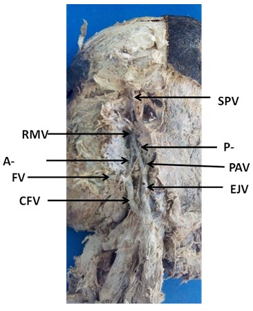
SPV: Superficial temporal vein; S-RMV: Short trunk of retromandibular vein; A-RMV: Anterior division of retromandibular vein; P-RMV: Posterior division of retromandibular vein; FV: Facial vein; PAV: Posterior auricular vein; CFV: Common facial vein; EJV: External jugular vein. Figure 1: Left side of the head & neck showing a very short trunk of the retromandibular vein, which immediately divided into anterior and posterior divisions. The anterior division joined with the facial vein to form a common facial vein which directly drained into external jugular vein instead of internal jugular vein.
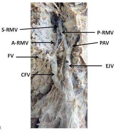
S-RMV: Short trunk of retromandibular vein; A-RMV: Anterior division of retromandibular vein; P-RMV: Posterior division of retromandibular vein; FV: Facial vein; PAV: Posterior auricular vein; CFV: Common facial vein; EJV: External jugular vein. Figure 2: Magnified Left side of the head & neck showing a very short trunk of the retromandibular vein, which immediately divided into anterior and posterior divisions. The anterior division joined with the facial vein to form a common facial vein which directly drained into external jugular vein instead of internal jugular vein.
STV: Superficial temporal vein; RMV: the retromandibular vein; FV: Facial vein; EJV: External jugular vein Figure 3: Right part of the head & neck showing retromandibular vein joined with the facial vein to form the external jugular vein.
Discussion
Different patterns of variations in the venous drainage have been observed. Many authors have reported anatomical variation of the venous drainage of head and neck. Unusual variations of the facial vein draining into the external jugular vein instead of draining into the internal jugular vein have been reported [5, 6]. The other variations include: the formation of the external jugular vein by the union of facial and lingual vein has been reported [7]; common facial vein terminating as a second external jugular vein has also been reported [8].
The external jugular vein is increasingly being utilized for cannulation to conduct diagnostic procedures or intravenous therapies [9]. These anomalous patterns may partly be explained by embryological occurrence of unusual retention and/or regression of venous anastomotic channels in the primitive pharyngeal region during development [10]. Prior knowledge of the existence of these variations is very essential for all clinical practitioners.
Acknowledgement
It is gratifying to express my indebtedness to all people who rendered notable assistance to bring this work to a successful completion.
References
-
Standring S (2006) Gray’s Anatomy: The Anatomical Basis of Clinical Practice. 39 (Edn.), Edinburgh, Elsevier Churchill Livingstone, pp: 273-274.
-
Povoski SP (2007) Eliminating the “Pitfalls” of chronic in- dwelling central venous access device placement in can- cer patients by utilizing a venous cut down ap- proach and by selectively and appropriately utilizing intraoperative venography. International Seminars in Surgical Oncology 4(1): 16.
-
Prasad VSV, Daharwal S, Bahe A (2016) External jugular venous access in children: a low cost and feasible route for emergency fluid resuscitation and inotropic therapy in resource poor clinical settings? J Pediatr Crit Care 3(3): 16-21.
-
Wong SS, Kwaan HC, Ing TS (2017) Venous air embolism related to the use of central catheters revisited: with emphasis on dialysis catheters. Clin Kidney J 10(6): 797- 803.
-
Gupta V, Tuli A, Choudhry R, Agarwal S, Mangal A (2003) Facial vein draining into external jugular vein in humans: its variations, phylogenetic retention and clinical relevance. Surg Radiol Anat 25(1): 36-41.
-
Chauhan NV, Rani A, Chopra J, Rani AK (2011) Anomalous formation of external jugular vein and its clinical implication. Natl J Maxillofac Surg 2(1): 51-53.
-
Abhinitha P, Rao MKG, Kumar N, Nayak SB, Ravindra SS, et al. (2013) Absence of external jugular vein and abnormal drainage pattern in the veins of the neck. OA Anatomy 1(2): 15.
-
Tellez Hernandez LV, Tibaduiza Rodriguez IA, Ferreira Arquez H (2019) Atypical unilateral venous drainage of head and neck. Int J Pharm and Research 11(1): 1140- 1142.
-
Chauhan NV, Rani A, Chopra J, Rani AK (2011) Anomalous formation of external jugular vein and its clinical implication. Natl J Maxillofac Surg 2(1): 51-53.
-
Bertha A, Rabi S (2011) Anatomical variations in termination of common facial vein. Journal of Clinical and Diagnostic Research 5(1): 24-27.
- Research Progress of Induced Pluripotent Stem Cells and Their Clinical Application Prospects
- Nishan Al-Kamal is the Starting Point of A Feminist Scientist
- Current Concepts and Future Perspectives of Stem Cell Therapy in Peripheral Arterial Disease
- Stem Cell and Oxidative Stress-Inflammation Cycle
- Adipose Derived Mesenchymal Stem Cells Origin, Characteristics and Promises
- Mitochondria Targeted Antioxidants can Improve In Vitro Embryo Production in Buffalo