Adult Stem Cell Therapy as an Alternative Treatment in Knee Osteoarthritis
Objective: Osteoarthritis (OA) of the knee, also known as degenerative joint disease of the knee because of progressive loss of articular cartilage, is the most common disabling conditions of the knee joint, which often presents with pain and stiffness and progresses to affect activity of daily living. The prevalence of knee OA will continue to rise due to factors like obesity and increased life expectancy. Roughly 13% of women and 10% of men 60 years and older have symptomatic knee osteoarthritis. Among those older than 70 years of age, the prevalence rises to as high as 40%. Our aim is to evaluate the importance of adult stem cell therapy (ADSC) as an alternative treatment in knee osteoarthritis. Patient, Materials and Methods: A case of an elderly patient with early right knee osteoarthritis (grade 2). Right knee severity was assessed using the Western Ontario and McMaster universities osteoarthritis index (WOMAC) scale. Radiologic studies (X-ray and MRI) were done. Patient had a session of autologous adult stem cell therapy - Mesenchymal stem cells retrieved from the fat and bone marrow along with platelet rich plasma (PRP) from blood which was administered intra-articularly via ultrasound guidance. Procedure was done on an outpatient basis and was well tolerated with no complications. Results: Patient was observed in the first 6 months under the instructions of no physiotherapy to preserve new maturing cells. The WOMAC assessed on the 0 and 6 months showed quality improvement from an initial score of 48/96 to a score of 8/96. Post procedure X-ray of the Right knee on the sixth month showed marked progressive changes. Conclusion: Adult stem cell therapy is promising to be an effective alternative treatment in knee osteoarthritis. It is minimally invasive, safe with no major treatment related adverse effect. Case study showed a positive outcome according to all the grading systems used in this study for the initial 6 months.
Introduction
Knee osteoarthritis (OA), also known as degenerative joint disease, is typically the result of wear and tear and progressive loss of articular cartilage (medial loss occurs in 70% of cases) [1]. The ligaments become lax, making the joint becomes less stable, with local pain arising from the ligaments and tendons [2]. It presents clinically with patient complaints of pain and impaired function, which are thought to result from cartilage degeneration and other skeletal changes. These changes can by examined radiographically and quantified using the semiquantitative grading scale known as the Kellgren-Lawrence (KL) scale. Knee osteoarthritis is classified as either primary or secondary, depending on its cause. Primary knee osteoarthritis is the result of articular cartilage degeneration without any known reason. This is typically thought of as degeneration due to age as well as wear and tear. Secondary knee osteoarthritis is the result of articular cartilage degeneration due to a known reason like trauma, autoimmune diseases.
Radiologic definition is symptomatic osteoarthritis of the knee classified as Kellgren-Lawrence Grade 0: no pathological features; Grade 1: doubtful narrowing of joint space and possible osteophytic lipping; Grade 2: definite osteophytes and possible narrowing of joint space; Grade 3: moderate multiple osteophytes, definite narrowing of joint space, some sclerosis, and possible deformity of bony ends; Grade 4: large osteophytes, marked narrowing of joint space, severe sclerosis, and definite deformity of bone ends. Treatment for knee osteoarthritis begins with conservative methods like patient education, activity modification, physical therapy, weight loss and medications like NSAIDS, Glucosamine and chondroitin sulfate, corticosteroid injections and progresses to surgical treatment options when conservative treatment fails [3]. Mesenchymal stem cells (MSCs) are known to have regenerative capacity and can be separated from a variety of tissue sources including adipose (fat) tissue and bone marrow. These cells have multi-potential abilities to differentiate into bone, cartilage, muscle, and fat and can be multiplied in culture [4, 5, 6, 7]. Adipose derived stem cells (ADSCs) and bone marrow derived stem cells (BMSCs) are MSCs which have been collected from fat and bone marrow respectively. Easily accessible subcutaneous fat from the abdomen or the flanks can be collected via mini-liposuction and contains high amounts of ADSCs [8]. These cells produce different cytokines and growth factors that when expressed is thought to be part of a healing cascade and new blood vessel formation by stimulating a local inflammatory process and immunomodulatory/paracrine response which enables a signaling that causes the repair of damaged tissue [9, 10]. MSCs (ADSC and BMSC) are anti-inflammatory and promote angiogenesis. Their complex signaling process causes the signaling of other cells to the area to repair damaged tissue which promotes regeneration. MSC has the potential to fully heal damaged tissues and organs and this ability has made MSC a potential game changer in respect to treatment of various diseases and injuries. The exploitation of stem cells’ ability to promote a healing cascade is a novel progressive shift in medicine.
The stromal vascular fraction (SVF) is the mixture of cells from adipose tissue collected via mini-lipoaspiration after the adipocytes (fat cells) have been depleted. The remaining cells include a large percentage of ADSCs. SVF contains a variety of regenerative cells and growth factors and can be offered as an out-patient surgical procedure, it represents a new therapeutic tool for many indications. Friedenstein et al, in a series of seminal studies in the 1960s and 1970s, showed that the osteogenic potential of bone marrow (BM) cells was associated with a minor subpopulation of cells in the BM. These cells were distinguishable from most hematopoietic cells by their rapid adherence to tissue culture vessels and the fibroblast-like appearance of their progeny in culture, pointing to their origin from the stromal compartment of BM. While now known to be technically incorrect, the current colloquial term “mesenchymal stem cell” dates to 1991. A work by Darwin Prockop and others further defined the cells and their multilineage capability. The ability to grow and expand them efficiently and relatively easily and the wide variety of functions they have now been described to perform led to the birth of an entire subfield of cell therapy. The marrow stromal cell field expanded very rapidly, and the potential use of these cells in therapy is being tested worldwide for many indications [11]. Local injection of MSCs (ADSCs and BMSCs) may reduce inflammation and promote healing/ normal tissue formation. SVF can be collected bed side after a simple adipose tissue collection making it a practical surgical procedure for physicians to provide to their patients. Some publications have demonstrated success of MSCs for specific conditions including neurodegenerative disc diseases, osteoarthritis, tendinopathy, xerostomia, psoriasis The following is a case report of a woman with right knee osteoarthritis [12, 13, 14].
Methods
The following case report started on the 13/11/2020. Diagnosed with Right knee osteoarthritis (Grade 2). The patient provided written informed consent to undergo the experimental clinical procedure as well as consent to publication of outcomes, images, and data. Right knee severity was assessed using the Western Ontario and McMaster universities osteoarthritis index (WOMAC) scale. Radiologic studies (X-ray and MRI) were done. The first session was a combination of MSCs {ADSCs (svf) and BMAC} plus PRP. Then a follow up was observed by 6months under the instructions of no physiotherapy [15].
First Treatment Session Proceeded after Consultation and all Necessary Requirements were Carried out.
The SVF was created using a standard tumescent liposuction solution. Tumescent was introduced into the subcutaneous fat tissue in the lower abdomen using an infiltration cannula to create local numbing. Adipose tissue was collected with a harvesting cannula. Using a commercially available collection kit and collagenase enzyme, the collected fat was processed to obtain the SVF for injection/transplantation – 2 ml SVF from the collected fat (1.96 x 10⁹ total viable cells per ml tested with 100% viability from Moxi Flow) for intra-articular injection, and 3 ml of SVF from the collected fat (3.15 x 10⁹ total viable cells per ml tested with 100% viability from MoxiFlow) for Intravenous transplantation.
The Platelet Rich Plasma concentrate required for injection was prepared at the Glory Wellness & Regenerative Centre in Lagos, Nigeria by venipuncture of the patient. Venous blood drawn from the patient was centrifuged at room temperature to separate the plasma, the blood, the buffy coat and residual red blood cells (RBCs) [16]. Using a 10 ml syringe, approximately 7 ml of PRP was collected. BMAC was obtained through an ultrasound guided bone marrow aspiration, the left PSIS was located and anesthetized. Using a trocar, bone marrow was aspirated using heparinized syringes. BMAC of 4.5 ml was processed. After local anesthesia, 5ml of blood-tinged effusion was aspirated from the right knee joint and washed with 8ml of normal saline that was totally aspirated then a total of 4ml mixture of SVF (1 ml – 1.96 x 10⁹ total viable cells per ml tested with 100% viability from MoxiFlow), BMAC (1ml) and PRP (2ml of the 7 ml PRP processed from the whole blood) was injected into the right knee joint. The patient was monitored for any safety events including adverse events (AEs) and severe adverse events (SAEs) before, during and after all the procedures [17]. SAEs are defined as any fatal or life-threatening events leading to hospitalizations or requiring major medical interventions. Baseline parameters collected included blood work, radiographic imaging, vitals (blood pressure, pulse, temperature, respiratory rate). Radiographic imaging was done at 6 months.
Results
The entire procedure from the mini-lipoaspiration, the blood collection, and the SVF + BMAC + PRP injection was well tolerated during the initial treatment session with no reported adverse effects. During the follow up period of 6months, the patient did not report any AEs or SAEs. Normal activities were resumed in under a week and the patient reported back after a month with much improvement noted and reported a willingness to repeat therapy should it be necessary. During the follow up period, the patient reported improvements in aspects to pain and function of the knee as assessed by the WOMAC, WOMAC assessment showed an improvement from a total score of 48 at initial presentation to 8 at 6months.
Radiologic Changes were progressive and Noted
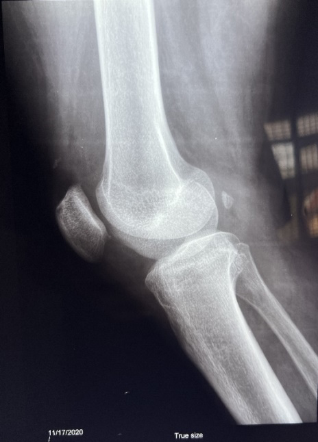
At presentation, an MRI and X-ray of the Right hip was done as shown in Figures 1 respectively. Reports by the radiologist noted. MRI of the right knee joint (November 2020) - showed features of mild degenerative changes evidenced by
- Articular cartilage loss.
- Small marginal osteophytes.
- Minimal joint effusion noted. Impression: Low grade partial thickness tear of mid substance of anterior cruciate ligament. Small radial tear of the body of medial meniscus Ruptured baker cyst with adjoining inflammation seen. Mild knee joint effusion seen. Mild degenerative changes seen in form of articular cartilage loss and small marginal osteophytes.
Small marginal osteophytes seen at the border of the patella.
- The tibiofemoral joint space is preserved.
- The distal femur and proximal tibia appear within normal limits. Impression: Early knee osteoarthritis.
- The tibiofemoral joint space and alignment are preserved.
- The distal femur, proximal tibia and patella appear normal.
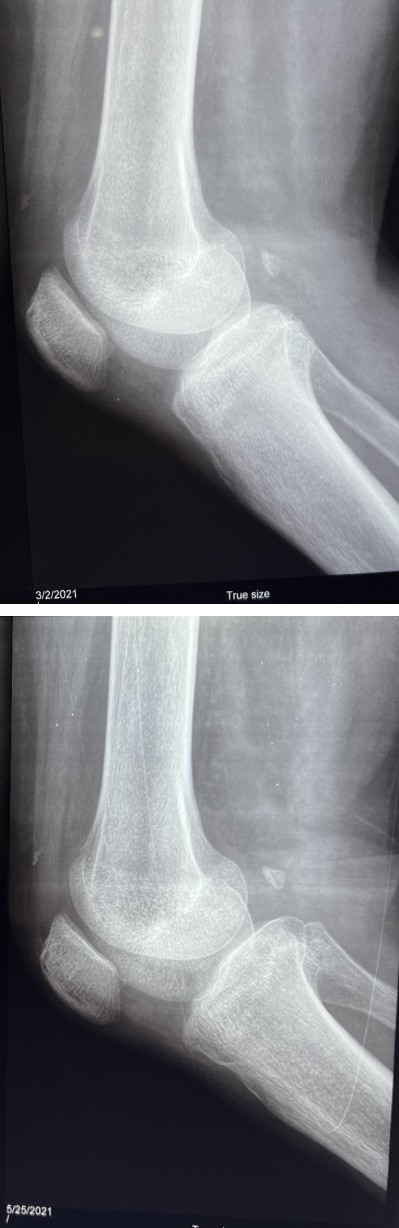
Figure 2a: X-ray of the right knee joint 6 months post procedure (May 2021).
The normal bone density is preserved.
- The tibiofemoral joint space is preserved.
- No bone or joint pathology seen. Impression: Essentially normal study.
Figure 2b: X-ray of the right knee joint 6 months post procedure (May 2021).
Discussion
Osteoarthritis is a chronic degenerative joint disease that has evidence based conservative treatment options like exercise, weight management, patient education and some medications for pain but this has not proved to reverse the progress of the degeneration but rather manage the damage already done. Definitive therapy known for advanced stages is usually total knee replacement, which is a surgical procedure, which is where adult stem cell therapy has it advantage as it is minimally invasive, safe with no reported treatment related adverse effect. Recent literature has revealed the anti-inflammatory and healing properties of stem cells. The use of autologous SVF and BMAC in conjunction with PRP could be a novel therapy. Future studies could help to establish appropriate clinical protocols including repeat dosing, quantity/volume of MSCs (BMAC and SVF) or concentration of PRP for specific indications. Also, future studies need to compare cases with and without intravenous transplantation of SVF to determine the extent of the role of systemic transplantation of autologous SVF in localized regeneration of the hip joints.
Proposed mechanisms of action in this patient could be a paracrine effect or engraftment of the injected cells. The injection of the MSCs (ADSCs and BMSCs) most probably stimulated a reduction of inflammation followed by the formation of new, healthy tissue. Complete tissue remodelling because of a complex sequence of events stimulated by expression of growth factors, chemokines, and cytokines.
Direct injection of MSCs (SVF and BMAC) plus PRP in severe knee osteoarthritis can be effectively and safely completed in an outpatient setting according to this case study. The patient tolerated the procedure with no reported adverse events. Radiographic imaging depicted the stages of healing after a single injection of the cellular mixture. Anticipated outcomes for healing processes and detailed protocols could be determined with larger clinical studies.
Ethics Approval and Consent to Participate
The patient provided written informed consent and agreed to participate in the study.
Consent for Publication
The patient provided written informed consent and agreed to have their data published.
Availability of Data and Materials
The datasets during and/or analysed during the current study are available from the corresponding author on request.
Authors’ Contributions
DI and OO designed the protocol. DI was responsible for clinical procedures and follow up of patient. DI and OO wrote the manuscript. All authors contributed toward critically revising the paper and agree to be accountable for all aspects of the work. All authors read and approved the final manuscript.
References
-
Hsu H, Siwiec RM (2022) Knee Osteoarthritis. StatPearls, Treasure Island.
-
Yaseen K (2024) Osteoarthritis. Merck Manual Professional Version.
-
Hayes B, Kittelson A, Loyd B, Wellsandt E, Flug J, et al. (2016) Assessing Radiographic Knee Osteoarthritis: An Online Training Tutorial for the Kellgren-Lawrence Grading Scale. MedEdPORTAL 12: 10503.
-
Comella K, Ikudayisi D (2018) Injection of Stromal Vascular Fraction Plus Platelet-Rich Plasma in a Non- Healing Decubitus Ulcer. J Med Cases 9(10): 323-327.
-
Caplan AI (2017) Mesenchymal Stem Cells: Time to Change the Name! Stem Cells Transl Med 6(6): 1445- 1451.
-
Hematti P, Keating A (2013) Mesenchymal Stromal Cells in Regenerative Medicine: A Perspective. Mesenchymal Stromal Cells, pp: 3-16.
-
Przybyt E, Harmsen MC (2013) Mesenchymal Stem Cells: Promising for Myocardial Regeneration? Curr Stem Cell Res Ther 8(4): 270-277.
-
Minteer D, Marra KG, Rubin JP (2013) Adipose-derived Mesenchymal Stem Cells: Biology and Potential Applications. Adv Biochem Eng Biotechnol 129: 59-71.
-
Gimble JM, AJ Katz, BA Bunnell (2007) Adipose-Derived Stem Cells for Regenerative Medicine. Circ Res 100(9): 1249-1260.
-
Rehman J, Traktuev D, Li J, Merfeld-Clauss S, Temm CJ, et al. (2004) The Secretion of Angiogenic and Anti- Apoptotic Factors by Human Adipose Stromal Cells. Circulation 109(10): 1292-1298.
-
Viswanathan S, Keating A, Deans R, Hematti P, Prockop P, et al. (2014) Soliciting Strategies for Developing Cell- based Reference Materials to Advance Mesenchymal Stromal Cell Research and Clinical Translation. Stem Cells and Development 23(11): 1157-1167.
-
Chahal J, Gomez-Aristizabal A, Shestopaloff K, Bhatt S, Chaboureau A, et al. (2019) Bone Marrow Mesenchymal Stromal Cell Treatment in Patients with Osteoarthritis Results in Overall Improvement in Pain and Symptoms and Reduces Synovial Inflammation. Stem Cells Transl Med 8(8): 746-757.
-
Usuelli FG, Grassi M, Maccario C, Viganoa M, Lanfranchi L, et al. (2017) Intratendinous Adipose-derived Stromal Vascular Fraction (SVF) Injection Provides a Safe, Efficacious Treatment for Achilles Tendinopathy: Results of a Randomized Controlled Clinical Trial at a 6-month Follow-up. Knee Surgery, Sports Traumatology, Arthroscopy 26: 2000-2010.
-
Comella K, Bell W (2017) First in Man Intraglandular Implantation of Stromal Vascular Fraction and Adipose Derived Stem Cells Plus Platelet Rich Plasma in Irradiation Induced Gland Damage: A Case Study. Int Med Case Rep J 10: 295-299.
-
Comella K, Parlo M, Daly R, Dominessy K (2018) First- in-Man Intravenous Implantation of Stromal Vascular Fraction in Psoriasis: A Case Study. Int Med Case Rep J 11: 59-64.
-
Kobolak J, Dinnyes A, Memic A, Mobasheri A, Khademhosseini A (2016) Mesenchymal Stem Cells: Identification, Phenotypic Characterization, Biological Properties and Potential for Regenerative Medicine through Biomaterial Micro-Engineering of Their Niche. Methods 99: 62-68.
-
Caplan AI, Correa D (2011) The MSC: An Injury Drugstore. Cell Stem Cell 9(1): 11-15.
- Psychogenic Erectile Dysfunction in Late Adulthood: A Case Report on Clinical Intervention and Intimacy Restoration
- Clinical Trials on COVID-19 in 2025: A New Chapter in Global Health Research
- Innovations and Challenges in Contemporary Medical Clinical Trials: An Editorial Perspective
- Innovations and Challenges in Contemporary Medical Clinical Trials: A Critical Perspective
- Reimagining Clinical Trials: The Power of Continuous Feedback from Medical Reports
- Factors Influencing Brain Drain: Perspectives from a Medical School in Turkey