Gar²De²Nia Risk Score as a New Scoring for Severe Coronary Artery Restenosis and It’s Introducing as a Web Score Calculator Pilot Study
Objectives: Gar²De²Nia score is designed to predict highly suspicious probability of significant coronary stenosis in patients without proven CAD or those who have already been revascularized. Background: There are a large number of scores that are used for cardiovascular risk assessment and different online calculators are used to perform it. Risk prediction models provide risk estimates that can assist our decision making and improve guiding for individual clinical outcomes and personalized preventive care. Methodology: Gar²De²Nia is an acronym that incorporates following parameters: Gender, Age, Renal impairement, Respiratory disease, Diabetes Melittus, Echo abnormalities, ECG abnormalities, new symptoms, Ischaemia, Atrial fibrillation. The pointing scale was formed (-1 to 2) on the personal interpretation of evidence based data and is applied and designed as web calculator. The established cut-off values for the risk of significant coronary stenosis are 4. For values below 4, the probability of significant CAD is low, below 0 is very low, equal or greater than 4 significant CAD is high and above 8 is very high. https://www.gardenia.estavela.in.rs Results: Standard statistical formulas for sensitivity, specificity, PPN and NPV were used (sensitivity 91, 43%, specificity 73, 33%, PPV 88, 89, NPV 78, 57). Estimated pilot sample size of 100 patients, was tested on patients cohort treated at the Institute for Cardiovascular Disease Dedinje in the year of 2022. Conclusion: This original score is to be checked throughout everyday clinical practice. It is easy to use. It can help in decision making in risk stratification and further treatment of the patients who has never been to cardiologist before, those who has already been revascularized and are symptomatic again or need any kind of non-cardiac surgery and the patients we have doubts about what to do next.
Full Text
https://www.gardenia.estavela.in.rs (direct link to the web score calculator).
There are a large number of scores that are used for cardiovascular (CV) risk assessment. Mostly have been done in primary care to predict the occurrence of coronary events in the general population. The most ESC widespread and most serious one is SCORE based on a large population Framingham study, that is used to predict the 10-year occurrence of coronary events based on the presence of established risk factors for CVD [1]. SCORE tables were modified to SCORE 2, SCORE2-OP tables, adjusted according to four subgroups of countries varying in low, moderate, high and very high age-standardized cardiovascular mortality rates. In apparently healthy people aged 40-69 years it is recommended to estimate the 10-year total atherosclerotic cardiovascular disease (ASCVD) risk with the SCORE 2 model and in those aged ≥70 years with the SCORE 2 O.P. model [2]. These models are calibrated Charts with the results that should be used to guide decisions and actions of preventive strategies in apparently healthy people [1, 2].
Different online calculators and smartphone applications perform cardiovascular risk assessments. You may also complete an assessment at your healthcare provider’s office. Each tool may ask for slightly different information, but there shouldn’t be much difference in the results [3]. One of the reputable ACC/AHA cardiac risk calculators is ASCVD risk calculator - also predicts your lifetime risk of a heart problem. Mainly heart risk calculators classify risk of cardiovascular disease as: Low: Less than a 5% risk, Borderline: 5% to 7.4% risk, Intermediate: 7.5% to 19.9% risk, High: More than a 20% risk [3]. Apart from this score, which has entered into daily practice and is based on the treatment and primary prevention of the modern age, there are a large number of official scores that are used as an aid for both primary and secondary prevention of CVD. All the set parameters of these scales like the isolated risk factors are based on the gender and age of each individual. These basic parameters also exist in my new model.
Standard risk factors for CVD have been proven by evidence base medicine as attributable, contributing to the occurrence of both primary and secondary CVD events [4]. The HEART score was developed in the Netherlands in 2008 by Six, Backus and Kelder as a rapid risk stratification tool for patients with chest pain according to their short-term risk MACE (defined as acute myocardial infarction, need for PCI /CABG and death within 6 weeks) to help identify low-risk patients, suitable for earlier discharge [4, 5]. Prior risk stratification tools include the GRACE and TIMI scores; these scoring systems, however, were derived for high risk patients examining the need for invasive therapy rather than the evaluation of individuals with undifferentiated chest pain [6, 7].
Risk prediction tools provide objective risk estimates that can assist the decision making of health professionals. Good risk prediction models should improve individual clinical outcomes and also resources allocation by avoiding both under- and over-treatment [7, 8]. Risk prediction tools will also be becoming increasingly important as clinical practice guidelines continue to move toward personalized preventive care. The integration of clinical risk prediction equations will be essential for guiding absolute risk assessment [9, 10]. Risk prediction can also impact the patient’s behavior and treatment decisions by improving insight into their cardiovascular prognosis and anticipating the potential impact of some therapies [11].
Over 20 years of clinical work with poly-vascular patients gave me the idea to try to create a simple, helpful template for stratifying these patients for referral to re- invasive procedures. There are daily dilemmas concerning the need to refer patients to invasive diagnostics. All of them include clinical findings, routine use of available functional tests, MDCT coronary angiography on the one hand, and the risk of possible complications on the other. Complicated scores are available that are not often used in everyday practice. This is how and why the idea for Gar²De²Nia score was born, to create a skeleton platform that serves only to rely on in clinical decision-making. To combine together the cardiological parameters of ischemia, atrial fibrillation and comorbidities, which are mostly easily accessible but at the same time are not classic CV risk factors. Gar²De²Nia score is designed to predict highly suspicious probability of significant coronary disease in patients who have not been proven to have CAD or the probability of new significant coronary stenosis ( resulting in PCI/CABG/vascular procedures) in those who have previously already been revascularized (cardiac surgery, percutaneous intervention or vascular surgery). Scoring system is -1, 0, 1. The minimum scale value is - 4. The maximum possible value is 11.
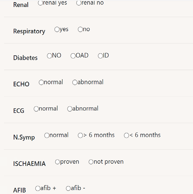
Gar²De²Nia is an acronym that incorporates the following parameters: G - Gender A - AGE R - RENAL impairment R- Respiratory disease - COPD, Asthma, ILD D - Diabetes Melittus E - ECHO abnormalities E - ECG abnormalities N- NEW SYMPTOMS I - ISCHAEMIA A - AFIB.
The established cut-off value for the risk of significant coronary stenosis is 4.
For values below 4, the probability of significant coronary disease is low (low risk), below 0 the risk is very low. For values equal to or greater than 4, the probability of having a significant CAD is high. For values over 6 the risk is very high.
The scale was formed on the personal interpretation of evidence based data from the literature. Standard formulas for sensitivity, specificity, PPN and NPV were used. (Sensitivity 91,43%, specificity 73,33%, PPV 88,89, NPV 78,57). 90% confidence interval (CI 1, 45-8,55) for estimation based on pilot sample size of 100 patients. The score was tested on patients cohort treated at the Institute for Cardiovascular Disease Dedinje in the year of 2022. Those patients were already diagnosed, all necessary data were available, so Gar²De²Nia score could be checked.
It is possible to recruit any target patient population for which there are adequate medical data. Randomization can be carried out according to the current rules, without additional aggravating circumstances. It is one time scoring system, though keeping the participants is not necessary because their monitoring can be carried out using different control examinations. That is the completely adequate way to get a good feedback. Those are common clinical examinations that do not require the implementation of any additional, complicated procedures, so patients are happy to respond. The used score cannot provide precise information about the way of further treatment, but it can give an insight into the prognosis. Estimates are simple. There is no objective medical reason why someone would not be willing to participate in the application of this score. The precise reliability of the Gar²De²Nia score needs to be proven on a larger number of patients, but on this sample seems to be accurate.
A positive point is given to the male gender because mortality from CHD and stroke remains higher among men than women until old age across a range of economically, socially and culturally diverse countries [12, 13]. While CHD and stroke mortality rates declined considerably between 1980 and 2010 in both sexes, there was some indication for stronger age-specific reductions in CHD in men than women. In 2019, total CVD DALYs were higher in men than women before age 80 to 84 years. After this age, the pattern reverses [14]. The sex differences in DALYs is most striking between ages 30 and 60 years (men greater) and age >80 years (women greater) [12, 13, 14, 15].
Cardiovascular diseases are the leading cause of death worldwide in 2019, and the majority of deaths from CVD are caused by ischemic heart disease, with most deaths occurring between the ages of 30 and 70 [14]. Coronary artery disease is proven to be associated with standard, established risk factors, and heredity is one of the strongest and non- modifiable factors [16]. Especially in younger people, the more pronounced the genetic component, the greater the possibility of CAD [17]. Consequently, coronary disease is more extensive. Nowadays, many other factors, among which the consumption of various intoxicating and illegal substances, various types of drugs along with the passionate consumption of cigarettes, also influence the emergence of a significant single-vessel CAD [16, 17]. Therefore, contrary to many tables within the scores, the older sex, over 70 years of age, was scored with fewer points than the younger age [14, 15, 16, 18]. Giving more importance to the severity of the coronary disease itself and not the age [18].
It is known that renal insufficiency is an independent risk factor for the onset, accelerated development and progression of the severity of coronary artery disease [19]. This also applies to moderate and severe forms of renal insufficiency, and patients on dialysis have a markedly increased risk of coronary vascular events.
The cardio-renal axis has been shown to influence cardio-metabolic changes through neurohumoral factors and represents a feedback and inseparable connection between these two organs with an impact on the progression of atherosclerotic disease [19, 20, 21]. The imbalance of mentioned mechanisms are leading to disorders of lipid metabolism, disorders and augmentation of oxidative processes at the sub-cellular level that affects the increase in CV morbidity [21, 22].
There are controversial data on the connection of respiratory disorders (COPD, asthma, interstitial lung diseases) with the existence of coronary artery disease.
Impaired lung function might increase cardiovascular risk through raised inflammation, ventilation–perfusion mismatch, impaired toxic waste removal [23]. The association could, on the contrary, also be driven by reverse causation, where (initially asymptomatic) cardiovascular disease worsens lung function, perhaps because of physical weakness or inadequate pulmonary circulation [23]. Chronic obstructive pulmonary disease (COPD) and coronary artery disease (CAD) are global epidemics that all by itself incur significant morbidity and mortality [24]. Both are frequently found in combination and also as independent diseases with the common causal factors, primarily smoking, and lately strongly over estimated air pollution [23, 24]. COPD was independently associated with ischemic heart disease (IHD), heart failure (HF), atrial fibrillation, and peripheral vascular disease, which contribute significantly to all-cause mortality [24, 25]. But, despite these findings, patients with COPD, asthma, and ILD were less likely to receive coronary revascularization than patients without these lung conditions [25]. And there was no difference in mortality in those who had both coronary artery disease and lung disease [25]. Due to all the available data from the literature, I decided to assign - 1 point for patients suffering from the mentioned respiratory disorders. The existence of severe coronary artery disease is less likely in serious pulmological patients (only in those where there is a need for inhalation therapy fall into this category).
Diabetes mellitus is one of the most tested and proven risk factors for the occurrence of significant coronary disease even since the Framingham study [26]. The present understanding of the epidemiological factors that influence cardiovascular disease risk (CVD-R) is based on the first report of study results at a 6-year median follow-up and numerous subsequent analyses of long-term follow-up data from the original Framingham cohort as well as their offspring [26, 27, 28]. Metabolic dis balance include not only hyperglycemia but insulin resistance that influence formation of coronary plaque among even newly diagnosed DM or IGT patients even when the CAG appeared normal, suggesting that preventive measures against atherosclerosis should be initiated prior to DM development [29, 30].
Even diabetic patients with intermediate coronary artery stenosis had the worse outcome, which was comparable to patients with severe stenosis [30].
Metabolic syndrome and its impact on all levels of micro and macro-circulation, in these patients, lead to the development of more progressive atherosclerosis, though significant coronary disease and an increase in CV morbidity and mortality [31].
Low insulin sensitivity underlies the metabolic syndrome that includes central obesity, dyslipidemia, hyperglycemia, hypertension, impaired fibrinolysis, and atherosclerosis [32]. Insulin resistance is the hallmark of type 2 diabetes and is associated with adiposity, hypertension, and dyslipidemia as part of the metabolic syndrome [33, 34]. Production of advanced glycation end-products, diabetic patients also have higher levels of sub clinical inflammation and develop endothelial dysfunction. The combination of hypercholesterolemia, inflammation, and endothelial dysfunction are the key mechanisms involved in progression of atherosclerotic coronary artery disease [34]. Patients with insulin-treated diabetes mellitus are usually patients with poor glycemic control and are expected to suffer more complications compared with patients with non-insulin- treated T2DM [35]. Large meta-analysis, including eleven studies involving a total of 64,152 patients with T2DM, showed that patients treated with insulin had a significantly higher rate of mortality and major adverse cardiac events compared with diabetic patients not on insulin (NITDM) after surgical revascularization [35]. Scale in my score was made considering this fact, the worse outcome coming from the more complex, combine anti-diabetic therapy.
Echocardiographic changes are considered as systolic thinning and abnormal changes in systolic wall thickening and wall motion abnormalities are seen commonly in patients with coronary events [36]. A long time ago and through the decades these parameters are been established as a very reliable marker of CAD.[36] Although the paradigm in diagnosing CAD has been evolved using multiplicity imaging, still echocardiograpy remained like gold standard for recognizing the possibility of having CAD as well as the staging of its severity [37]. Trans thoracic echocardiography can visualize and aid in the differential diagnosis of chest pain and it is always included in the approach for risk stratification and the diagnostic workup for the evaluation of chest pain [38, 39]. Back to Strong Heart Study left ventricular wall motion abnormalities even without overt cardiovascular disease are associated with 2.4- to 3.4-fold higher risks of cardiovascular morbidity and mortality, independent of established risk factors [40]. Consequently, that is the reason why only left ventricular segmental systolic abnormalities were incorporated in Gar²De²Nia score as abnormal. Globally decreased systolic ejection fraction can be influenced by different causes besides CAD [36]. If we count poor ejection fraction and valvular abnormalities, broader spectrum of CV diseases would be included, but the main cause would be lost [37, 38]. Significant valvular diseases can influence heart failure and vice versa. Also, the whole pallet of heart failure ( HFrEF/mrEF/pEF) is influenced by other risk factors, comorbidities, not only ( although the mostly) ischemic heart disease [37].
Abnormal resting ECG is common in patients with known or suspected chronic CAD. ECG had important diagnostic and prognostic values [41]. ECG done at rest is considered pathological in Gar²De²Nia score only if there are any ST segment abnormalities (mainly depression, rarely elevation in any of ECG leads but not STEMI ). Resting ST segment depression is a marker for a higher prevalence of severe coronary artery disease with a poor prognosis, but not due to left ventricular hypertrophy, conduction defects or drug effect [42]. Altered precordial T-wave balance (T1 > T6, T-wave in V1 > 1.5 mm and upright T-wave in V1) may be predictive of significant coronary artery disease (CAD) [43]. Having in mind that in patients with NSTEMI the sum of ST- segment depression in all ECG leads is a powerful predictor of all-cause mortality at 30 days, independent of clinical variables and correlates with the extent and severity of coronary artery disease [44]. The presence of even minimal (<1 mm) ST-segment elevation in anterior or inferior leads is independently associated with adverse outcomes [44]. That means even discrete ST and T disturbances in one lead can be marked as abnormal in this score. The use of stress exercise ECG and pharmacological testing is well established and precisely defined, for risk stratification and high suspicion of CAD, but it is not used in this score. Echocardiographic segmental changes are more sensitive even at rest, so those echo changes at rest sometimes may point out more severe coronary disease [42]. If we have ischemic changes at low heart rate on rest ECG, it has prognostic implications [42, 43], although existence of micro-vascular disease cannot be excluded [44].
There are typical and atypical angina symptoms, especially in diabetic patients with metabolic senzorial alterations and neuropathies [45]. Regardless of the presence of cardiovascular risk factors, the manifestation of chest pain usually is the beginning of a diagnostic evaluation for the presence of significant CAD [45]. The presence of typical angina symptoms in a contemporary cohort of patients referred for coronary angiography for stable CAD was the most important independent predictor of obstructive CAD [46]. In large group of nearly 10000 patients in PROMISE trial typical presenting symptoms were evaluated (substernal/ left-sided chest pain, other chest/neck/arm pain, dyspnea, and other symptoms) among other diagnostic methods. In low-risk outpatients evaluated for coronary artery disease, typicality of symptoms was not closely associated with higher baseline risk but was related to differences in processes of care and the prognostic value of a positive test. Adverse events were not associated with clinician risk estimates or symptoms alone [47]. Previously called stable angina and new classification has made it chronic coronary syndrome, incorporate patients with a long history of having chest pain and their prognosis is not worse if revascularization is not done ( trials as COURAGE, ISCHEMIA, ORBITA ) [48, 49]. So, the presence of new onset angina, symptoms ( de novo) or worsening of previously mild symptoms within six months are supposed to have higher CV risk, hence the higher impact on this score scale [48, 49, 50].
The proven ischaemia is considered to be in Gar²De²Nia score if: - any of available tests for the functional significance of CAD, myocardial ischemia tests are positive ( diagnostic options are enhanced by advances in technology and feasibility-ergometry, exercise and dobutamin stress echo test, CMR stress tests, SPECT perfusion tests, previously MDCT or coronary angio with described coronary stenosis, any myocardial revscularization (PCI/CABG), any myocardial infarction (NSTEMI/STEMI) documented by hospitalization.
The possibility of including any of these data can help in risk stratification both of revascularized and those who were not. The existence of significant coronary stenosis or re-stenosis to be revealed [51].
Coexistence of CAD and atrial fibrillation (AF) is associated with more complex management strategies characterized by increased bleeding risk from anti- coagulation. Furthermore, the presence of both diseases is associated with higher mortality [52]. Future studies should focus on studying the common molecular pathways [52]. The prevalence of CAD in AF patients can be wide ranging and reflects the population being studied, but the large multi center international clinical trials investigating efficacy of direct oral anticoagulant agents (ROCKET-AF, RELY) likely represent the typical AF patient population [53, 54]. Within these studies, CAD prevalence was reported to be 15-20% [54]. In the LIFE‐Heart Study, coronary artery sclerosis defined as non-critical plaque ( <75%) was associated with increased risk of AF, but not coronary artery disease (>75%) [55, 56]. The decision made for scoring is based on these data, having atrial fibrillation assigned negative point.
This original score is to be used and checked throughout everyday clinical practice. It is easy to use, the explanation is written in this article. It can help in decision making in risk stratification and further treatment of the patients who has never been to cardiologist before, those who has already been revascularized and now need any other kind of non- cardiac surgery and the patients we have doubts about what to do next.
References
-
SCORE2 working group and ESC Cardiovascular risk collaboration (2021) SCORE2 risk prediction algorithms: new models to estimate 10-year risk of cardiovascular disease in Europe. Eur Heart J 42(25): 2439-2454.
-
Visseren FLJ, Mach F, Smulders YM, Carballo D, Koskinas KC, et al. (2021) ESC National Cardiac Societies; ESC Scientific Document Group. 2021 ESC Guidelines on cardiovascular disease prevention in clinical practice. Eur Heart J 42(34): 3227-3337.
-
Goff DC Jr, Lloyd Jones DM, Bennett G, Coady S, D’Agostino RB, et al. (2014) 2013 ACC/AHA guideline on the assessment of cardiovascular risk: a report of the American College of Cardiology/American Heart Association Task Force on Practice Guidelines. Circulation 129(25): 49-73.
-
Six AJ, Backus BE, Kelder JC (2008) Chest pain in the emergency room: value of the HEART score. Neth Heart J 16: 191-196.
-
Backus BE, Six AJ, Kelder JC, Mast TP, Van den Akker F, et al. (2010) Chest pain in the emergency room: a multicenter validation of the HEART Score. Crit Pathw Cardiol 9(3): 164-169.
-
Six AJ, Cullen L, Backus BE, Greenslade J, Parsonage W, et al. (2013) The HEART score for the assessment of patients with chest pain in the emergency department: a multinational validation study. Crit Pathw Cardiol 12(3): 121-126.
-
Mahler SA, Miller CD, Hollander JE, Nagurney JT, Birkhahn R, et al. (2013) Identifying patients for early discharge: performance of decision rules among patients with acute chest pain. Int J Cardiol 168(2): 795-802.
-
Rosselo X, Dorresteijn JAN, Janssen A, Lambrinou E, Scherrenberg M, et al. (2019) Risk prediction tools in cardiovascular disease prevention: A report from the ESC Prevention of CVD Programme led by the European Association of Preventive Cardiology (EAPC) in collaboration with the Acute Cardiovascular Care Association (ACCA) and the Association of Cardiovascular Nursing and Allied Professions (ACNAP). European Journal of Preventive Cardiology 18(7): 534-544.
-
Karmali KN, Lloyd Jones DM (2017) Implementing Cardiovascular Risk Prediction in Clinical Practice: The Future Is Now. J Am Heart Assoc 6(4): e006019.
-
Hedberg B, Malm D, Karlsson JE, Årestedt K, Broström A (2018) Factors associated with confidence in decision making and satisfaction with risk communication among patients with atrial fibrillation. Eur J Cardiovasc Nurs 17(5): 446-455.
-
Bots SH, Peters SAE, Woodward M (2017) Sex differences in coronary heart disease and stroke mortality: a global assessment of the effect of ageing between 1980 and 2010. BMJ Glob Health 2(2): e000298.
-
Roth GA, Mensah GA, Johnson CO, Addolorato G, Ammirati E, et al. (2020) Global Burden of Cardiovascular Diseases and Risk Factors, 1990–2019: Update from the GBD 2019 Study. J Am Coll Cardiol 76(25): 2982-3021.
-
Fuster V (2014) Global burden of cardiovascular disease: time to implement feasible strategies and to monitor results. J Am Coll Cardiol. 64(5): 520-522.
-
Roth GA, Mensah GA, Johnson CO, Addolorato G, Ammirati E, et al. (2020) Global Burden of Cardiovascular Diseases and Risk Factors, 1990–2019: Update from the GBD 2019 Study. J Am Coll Cardiol 76(25): 2982-3021.
-
GBD 2016 Causes of Death Collaborators (2017) Global, regional and national age-sex specific mortality for 264 causes of death, 1980-2016: A systematic analysis for the global burden of disease study 2016. Lancet 390(10100): 1151-1210.
-
Michos ED, Choi AD (2019) Coronary Artery Disease in Young Adults: A Hard Lesson but a Good Teacher. Editorial Comment. J Am Coll Cardiol 74 (15): 1879- 1882.
-
Yang J, David B, Singh A, Divakaran S, De Filippis EM, et al. (2019) Risk factor profiles and outcomes of very young adults with myocardial infarction: results from the YOUNG-MI registry. Am J Med 133(5): 605-612.
-
Carroll AJ, Huffman MD, Zhao L, Jacobs DR, Stewart JC, et al. (2020) Associations between depressive symptoms, cigarette smoking, cardiovascular health: longitudinal results from CARDIA. J Affect Disord 260: 583-591.
-
Chade AR, Lerman A, Lerman LO (2005) Kidney in Early Atherosclerosis. Hypertension 45: 1042-1049.
-
Jankowski J, Floege J, Fliser D, Böhm M, Marx N (2021) Cardiovascular Disease in Chronic Kidney Disease: Pathophysiological Insights and Therapeutic Options. Circulation 143(11): 1157-1172.
-
Cachofeiro V, Goicochea M, Vinuesa SG, Oubiña P, Lahera V, et al. (2008) Oxidative stress and inflammation, a link between chronic kidney disease and cardiovascular disease. Kidney Int 74(111): S4-S9.
-
Carracedo J, Alique M, Vida C, Bodega G, Ceprián N, et al. (2020) Mechanism of Cardiovascular disorders in patients with kidney disease: A process related to accelerated senescence. Front Cell Dev Biol 8: 185.
-
Nowak C (2018) Lung Function and Coronary Artery Disease Risk. Circulation Genomic Precision Medicine 11(4): e002137.
-
Boschetto P, Beghe B, Fabbri L, Ceconi C (2012) Link between chronic obstructive pulmonary disease and coronary artery disease: Implication for clinical practice. Respirology 17(3): 422-431.
-
Carter P, Lagan J, Fortune C, Bhatt DL, Vestbo J, et al. (2019) Association of Cardiovascular Disease With Respiratory Disease. J Am Coll Cardiol 73(17): 2166- 2177.
-
Kannel WB, McGee DL (1979) Diabetes and Cardiovascular Diseases. The Framingham Study. JAMA 241(19): 2035-2038.
-
Qazi MU, Malik S (2013) Diabetes and Cardiovascular Disease: Insights from the Framingham Heart Study. Global Heart 8(1):43-48.
-
Abohelwa M, Kopel J, Shurmur S, Ansari MM, Awasthi Y, et al. (2023) The Framingham Study on Cardiovascular Disease Risk and Stress-Defenses: A Historical Review. J Vasc Dis 2(1): 122-164.
-
Nemoto N, Nakajima R, Ymazaki K, Utsunomiya M, Hori M, et al. (2015) Relationship between Insulin Levels and Coronary Atherosclerosis in Newly Diagnosed Diabetes Mellitus and Impaired Glucose Tolerance. Int J Clin Cardio 2(1).
-
Zhang HW, Jin JL, Cao YX, Guo YL, Wu NQ, et al. (2021) Association of diabetes mellitus with clinical outcomes in patients with different coronary artery stenosis. Cardiovasc Diabetol 20(1): 214.
-
Alfaddagh A, Welty FK (2020) Management of Stable Coronary Artery Disease in Patients with Diabetes Mellitus. ACC.
-
Cho YR, Ann SH, Won KB, Park GM, Kim YG, et al. (2019) Association between insulin resistance, hyperglycemia, and coronary artery disease according to the presence of diabetes. Sci Rep 9(1): 6129.
-
Reaven GM (1988) Role of insulin resistance in human disease. Diabetes 37: 326-337.
-
DeFronzo RA, Ferrannini E (1991) Insulin resistance: a multifaceted syndrome responsible for NIDDM, obesity, hypertension, dyslipidemia, and atherosclerotic cardiovascular disease. Diabetes Care 14(3): 173-194.
-
Krishna M, Pravesh KB, Hongzhi Q, Zhangui AT (2016) Comparing the Clinical Outcomes Between Insulin- treated and Non-insulin-treated Patients with Type 2 Diabetes Mellitus after Coronary Artery Bypass Surgery: Systematic Review and Meta-analysis. Medicine 95(10): e3006.
-
Drăgulescu SI, Streian C, Mourtada A, Ioan M, Sanbouskani A (1981) Evaluation of segmental wall motion disturbances by echocardiography in ischemic heart disease. Med Interne 19(2): 167-172.
-
Baggiano A, Italiano G, Guglielmo M, Fusini L, Guaricci AI, et al. (2022) Changing paradigms in the diagnosis of ischemic heart disease by multimodality imaging. J Clin Med 11(3): 477.
-
Levine GN, O Gara PT, Beckman JA, Al Khatib SM, Birtcher KK, et al. (2019) Recent innovations, modifications, and evolution of ACC/AHA Clinical Practice Guidelines: an update for our constituencies: a report of the American College of Cardiology/American Heart Association Task Force on Clinical Practice Guidelines. Circulation 139(17): 879-886.
-
Gulati M, Levy PD, Mukherjee D, Amsterdam E, Birtcher KK, et al. (2021) 2021 AHA/ACC/ASE/CHEST/SAEM/ SCCT/SCMR Guideline for the Evaluation and Diagnosis of Chest Pain: A Report of the American College of Cardiology/American Heart Association Joint Committee on Clinical Practice Guidelines. Circulation 144(22): 368-454.
-
Cicala S, de Simone G, Roman MJ, Best LG, Lee ET, et al. (2007) Prevalence and Prognostic Significance of Wall-Motion Abnormalities in Adults Without Clinically Recognized Cardiovascular Disease: The Strong Heart Study. Circulation 116(2): 143-150.
-
Cres PM, Lehmann KG, Froelicher VF (1991) Correlation between resting ST segment depression, exercise testing, coronary angiography, and long-term prognosis. Am Heart J 122(6): 1617-1628.
-
Kharel H, Pokhrel NB, Pokhrel B, Chapagain P, Poudel CM (2020) Implications of Localized ST Depression in a Vascular Territory and Altered Precordial T-Wave Balance in Ischemic Heart Disease. Cureus 12(6): 8580.
-
Savonitto S, Cohen MG, Politi A, Hudson MP, Kong DF, et al. (2005) Extent of ST-segment depression and cardiac events in non-ST-segment elevation acute coronary syndromes. Eur Heart J 26(20): 2106-2113.
-
Kaolawanich Y, Thongsongsang R, Songsangjinda T, Boonyasirinant T (2021) Clinical values of resting electrocardiography in patients with known or suspected chronic coronary artery disease: a stress perfusion cardiac MRI study. BMC Cardiovasc Disord 21(1): 621.
-
Kontos M, de Lemos J, Deitelzweig SB, Diercks DB, Gore MO, et al. (2022) 2022 ACC Expert Consensus Decision Pathway on the Evaluation and Disposition of Acute Chest Pain in the Emergency Department. J Am Coll Cardiol 80(20): 1925-1960.
-
Nakas G, Bechlioulis A, Marini A, Vakalis K, Bougiakli M, et al. (2019) The importance of characteristics of angina symptoms for the prediction of coronary artery disease in a cohort of stable patients in the modern era. Hellenic Journal of Cardiology 60 (4): 241-246.
-
Lowenstern A, Alexander KP, Pagidipati NJ, Hill CL, Pellikka PA, et al. (2022) Presenting Symptoms in Patients Undergoing Coronary Artery Disease Evaluation: Association With Noninvasive Test Results and Clinical Outcomes in the PROMISE Trial. Circulation Cardiovascular Quality and Outcomes 15(5): e008298.
-
Maron DJ, Hochman JS, Reynolds HR, Bangalore S, O Brien SM, et al. (2020) Initial invasive or conservative strategy for stable coronary artery disease. N Engl J Med 382: 1395-1407.
-
White LD, Lee TH, Cook F, Weisberg MC, Rouan GW, et al. (1990) Comparison of the natural history of new onset and exacerbated chronic ischemic heart disease. JACC 16(2): 304-310.
-
Albuquerque LC, Gomes WJ (2018) ORBITA Trial: Redefining the Role of Intervention in the Treatment of Stable Coronary Disease?. Braz J Cardiovasc Surg 33(1): 3-5.
-
Min JK, Leipsic J, Pencina MJ, Berman DS, Koo BK, et al. (2012) Diagnostic accuracy of fractional flow reserve from anatomic CT angiography. JAMA 308(12): 1237- 1245.
-
Mekhael M, Marrouche N, El Hajjar AH, Donnellan E (2022) The relationship between atrial fibrillation and coronary artery disease: Understanding common denominators. Trends in Cardivascular Medicine.
-
Dixit S (2022) Clarifying the association between atrial fibrillation and coronary artery disease. Trends in Cardiovascular Medicine.
-
Patel MR, Mahaffey KW, Garg J, Pan G, Singer DE, et al. (2011) Rivaroxaban versus warfarin in patients with non-valvular atrial fibrillation. N Engl J Med 365(10): 883-891.
-
Kornej J, Henger S, Seewöster T, Teren A, Burkhardt R, et al. (2020) Prevalence of atrial fibrillation dependent on coronary artery status: Insights from the LIFE-Heart Study. Clin Cardiol 43(12): 1616-1623.
-
Beutner F, Teupser D, Gielen S, Holdt LM, Scholz M, et al. (2011) Rationale and design of the Leipzig (LIFE) Heart Study: phenotyping and cardiovascular characteristics of patients with coronary artery disease. PLoS One 6(12): e29070.
- Anomalous Origin of the Left Coronary System from the Right Coronary Cusp: A Rare Coronary Anomaly in a Patient Undergoing Aortic Stenosis Workup
- Optimizing the Therapeutic Potential of Sacubitril/Valsartan: The Promise of Co-Crystal Engineering
- Association between Mortality and Topography of Peripheral Arterial Disease due to Atherosclerosis Obliterans
- Mitral Valve Replacement vs Mitral Valve Repair
- Characteristics and Evolution of Patients with Heart Failure Hospitalized in the Cardiology Department of Dalal Jamm Hospital
- Distribution of Association between Basic Knowledge of Chest Pain and Myocardial Infarction (Heart Attack) and Demographic Variables: A Survey-Based Study