Cutaneous Squamous Cell Carcinoma on Permanent Tattoo of the Face: Diagnostic that should be Prevent by Dermoscopy and Local Medical Care
Many skin lesions have been reported in connection with tattoos, both benign and malignant. The pathogenesis of malignant transformation in tattoos is unknown, although hypotheses include the toxic effects of the pigments. To date, we have notice 31 case reports and series (17 men, median age: 50.5 years) of KA and CSC on tattoos. Other Factors: UV exposure, fair skin, and immunosuppression increase incidence of cSCC. Recurrence and metastasis are associated with tumor diameter greater than 2 cm, depth greater than 2 mm or beyond subcutaneous fat, extensive or largecaliber perineural involvement, and poor differentiation on histopathology. AJCC 8 is used for TNM staging for cSCC of the head and neck. BWH offers an alternative T staging system. Management is primarily surgical with rare indications for adjuvant chemoradiation.
Background
Cutaneous squamous cell carcinoma is the second most common malignity. It comprises 20% of all cases of nonmelanoma skin cancer, and results in 1 million cases in the United States each year resulting in up to 9000 approximed deaths. This incidence continues to growth annually with an estimated 50% to 200%, increasing with age, with an average age of onset in the mid-60s,in the last three decades [1] cSCC is more frequent in men than women (3:1 ratio) [1].
The disease has been linked to immunosuppression, Human Papilloma Virus (HPV) infection, arsenic exposure, radiation and chronic ulceration; injure such tattoo [2]. While it usually exhibits benign clinical behavior, some aggressive forms can lead to serious complications. Skin biopsy deep enough is crucial for making the diagnostic. Ten- year survival post-surgery exceeds 90% but falls drastically in case of metastases which makes early detection an essential prognostic factor [3]. Here we represent a case of young women with pigmented cutaneous squamous cell carcinoma on permanent tattoo of the face that had remained undiagnosed for a prolonged period of time.
Case Report
A ninety-year-old woman was referred to our Dermatology department for investigation of a solitary facial skin lesion that has been evolving for more than two years. She reported no medical history apart from multiple painful sunburns in childhood, took no medication, and family history for dermatological disease was negative.
The clinical examination showed a developed ulcerative tumor with a seropurulent surface, roughly rounded, 4cm long axis, flesh-colored with beaded border, sitting at the level of the right cheek with extension towards the lower eyelid and the nose (Figure 1).
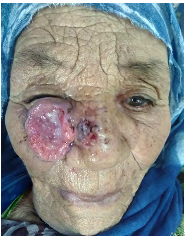
The tumor was located on the upper half of the cheek on the right with extension to the right eye that had caused vision impairment. The lesion was not painful. Lymph nodes examination revealed no anomalies, especially Local lymph nodes and parotid. Skin biopsy of the affected area showed nests of epithelial cells arising from the epidermis and extending to the dermis with important keratinization. The diagnostic of cutaneous squamous cell carcinoma was given. The patient underwent surgery for total tumor resection.
Discussion
Squamous cell carcinoma also called epidermoid carcinoma is the second most common type of skin cancer, with basal cell carcinoma being the first [4]. SCC commonly affects the scalp, neck region, back of hands, superior surface of the pinna, and the lip. SCC lesions may have a scaly, erythematous macule or plaque; Telangiectasia, central ulceration may also be present. The ulcer may be superficial and hidden by a crust. Removal of the crust may reveal a well-defined papillary base [4].
SCC arising in areas of prior radiation, thermal injury areas of chronic ulcers and chronic draining sinuses or permanent tattoos. Also The presence of rare familial syndromes (including xeroderma pigmentosum, albinism, epidermolysis bullosa, epidermolysis verruciformis, Ferguson Smith epithelioma, Rothmund-Thomson syndrome, Bloom syndrome) can predispose an individual to multiple cSCCs at a young age [1]. Many skin lesions have been reported in connection with tattoos, both benign and malignant. The pathogenesis of malignant transformation in tattoos is unknown, although hypotheses include the toxic effects of the pigments [5].
Although most SCCs are relatively slow growing and nonaggressive, some (2% to 5%) can show rapid growth and metastases. Aggressive tumors have a higher frequency of metastasis in immunocompromised patients and when develop from scars, burns, prior injury (Marjolin ulcer) or tattoos. Than those originating in actinic damaged skin [4]. Also, Existence of SCC on ears, lips, or size >2 cm are high- risk features of SCC [4].
It commonly arises in the sun-exposed areas. The tumor can present as hard lump with overlying scale, raised growth with central depression, open sore, thick wart like skin and can also form an ulce. The dermoscopy is very usefull; In early SCCs, the vesselsare predominantly polymorphous, mainly consisting of dotted/glomerular, linear, and hairpin. Moreover, early SCCs mainly displayed scales, white structure less areas, ulceration/bleeding, white halos surrounding vessels, erythema background, blood spots, and white circles surrounding follicles, findings in line with previous evidence.
The diagnostic requiert a skin biopsy deep enough to permit the pathologist to observ on depth of invasion, perineural or lymphovascular penetration, differentiation, and relation with the overlying epidermis [1] that can show nest of epithelial squamous cells deep into the dermis. The malignant cells are often large with eosinophilic cytoplasm and a vesicular nucleus with variable degree of keratinization. Cutaneous SCC has more potential to metastasize than basal cell carcinoma, with tumor diameter being the most important prognostic factor [6]. Tumor with a size >2 cm and a significant perineural invasion has local recurrence and metastatic risk as high as 47% and 35%, respectively [7].
Mostly, imaging is not needed unless the clinical examination is revealing an involvement of large-caliber nerves, muscle or bone, lymph node involvement, or when high-risk properties are existing. Compute tomography with contrast is helpful for appraisal of lymph node, soft tissue, or bone involvement. MRI is favorite to measure perineural invasion or orbital and intracranial extension [1]. On the molecular level, several pathways have been implicated in the disease. Genomic instability caused by P53 mutation represent one of the earliest even in the development of squamous cell carcinoma [8].
Other changes in tumor suppressor genes like NOTCH and CDKN2A has also been reported and oncogenes like RAS. The collection of all those mutations eventually distorts important signaling pathways including the activation of NF-KB, MAPK which mediates Epidermal growth factor overexpression [9, 10]. Treatment of cutaneous squamous cell carcinoma is mainly surgical. Complete removal of the tumor with histopathological control of excision margins is the gold standard [11].
Other alternatives Includes radiotherapy and cryotherapy. The use of oral tretinoin (Acitretin) may help decrease tumor size and overall tumor load in case of multiple cutaneous squamous cell carcinoma [11]. Measures of prevention are helpful for very high-risk, immunosuppressed patients Including those aimed at managing field cancerization (large defects of DNA damaged skin 5-fluorouracil, imiquimod, topical retinoids, diclofenac sodium, ingenol mebutate, chemotherapy wraps, photodynamic therapy, nicotinamide and acitretin or capecitabine. But Photoprotective measures including sunscreen application remains primordially, it have been shown to decrease SCC by 40% [1].
Conclusion
To date, we have notice 31 case reports and series (17 men, median age: 50.5 years) of KA and CSC on tattoos. Lesions usually develop rapidly after completion of the tattoo, between one week and several months. Exceptional cases have been described in old tattoos.Additional studies on tumor specimens are warranted to identify the possible causative agents in tattoo ink that may be responsible for these reactions.
References
-
Waldman A, Schmults C (2019) Cutaneous Squamous Cell Carcinoma. Hematol Oncol Clin N Am 33(1): 1-12.
-
Rinker MH, Fenske NA, Scalf LA, Frank LG (2001) Histologic Variants of Squamous Cell Carcinoma of the Skin. Patholgy update 8(4): 363.
-
Varra V, Woody NM, Reddy C, Joshi NP, Geiger J, et al. (2018) Suboptimal Outcomes in Cutaneous Squamous Cell Cancer of the Head and Neck with Nodal Metastases. Anticancer Res 38(10): 5825–5830.
-
Ferri FF (2022) Squamous Cell Carcinoma Ferri’s Clinical Advisor 1410-1411
-
Papageorgiou C, Lallas A, Manoli MS, Longo C, Liopyris K (2021) Evaluation of dermatoscopic criteria for early detection of squamous cell carcinoma arising on an actinic keratosis. J Am Acad Dermatol 86(4): 791-796.
-
Keena TYQ, Zwald FO, Chrysalyne DS (2018) Cutaneous squamous cell carcinoma Incidence, risk factors, diagnosis and staging. J Am Acad Dermatol 78(2): 237- 247.
-
Goepfert H, Dichtel WJ, Medina JE, Lindberg RD, Luna MD (1984) Perineural invasion of squamous cell carcinoma of the head and neck. AmJ surg 8(4): 542-547.
-
Brash DE , Rudolph JA, Simon JA, Lin A, McKenna GJ (1991) A role for sunlight in skin cancer: UV-induced p53 mutations in squamous cell carcinoma. Proc Natl Acad Sci USA 88(22): 10124-10128.
-
South AP, Purdie KJ, Watt AS , Haldenby S, Breems ND (2014) NOTCH1 mutations occur early during cutaneous squamous cell carcinogenesis. J Invest Dermatol 134(10): 2630-2638.
-
Brown VL, Harwood CA, Crook T, CroninGJ, Kelsell DP, et al. (200) 4p16INK4a and p14ARF tumor suppressor genes are commonly inactivated in cutaneous squamous cell carcinoma. J Invest Dermatol 122(5): 1284-1292.
-
Stratigos A, Garbe C, Lebbe C, Malvehy J, Marmol VD (2015) Diagnostic and treatment of invasive squamous cell carcinoma of the skin :European consensus–based interdisciplinary guidelines. Eur J cancer 51(14): 1989- 2007.
- Cancer Diagnosis from RNA Sequence of Blood Cells by Using AI
- Field Cancerization in Oral Cavity, Case Report and Review of Literature. Oncologic Program Salud Integral Hospital, Managua, Nicaragua
- Identification of B Lymphocytes in Cancer Patient’s Blood
- A Case Report of a Breast Cancer Patient Developing Pneumonitis as a Result of Abemaciclib Therapy
- Immune Checkpoint Therapeutics for Today’s Fight and Beyond
- The Amalgamated Sophomore-Gonadoblastoma