Molecular Detection and Genotyping of Group A Rotavirus by Multiplex Semi-Nested RT-PCR in Sewage Water and Sludge
Group A rotavirus (RV-A) infections are a major cause of morbidity and mortality in human around the world. The aim of this research was to conduct a molecular characterization of RV-A glycoprotein (G) and protease-sensitive (P) in urban sewage (n=54; 27 raw sewage and 27 treated sewage) and sewage sludge (n=27) from Egypt by multiplex semi-nested RT-PCR method. RV-A was detected in 29.9% of raw sewage, 7.4% of treated sewage, and 18.5% of sewage sludge samples. In the positive samples, the RV-A G genotypes were as follow: G3 (n=6), G9 (n=3), G1 (n=2), G4 (n=1), G10 (n=1), G1+G3 (n =1), and G1+G3+G10 (n=1) whereas P types detected included: P[4] (n=1), P[6] (n=3), P[8] (n=8), and nontypeable P (n=3). The most detected G-P combination was G3P [8] (n=6). Other G-P combinations such as G1P [8] (n=2), G9P [6] (n=2), G4P [6] (n=1), and G10P[4] (n=1) were also detected. The highest detection rates of RV-A in sewage samples were found in winter (50%), followed by summer (25%), then in spring and autumn (12.5%). The results showed not only that sewage analysis contains dramatic information concerning enteric viruses, but also that environmental monitoring is an important approach in describing the local circulation of specific viruses among population.
Introduction
Group A rotavirus (RV-A) is the major pathogen of sever gastroenteritis in both infants and young children in developing and developed countries, causing about 30– 50 % of these diseases, with a high rates of morbidity and mortality in the population of developing countries [1]. Although, morbidity and mortality due to RV infections has reduced due to actual universal mass vaccination program against this virus, it still causes about 192,700 deaths per year in children under 5 years old [2].
RV-A, belonging to Rotavirus genus and Reoviridae family, are segmented and non-enveloped double- stranded RNA viruses. Based on the differences in the nucleotide sequences of the two outer capsid proteins (VP4 and VP7 genes) of RV-A, it has been subdivided into 27 glycoprotein (G) and 23 protease-sensitive (P)- genotypes [1, 3]. RV-A strains bearing the combinations such as G1P[8], G9P[8], and G2P[4] are considered as the most prevalent genotypes in humans in the Middle Eastern and North African, accounting for 37.7%, 22.5%, and 8.1% of all RV positive samples, respectively [4].
Currently, RV-A monitoring was mostly performed in clinical cases. However, asymptomatic infection of RV-A is common [5], and case-based monitoring can only focus on the symptomatic patients. The epidemiology of RV-A in asymptomatic persons was not well studied. Because the particles of RV-A can be released from both asymptomatic and symptomatic persons to sewerage system, we can get more comprehensive data on the molecular characterizations of RV-A by sewage analysis [6]. The objective of this study was to investigate the prevalence of VP4 and VP7 genotypes of human rotavirus in sewage water and sewage sludge using semi-nested multiplex RT- PCR.
Materials and Methods
Sample Collection and Concentration
Twenty seven raw sewage, 27 treated sewage, and 27 sludge samples were collected monthly, from June 2015 to August 2017, from Zenin wastewater treatment plat (WWTP). The sewage samples were concentrated by the adsorption–elution technique, using an electronegative membrane (0.45µm pore size, and 142mm diameter), and organic flocculation method [7, 8] whereas the sewage sludge samples were concentered by method described previously by EPA (1984) [9].
RNA Extraction and cDNA Synthesis
Total RNA was extracted from 1ml aliquots of the concentrated sewage and sludge samples with QIAamp Viral RNA Mini Kit (Qiagen, Germany). RT of the viral RNA was conducted using a reverse primer VP7-R (50μg/μl) and Con 3 (50μg/μl) for G and P types, respectively. Initially, 5μl of the viral RNA with 1μl of the reverse primer was heated at 65°C for 10 minutes in Biorad PCR machine (USA), then immediately snap chilled on ice. Next, 4 µl of 10 mM dNTP (Bioline, USA), 0.5 µl of Maloney murine leukemia virus reverse transcriptase (MMLV, Promega), 10µl of 5x RT buffer, 0.5µl RNase inhibitor, 4 µl DEPC-treated water. The mixture was incubated at 30°C for 1 hr, 42°C for 30 min, then 95°C for 5 min at the end of the incubation step.
G and P Genotyping
Amplifying of a VP7 gene was performed using two steps according to protocol described previously by Iturriza Gomara et al. (2004) [10]. The first-round of PCR amplification of the VP7 gene was performed with 5 of cDNA, 10 μl of M-MLV 5X reaction buffer, 4 μl of 25 mM MgCl2, 4 μl of 10 mM dNTPs, 0.25 μl of 5 U/ ml Go Taq DNA polymerase (Promega, USA) and 1 μl of 25 pmol of each primer (Table 1), 24.75 μl DEPC-treated water. PCR conditions on the thermocycler (Bio-Rad, Singapore) were as follows: 95°C (5 min); followed by 35 cycles consisted of 95°C (1 min), 52°C (1 min), and 72°C (1 min); then a final extension step at 72°C (10 min). The second round VP7 multiplex was performed in 50 μl total volume containing a 2.5 μl of the first PCR product as a template with 1 μl of each G-type-specific primer or P-type-specific primer (Table 1), 10 μl of M-MLV 5X reaction buffer, 4 μl of 25 mM MgCl2, 4 μl of 10 mM dNTPs, 0.25 μl of 5 U/ml Go Taq DNA polymerase (Promega, USA), 23 μl DEPC- treated water. The thermal cycling conditions were as follows: 95°C (5 min); followed by 30 cycles at 94°C (1 min), 42°C (2 min), 72°C (1 min); then a final extension step at 72°C (10 m).
Ref. nt. Position Amplicon (bp) Sequence (5-3) Primer
G-typing
[11] 51-71 881 ATG TAT GGT ATT GAA TAT ACC AC VP7-F 1st round 914-932 AAC TTG CCA CCA TTT TTT CC VP7-R
250-269 682 ACG AAC TCA ACA CGA GAG G G3 2nd round
757-776 179 CTT GAT GTG ACT AYa A AAT AC G9
666-687 266 ATG TCA GAC TAC ARb A TAC TGG G10
178-198 754 GTC ACA CCA TTT GTA AAT TCG G8
480-499 452 CGT TTC TGG TGA GGA GTT G G4
411-435 521 CAA TGA TAT TAA CAC ATT TTC TGT G G2
| G1 | CAA GTA CTC AAA TCA ATG ATG G | 618 | 314-335 | ||
|---|---|---|---|---|---|
| VP7-R | As above | ||||
| P-typing | |||||
| 1st round | Con3 | TGGCTTCGCCATTTLATAGACA | 876 | 11-32 | [13] |
| Con2 | ATTTCGGACCAT'lTATAACC | 868-887 | |||
| 2nd round | P[8] | TCTACTTGGRTTRACNTGC | 345 | 339-356 | [10,13, 14] |
| P[4] | CTATTGTTAGAGGTTAGAGTC | 483 | 474-494 | ||
| P[6] | TGTTGATTAGTTGGATTCAA | 267 | 259-278 | ||
| P[9] | TGAGACATGCAATTGGAC | 391 | 385-402 | ||
| P[10] | ATCATAGTTAGTAGTCGG | 583 | 575-594 | ||
| P[11] | GTAAACATCCAGAATGTG | 312 | 305-323 | ||
| Con3 | As above |
Table 1: Primers used for detection and G and P typing of RV
Ya = C or T; Rb = A or G; N= A, G, C or T Table 1: Primers used for detection and G and P typing of RV
Results
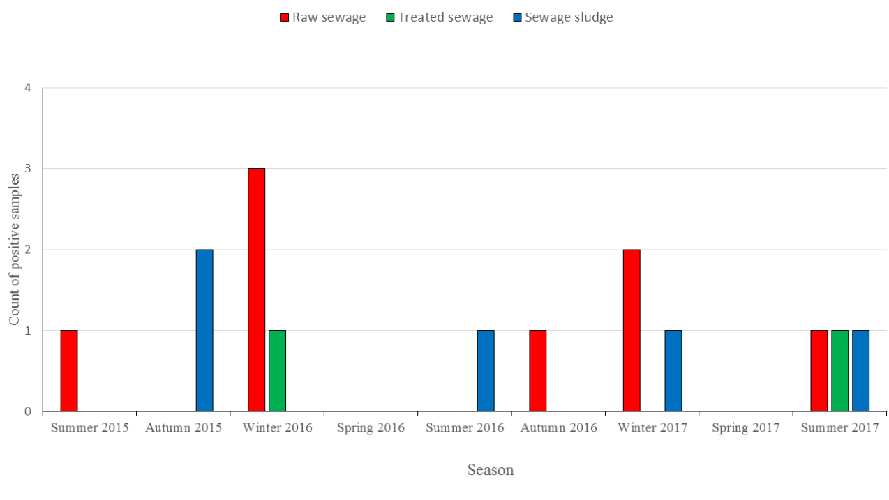
December to February. In the other seasons, the detection rate of RV-A was 25% (2/8) in summer season (June- August) whereas it was 12.5% (1/8) in autumn (September-November) and 0% in spring (March-April). In treated sewage samples, the detection rates of RV-A was 50% (1/2) in winter 2016, 50% (1/2) in summer 2017 whereas no virus was detected in spring and autumn. In sewage sludge, RV-A was found in 40% (2/5) of autumn, 40% (2/5) summer, 20% (1/5) of winter, and 0% (0/4) (Figure 1).
Prevalence and Seasonal Variation of Group A Rotavirus
RV-A was detected in 29.9% (8/27) of raw sewage, 7.4% (2/27) of treated sewage, and 18.5% (5/27) of sewage sludge. The highest detection rate of rotavirus in raw sewage was found in winter season (5/8, 62.5%), from
Molecular Characterization of the Detected RV
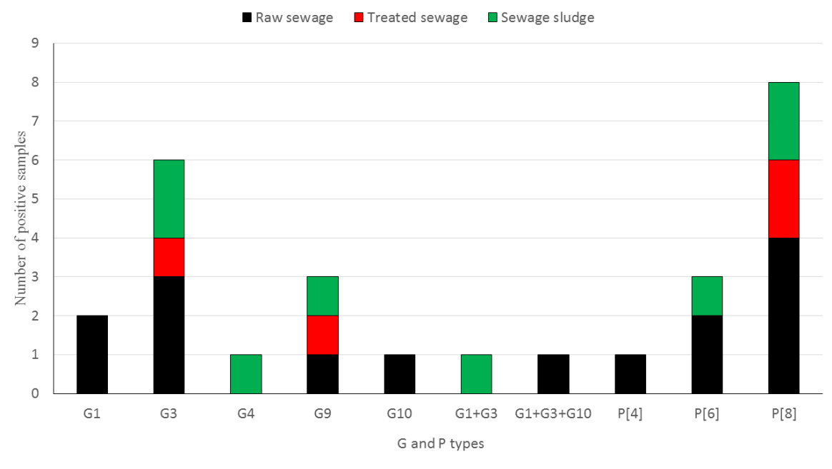
Analysis VP7 gene by multiplex semi-nested RT-PCR showed that G3 (6/15, 40%) was the most genotype strain, followed by G9 (3/15, 20%), and G1 (2/15, 13.3%). G4 and G10 genotypes were identified in the same percentage (1/15, 6.6%) of strains. Two mixed infections with two (G1+G3; 1/15, 6.6%) or three (G1+G3+G10; 1/15, 6.6%) G genotypes were also detected. Analysis VP4 gene showed that P[8] is the most common strain (53.3%; 8/15), followed by P[6] 20%; 3/15, and P[4] 6.6%; 1/15.
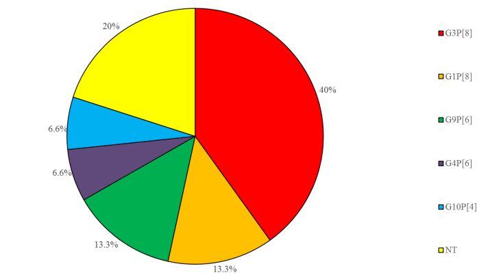
P[9], P[10], and P[11] strains were not detected in this study. Some positive samples (20%; 3/15) were non- typeable for G-P combination (Figure 2). The most common genotype combination was G3P[8] (6/15; 40%). G1P[8] and G9P[6] genotype combinations were detected in the same percentage 13.3%, 2/15. Other genotype combinations such as G4P[6] and G10P[4] were also detected in this study (6.6%, 1/15). The genotype combinations were not detected in some rotavirus- positive environmental samples (20%, 3/15) (Figure 3).
Discussion
Environmental sewage monitoring has been successfully performed to get data on molecular characterizations of various enteric viruses in Egypt [15, 16, 17, 18]. In this research, we investigated the prevalence, genotypes, and seasonality of RV-A in Giza government, Egypt. During the 27-month period, a total of 27 raw sewage, 27 treated sewage, 27 sewage sludge samples were collected monthly. In order to improve the sensitivity of detection method, we applied semi-nested PCR for the collected samples in this study. The high sensitivity of the nested RT-PCR detection has been previously described for double stranded RNA RV-A that could detect a few viral particles (10-100 particles) [13].
After conducting of semi-nested multiplex RT-PCR detection, rotavirus was identified in 29.9% (8/27), 7.4% (2/27), and 18.5% (5/27) of influent, effluent, and sewage sludge, respectively. Although the number of RV-A positive specimens was decreased upon treatment in the current wastewater treatment plant, the treated effluent still contained RV-A nucleic acids. This finding highlights the high risk of RV-A infection among the population when this effluent is reused in agricultural production and/or its discharge in bathing and recreational water.
In two previous studies conducted on the same wastewater treatment, RV-A was detected in 5/12(41.6%) and 16.7% of raw sewage and in 0% and 16.7 of treated sewage during 2006-2007 and 2009-2011, respectively [18, 19]. Moreover, the current viral prevalence is relatively high when compared with other previous reports. Differences in the prevalence of RV-A might be due to the variations in sensitivity of virus recovery and detection methods. For example, two reports from Bangkok documented that 8% and 0% of untreated sewage specimens were positive for rotavirus by ELISA technique [20, 21]; while by using indirect immunofluorescence, 21% of the samples were positive for rotavirus in a study from Sao Paulo [22]. Moreover, although a semi nested RT-PCR was applied in a study conducted in Barcelona, rotavirus was identified in only 4 of 15 sewage samples [23]. A similar study from France, however, documented a prevalence of rotavirus in 42% of untreated sewage [24, 25], which is higher than our current results. Two studies from Tunisia and China, found RV in 72·4% and 93.5% of sewage samples using real time PCR, respectively [6, 26]. Moreover, our detection rates of RV in sewage sludge is lower than those detected in study from Brazil [26].
Group A rotavirus should be recognized as potential marker of fecal pollution present in the aquatic environment since RV-A movement appears to be greater than the adenovirus, which is recognized as a potential indicator of the occurrence of enteric viruses in the environment [27]. RV-A stability in water environment, and its resistance to physico-chemical processes applied for sewage treatment promotes its dissemination and transmission in the water environment [28].
Genotyping was conducted on fifteen rotavirus positive sewage and sludge samples by multiples semi- nested RT-PCR. The most prevalent genotypes were G3, G9, P[8], P[6], with G3 P[8] being the most prevalent genotype combination in the positive samples. El-Senousy et al, (2014) [16] stated that G3 and P[8] were the second most common genotype after G1 and P[4] in water samples from Egypt. In agreement with our finding, G9 was the second most common genotype in sewage samples [29]. Indeed, G1 and P[8] were documented as the most common genotypes in Spain and Brazil [28, 30] as well as G1 and P[8] in Venezuela [31]. In agreement with our findings, genotype G3P[8] was found as the most frequent genotype in wastewater samples from Tunisia [32]. However, G1P[8] combination was documented as the most common genotype in the clinical and environmental samples from several countries, including the Middle Eastern and North African Region [4, 28, 30, 33].
This study is limited due to absence of clinical data. However, our previous studies demonstrated that genotypes G3 [34] and P[8] (unpublished data) were the most predominant genotypes in clinical samples from children with severe diarrhea which agree with this study. Thus, G3 and P[8] detected in the population might be originated from contaminated sewage. Sewage monitoring system has been observed to be more accurate than reporting of clinical cases of serious diseases in a population [35]. Data from the presence of enteric viruses in untreated sewage may provide a useful data on the epidemiology of enteric virus infections circulating in the population, including asymptomatic infections [36, 37].
The present research showed a higher detection rates of RV-A during the winter season. The same seasonality pattern has been reported in our previous study on clinical surveillances [34]. Seasonal occurrence of group A rotavirus in sewage with a prevalence peak in winter has been stated in other studies [6, 15, 38, 39]. The predominance of viral infections during the winter months, which refer to the possible transmission of viral gastroenteritis via a respiratory route, still not fully understood. However, increased virus stability in the environment at the lowered temperature was found in reports on astrovirus, poliovirus, and HAV [40, 41], could contribute to waterborne viruses during winter season, and therefore a higher viral concentrations in sewage.
The resistance of group A rotavirus to the active sludge treatment process agrees with previous reports that also found human sapovirus, astrovirus, and norovirus, in influent and effluent samples by using different types of sewage treatment, including chlorination treatment [42, 43, 44, 45, 46]. Although RT-PCR-based method is a fast and accurate tool for the detection of group A rotavirus, it does not differentiate between viable and non-viable virus. However, the presence of viral nucleic acids in water specimens can be of significant value as a marker of recent viral pollution because of the low stability of free genomes in water environments, especially RNAs [47].
Conclusion
Our study has characterized VP7 and VP4 genes of group A rotavirus from sewage samples demonstrating that G3 and P[8] strains are the most prevalent in the environment and therefore this study suggests that environmental surveillance of sewage provides a good assessment of group A rotavirus genotypes circulating in the local human community. Future studies should include both clinical and environmental samples and investigate the occurrence of additional enterically transmitted viruses to obtain powerful data for both epidemiological purposes and outbreak early warning.
References
-
Estes M, Greenberg H (2013) Rotaviruses. In: D M Knipe, et al. (Eds.), Fields virology 6th (Edn). Philadelphia: Wolters Kluwer business/Lippincott Williams and Wilkins.
-
Walker CL, Rudan I, Liu L, Nair H, Theodoratou E, et al. (2013) Global burden of childhood pneumonia and diarrhoea. The Lancet 381(9875): 1405-1416.
-
Trojnar E, Sachsenröder J, Twardziok S, Reetz J, Otto PH, et al. (2013) Identification of an avian group A rotavirus containing a novel VP4 gene with a close relationship to those of mammalian rotaviruses. J Gen Virol 94(1): 136-142.
-
Shaheen MNF (2018) Rotavirus gastroenteritis among hospitalized children under 5 years of age in the Middle Eastern and North African Region: a review. Eastern Mediterranean Health Journal.
-
Phillips G, Lopman B, Rodrigues LC, Tam CC (2010) Asymptomatic rotavirus infections in England: prevalence, characteristics, and risk factors. Am J Epidemiol 171(9): 1023-1030.
-
Zhou N, Lv D, Wang S, Lin X, Bi Z, et al. (2016) Continuous detection and genetic diversity of human rotavirus A in sewage in eastern China, 2013–2014. Virol J 13(1): 153.
-
Katzenelson E, Fattal B, Hostovesky T (1976) Organic flocculation: an efficient second-step concentration method for the detection for viruses in tap water. Appl Environ Microbiol 32(4): 638-639.
-
USEPA (2001) Manual of methods for virology. EPA/600/4-84/013. USEPA, Cincinnati, USA, pp: 6- 62.
-
EPA (Enviromental Protection Agency) (1984) Virus adsorption-elution (Viradel) disc filter procedures for recovering viruses from sewages, effluents, and waters. In manual methods for virology. Chapter 5, Pp: 1- 20.
-
Iturriza Gomara M, Kang G, Gray J (2004) Rotavirus genotyping: keeping up with an evolving population of human rotaviruses. J Clin Virol 31(4): 259-265.
-
Iturriza Gomara M, Isherwood B, Desselberger U, Gray J (2001) Reassortment in vivo: driving force for diversity of human rotavirus strains isolated in the United Kingdom between 1995 and 1999. J Virol 75(8): 3696-3705.
-
Gouvea V, Glass RI, Woods P, Taniguchi K, Clark HF, et al. (1990) Polymerase chain reaction amplification and typing of rotavirus nucleic acid from stool specimens. J Clin Microbiol 28(2): 276-282.
-
Gentsch JR, Glass RI, Woods P, Gouvea V, Gorziglia M (1992) Identification of group A rotavirus gene 4 types by polymerase chain reaction. J Clin Microbiol 30(6): 1365-1373.
-
Iturriza-Gómara M, Green J, Brown DW, Desselberger U, Gray JJ (2000) Diversity within the VP4 gene of rotavirus P[8] strains: implications for reverse transcription-PCR genotyping. J Clin Microbiol 38(2): 898-901.
-
El-Senousy WM, Barakat AB, Ghanem HE, Kamel MA (2013) Molecular epidemiology of human adenoviruses and rotaviruses as candidate viral indicators in the Egyptian sewage and water samples. World Applied Sciences Journal 27(10): 1235-1247.
-
El-Senousy WM, El-Gamal MS, Kamel MM, Elmahdy M Elmahdy (2014) Prevalence of Human and Animal Rotaviruses and HEV in Egyptian Nile Water Resources. World Applied Sciences Journal 32(11): 2218-2228.
-
Kamel AH, Ali MA, El-Nady HG, De Rougemont A, Pothier P, et al. (2009) Predominance and circulation of enteric viruses in the region of Greater Cairo, Egypt. J clin microbiol 47(4): 1037-1045.
-
Kamel AH, Ali MA, El‐Nady HG, Aho S, Pothier P, et al. (2010) Evidence of the co‐circulation of enteric viruses in sewage and in the population of Greater Cairo. Journal of applied microbiology 108(5): 1620- 1629.
-
El-Senousy WM, Ragab AMES, Handak EMAEH (2015) Prevalence of rotaviruses groups A and C in Egyptian children and aquatic environment. Food and environmental virology 7(2): 132-141.
-
Kittigul L, Raengsakulrach B, Siritantikorn S, Kanyok R, Utrarachkij F, et al. (2000) Detection of poliovirus, hepatitis A virus and rotavirus from sewage and water samples. Southeast Asian J Trop Med Public Health 31(1): 41-46.
-
Kittigul L, Khamoun P, Sujirarat D, Utrarachkij F, Chitpirom K, et al. (2001) An improved method for concentrating rotavirus from water samples. Memorias do Instituto Oswaldo Cruz 96(6): 815-821.
-
Mehnert DU, Stewien KE (1993) Detection and distribution of rotavirus in raw sewage and creeks in Sao Paulo, Brazil. Appl Environ Microbiol 59(1): 140- 143.
-
Gajardo R, Bouchriti N, Pinto RM, Bosch A (1995) Genotyping of rotaviruses isolated from sewage. Appl Environ Microbiol 61(9): 3460-3462.
-
Dubois E, Le Guyader F, Haugarreau L, Kopecka H, Cormier M, et al. (1997) Molecular epidemiological survey of rotaviruses in sewage by reverse transcriptase seminested PCR and restriction fragment length polymorphism assay. Appl Environ Microbiol 63(5): 1794-1800.
-
Hassine‐Zaafrane M, Kaplon J, Ben Salem I, Sdiri‐Loulizi K, Sakly N, et al. (2007) Detection and genotyping of group A rotaviruses isolated from sewage samples in Monastir, Tunisia between April 2007 and April 2010. Journal of applied microbiology 119(5): 1443-1453.
-
Prado T, Gaspar AM, Miagostovich MP (2014) Detection of enteric viruses in activated sludge by feasible concentration methods. Braz J Microbiol 45(1): 343-349.
-
Fumian TM, Vieira CB, Leite JP, Miagostovich MP (2013) Assessment of burden of virus agents in an urban sewage treatment plant in Rio de Janeiro, Brazil. Journal of water and health 11(1): 110-119.
-
Miagostovich MP, Ferreira FF, Guimarães FR, Fumian TM, Diniz-Mendes L, et al. (2008) Molecular detection and characterization of gastroenteritis viruses occurring naturally in the stream waters of Manaus, central Amazonia, Brazil. Appl Environ Microbiol 74(2): 375-382.
-
Ibrahim C, Cherif N, Hammami S, Pothier P, Hassen A (2016) Quantification and genotyping of Rotavirus A within two wastewater treatment processes. CLEAN– Soil Air Water 44(4): 393-401.
-
Villena C, El-Senousy WM, Abad FX, Pintó RM, Bosch A (2003) Group A rotavirus in sewage samples from Barcelona and Cairo: emergence of unusual genotypes. Appl Environ Microbiol 69(7): 3919-3923.
-
Rodríguez-Díaz J, Querales L, Caraballo L, Vizzi E, Liprandi F, et al. (2009) Detection and characterization of waterborne gastroenteritis viruses in urban sewage and sewage-polluted river waters in Caracas, Venezuela. Appl Environ Microbiol 75(2): 387-394.
-
Sdiri‐Loulizi K, Hassine M, Aouni Z, Gharbi‐Khelifi H, Chouchane S, et al. (2010) Detection and molecular characterization of enteric viruses in environmental samples in Monastir, Tunisia between January 2003 and April 2007. Journal of applied microbiology 109(3): 1093-1094.
-
Shaheen MNF (2018) Burden of Adenovirus, Astrovirus, Norovirus and Rotavirus Gastroenteritis in Egyptian Children during 2000-2017. J Med Microb Diagn 7(3): 283.
-
Shaheen M, El-Daim ASE, Hosseney EN, Shoeib AR, Ali MA (2017) Molecular characterization of Rotavirus Strains Causing Gastroenteritis in Children under 5 Years in Cairo, Egypt. MOJ Public Health 6(5): 00187.
-
Sinclair RG, Choi CY, Riley MR, Gerba CP (2008) Pathogen surveillance through monitoring of sewer systems. Adv Appl Microbiol 65: 249-269.
-
Clemente-Casares P, Pina S, Buti M, Jardi R, MartIn M, et al. (2003) Hepatitis E virus epidemiology in industrialized countries. Emerg Infect Dis 9(4): 448- 454.
-
Pinto R, Alegre D, Dominguez A, El-Senousy W, Sanchez G, et al. (2007) Hepatitis A virus in urban sewage from two Mediterranean countries. Epidem Inf 135(2): 270-273.
-
Myrmel M, Berg EM, Grinde B, Rimstad E (2006) Enteric viruses in inlet and outlet samples from sewage treatment plants. Journal of Water and Health 4(2): 197-209.
-
Tort LF, Victoria M, Lizasoain A, García M, Berois M, et al. (2015) Detection of common, emerging and uncommon VP4, and VP7 human group A rotavirus genotypes from urban sewage samples in Uruguay. Food and environmental virology 7(4): 342-353.
-
Bosch A (1995) The survival of enteric viruses in the water environment. Microbiologia 11(3): 393-396.
-
Abad FX, Pinto RM, Villena C, Gajardo R, Bosch A (1997) Astrovirus survival in drinking water. Appl Environ Microbiol 63(8): 3119-3122.
-
Nadan S, Walter JE, Grabow WO, Mitchell DK, Taylor MB (2003) Molecular characterization of astroviruses by reverse transcriptase PCR and sequence analysis: comparison of clinical and environmental isolates from South Africa. Appl Environ Microbiol 69(2): 747-753.
-
Le Cann P, Ranarijaona S, Monpoeho S, Le Guyader F, Ferre´ V (2004) Quantification of human astroviruses in sewage using real-time RT-PCR. Res Microbiol 155(1): 11-15.
-
Meleg E, Jakab F, Kocsis B, Ba´ nyai K, Melegh B, et al. (2006) Human astroviruses in raw sewage samples in Hungary. J Appl Microbiol 101(5): 1123-1129.
-
Haramoto E, Katayama H, Oguma K, Yamashita H, Tajima A., et al. (2006) Seasonal profiles of human noroviruses and indicator bacteria in a wastewater treatment plant in Tokyo, Japan. Water Sci Technol 54(11-12): 301-308.
-
Katayama H, Haramoto E, Oguma K, Yamashita H, Tajima A, et al. (2008) One-year monthly quantitative survey of noroviruses, enteroviruses, and adenoviruses in wastewater collected from six plants in Japan. Water Res 42(6-7): 1441-1448.
-
Carducci A, Casini B, Bani A, Rovini E, Verani M, et al. (2003) Virological control of groundwater quality using biomolecular tests. Water Sci Technol 47(3): 261-266.
- Antifungal Activity of New Acetophenone Derivatives
- Interconnected Microbiomes Human Health Within an Environmental Framework
- Silkworm-Based Vaccine Production for H5N1: A One Health Approach to Pandemic Preparedness
- Microbial Diversity and Lipolytic Activity of Bacteria and Fungi from Oil-Contaminated Sites in Makurdi Metroplois
- Antibiotic Resistance Profile of Bacteria Isolated at the Central Laboratory of the National Hospital Center of Nouakchott
- Epidemiology and Sensitivity to Antibiotics of Germs Isolated from Blood Cultures in the Laboratory of the National Hospital Center of Nouakchott-Mauritania