Nanobiotechnological Method for Studying Metabolically Active Natural Microbial Communities
An innovative nanobiotechnological express method for the detection of metabolically active microorganisms is proposed. The method is based on the inherent feature of microbial cells to generate metal nanoparticles during their metabolic activity in the process of reducing cations added in the system. The resulting nanoparticles are a new solid crystalline phase and can be detected with high accuracy by various physical methods. The technique was successfully tested and proved to be effective in the study of microbial activity in both samples from various cold ecosystems and pure psychoactive cultures from the genera Cryobacterium Methylophilus, Mycobacterium and Rhodococcus. This methodology can be used in ecology for monitoring the ecological state of natural ecosystems, in biotechnology for screening active samples when isolating industrially important microorganisms, and in astrobiology for identifying living metabolizing microbial cells in extraterrestrial environments.
Introduction
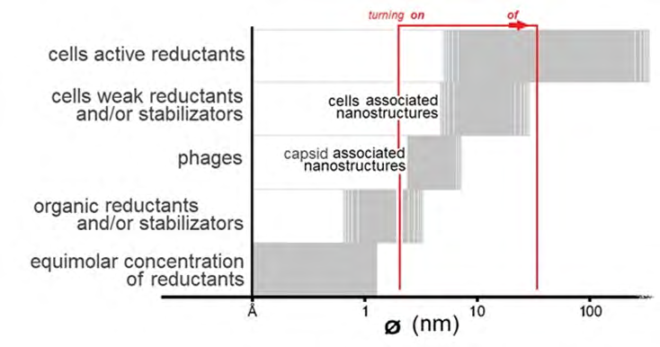
The ability of microorganisms to reduce metal cations (Men+) to a zero-valent state (Me0) with subsequent in situ formation of nanoparticles of zero-valent atoms (Me0NPs) is a well-documented fact [1, 2, 3, 4, 5, 6]. The formation of nanoparticles occurs in the presence of metabolically active cells as a result of contacts of cations with excreted biogenic compounds acting as reducing agents. This process also allows microorganisms to compensate for the negative effect on the cell of an excess amount of cations present in the environment. The reduced uncharged atoms start the process of their clustering by self-assembly, which leads to the very rapid formation of nanoclusters (Me0NCs) up to 1.5 nm in size. Such bioconversion of soluble toxic salts by reducing cations and precipitating zero-valent form to the insoluble non-toxic nanoparticles underlies the natural resistance of bacteria to metals [7, 8]. Under conditions of maintaining a high reductive activity of cells, self-assembly of nanoclusters continues and leads to the formation of larger and larger nanoparticles (Me0NPs) [9, 10, 11, 12]. Thus, the entire process of nanoparticle formation can be roughly divided into three stages (Table 1).
| Stages | Initial/final components | Transformation | Component size |
|---|---|---|---|
| I | Cations/Atoms | Ag+ → Ag° | 0,05 |
| II | Atoms/Nanoclusters | Ag° →Ag°NCs | up to 1-1,5 |
| III | Nanoclusters/Nanoparticles | Ag°NCs → Ag°NPs | 2 ~ 900 |
Table 1: Three stages of nanoparticle formation as a result of the reduction of metal cations on the example of silver.
Importantly, only the presence of metabolically active cells (capable of acting as donors of electrons and cation reducers) in the samples leads to the specific and rapid formation of biogenic nanoparticles. To assess the level of metabolic activity of cells, we developed a protocol called DBNG (Detection of Biogenic Nanoparticles Growth/ Generation), according to which the above parameter can be determined by the ability of the studied cell suspensions to form nanoparticles while adding a sterile salt solution with metal cations, for example, silver cations in the system. In the presence of active cells, de novo formation of biogenic nanoparticles that usually characterized by a specific size distribution occurs in 10–20 minutes (Figure 1). Concurrently, it is clearly proven that the formation of nanoparticles does not occur in sterile samples [12, 13, 14].
![Figure 1: Changes in the characteristic peak of silver nanoparticles formed in a suspension of cells M. smegmatis (in LB growth medium) after the addition of Tollens’ solution (Ag(NH3)2NO3) depending on the exposure time [15].](/fulltextimages/9384/fig_1.png)
The detected size distribution of nanoparticles makes it possible to characterize the ability of the studied microorganisms to act as cation reducers. In particular, it has been experimentally shown that the rate of nanoparticle generation correlates with the temperature range of microbial growth, and its maximum value corresponds to the optimum temperature for the microorganism [12, 16]. By summarizing the currently available data of studies on the biogenic formation of nanoparticles [4, 6, 17] and our own results on the size distribution of biogenic Ag°NPs obtained according to the protocol of the DBNG method, we developed the innovative algorithm for comparing the tested microorganisms in relation to their metabolic status. The comparison is based on two criteria: 1) The size of Ag°NPs, which should exceed 2 nm, indicating that it is nanoparticles that are generated, and not their precursors - nanoclusters; 2) The presence or absence of a pronounced peak in the size distribution of nanoparticles.
Based on the type of size distribution of nanoparticles, components of a biogenic or abiogenic nature affecting the dynamics of the NP formation can be additionally identified in the system. If the enlargement of nanoparticles slows down, it means that there are organic compounds in the medium that interact with nanoparticles inhibiting their growth and stabilizing their size [12, 13, 18, 19]. In general, the size distribution of nanoparticles depends on the level of metabolic activity of the cells as well as on the species of microorganism. An example is the different type of size distribution of Ag°NPs in all six microbial isolates from the same sample of stream water in the permafrost zone [16]. For one of these isolates (actinobacteria Serinibacter sp. PS306), a previously undescribed ability to massively form dimeric silver nanoparticles was shown. This phenomenon was proved to occur due to specific surface biopolymers of cells of this microorganism.
Testing of microbial activity according to the DBNG protocol can be performed directly in natural water samples. To study soil samples (with a predominance of a solid phase of various types), it is necessary to preliminarily carry out the extraction of microbial communities into the liquid phase. The inherent capacity of microbial cells to reduce cations allow for using different metals to form nanoparticles. According to the DBNG protocol, sterile solutions of silver salts, in particular, Tollens Ag(NH3)2NO3 solution, are most often used as the main model type of cations. Nevertheless, the use of several types of cations (salts of various metals) in parallel experiments can be quite informative, since different cells often exhibit specificity in relation to the reduction of certain types of cations allowing for a more detailed assessment of the properties of the studied microbial systems [3, 5, 8, 11].
Since the level of metabolic activity is determined precisely by the parameters of biogenic metal nanoparticles formed in situ over a fixed short time, measurements of size of nanoparticles should be made directly in the process of their self-assembly and enlargement. It is known that nanoparticles of many metals have the property of fluorescence at certain sizes, which in turn is associated with a certain ratio of their surface area and volume [18, 20]. This ability of nanoparticles to fluoresce makes it possible to evaluate the dynamics of their formation during the cation reduction reaction (Figure 2).
At the early stages of the formation and self-assembly of nanoclusters, some of the resulting nanoparticles reach a size sufficient for the onset of autofluorescence, which can be detected by spectrometry. As the reduction reaction continues, fluorescence persists in the test sample until all the nanoparticles present in the reaction mixture overcome the size corresponding to the cessation of fluorescence. This feature of the fluorescence of metal nanoparticles can be used for more accurate tracking of dynamics of their de novo formation, and hence makes fluorescence spectrometry a convenient tool in experiments to assess the metabolic activity in microbial systems [20].
The formation of biogenic nanoparticles in the medium inoculated with bacterial cultures occurs fast enough, typically within 10-20 minutes and is characterized by a peak at λ400-405 nm in the spectrophotometric study [1, 13, 21]. It is important to note that such duration of the NP formation does not exceed the cell doubling time for most microbial cultures, as well as the typical time of the intracellular metabolic response to external influences [7, 10]. In other words, the generation rate and features of the nanocrystalline particles reflect the actual physiological state of the cells at the time of the introduction of a sterile metal salt solution into the medium.
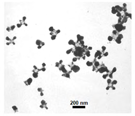
Unlike microbial cells, viral particles do not metabolize; however, the reduction of cations and the formation of metal nanoparticles are possible in their presence. Reducing agents in these cases are those amino acid residues of viral capsids that are capable of acting as electron donors. Since the reduction of cations occurs exclusively upon their contact with the surface of viruses, the clustering of reduced atoms leads to the relatively slow formation of small nanoparticles associated with capsid molecules [12, 22]. The expression “viral particles encrusted with metal nanoparticles” is widely used in the literature (Figure 3).
The sensitivity of the DBNG method in relation to living bacterial and viral particles, as established in preliminary studies (TEM), is approximately 10 cells x ml-1 [13]. Methodologically, studies of metabolic activity by the DBNG method can be carried out with an aqueous suspension of the test sample containing cells of various microorganisms or with cells of a pure culture. The process is initiated by adding a sterile solution of a salt of metal chosen for the experiment as a source of cations. The formation of nanoparticles can be detected by a large number of analytical methods, such as vis-spectroscopy, transmission electron microscopy (TEM), surface enhanced Raman scattering SERS, X-ray powder diffraction (XRD), energy dispersive X-ray spectroscopy (EDXS), dynamic light scattering (DLS), Zeta potential measurement (ZP) and others [1, 9, 22, 23, 24, 25, 26]. Importantly, the absence of nanoparticle formation should be confirmed in the control, free from cells for each experiment. The control is prepared mechanically (by filtration or centrifugation) in order to possibly preserve the initial chemical composition of the medium.
The developed DBNG method can be successfully used as a methodologically simple and low-cost alternative/ addition to classical microbiological methods and methods based on the determination of mRNA in the study of in situ metabolic activity of microorganisms. The DBNG method can be effectively applied in cases where the main task is to determine the actual activity of microorganisms or the metabolic potential of microbial communities in various ecosystems. It can also be used for the purpose of screening microorganisms of biotechnological importance. In general, the DBNG approach reduces the duration and complexity of the detection of metabolically active microorganisms and eliminates the need to use expensive preparations of functionalized nanoparticles. For the effective organization of such express in situ analysis, the necessary instrumentation has been developed, which implies the presence of a compact microchip, in which the reaction of reduction of cations (automatically added salt solution) takes place. The formation of nanoparticles is detected by a portable spectrophotometer.
On Figure 4 a generalized scheme of the protocol of the DBNG method and interpretation of the results obtained for various types of systems in which the biogenic formation of nanoparticles occurs are presented.
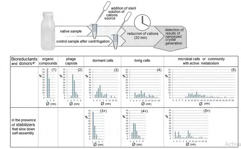
Figure 4: Scheme of the DBNG protocol and algorithm for interpreting the size distribution of generated biogenic nanoparticles for assessing the metabolic activity of microorganisms. Typical histograms of the size distribution of silver nanoparticles are shown in cases where the formation of nanoparticles is carried out: 1 - in a complete sterile medium, 2 - in a suspension of viral particles, 3 - in a suspension of resting bacterial cells, 4 - in a suspension of bacterial cells in an early logarithmic growth phase, 5 – in a suspension of an actively metabolizing microbial community. In variants 3+, 4+, and 5+ cells secrete stabilizers of nanoparticles that prevent their enlargement.
A wide variety of options for the size distribution of nanoparticles gives insights into determining the features of the studied biological objects.
Conclusion
The inherent ability of living cells to form nanoparticles by reducing metal cations can be used as the basis for the detection and study of metabolically active microorganisms in aqueous environments, including those containing various organics. The parameters of metal nanoparticles formed in the presence of microorganisms from metal cations added to the samples clearly reflect the physiological and biochemical state of the cells (high level of metabolic activity of microorganisms is associated with larger nanoparticles generated, and the size distribution of nanoparticles can provide additional information regarding the microbial system and chemistry environment). Viral particles can also be detected by the reducibility of capsid amino acid residues. However, in such cases, metal nanoparticles are generated slowly and their sizes rarely exceed 7–10 nm. Detection of de novo formed nanoparticles can be carried out by many analytical methods, which allows this innovative approach to be widely used in practice.
References
-
Angelikopoulos P, Sarkisov L, Cournia Z, Gkeka P (2017) Self-assembly of anionic, ligand-coated nanoparticles in lipid membranes. Nanoscale 9(3): 1040-1048.
-
Anh-Tuan L, Huy PT, Tam PD, Huy TQ, Cam PD, et al. (2010) Green synthesis of finely-dispersed highly bactericidal silver nanoparticles via modified Tollens technique. Cur Appl Physics 10(3): 910-916.
-
Buchmann B, Hecht FM, Pernpeintner C, Lohmueller T, Bausch AR (2017) Controlling non-equilibrium structure formation on the nanoscale. Chemphyschem 18(23): 3437-3442.
-
Gadd GM (2010) Metals, minerals and microbes: geomicrobiology and bioremediation. Microbiology (Reading) 156(3): 609-643.
-
Hilger A, Guppers N, Tenfelde M, Kreibig U (2000) Surface and interface effects in the optical properties of silver nanoparticles. Eur Phys J 10: 115-118.
-
Kumar D, Parashar A, Chandrasekaran N, Mukherjee A (2017) The stability and fate of synthesized zero-valent iron nanoparticles in freshwater microcosm system. Biotech 7(3): 227.
-
Le AT, Huy PT, Tam PD, Huy TQ, Cam PD, et al. (2010) Green synthesis of finely-dispersed highly bactericidal silver nanoparticles via modified Tollens technique. Curr Appl Phys 10(3): 910-916.
-
Li W, Zhang G, Liu L (2021) Near-infrared inorganic nanomaterials for precise diagnosis and therapy. Front Bioeng Biotechnol 9: 768927.
-
Lloyd JR, Yong P, Macaskie LE (1998) Enzymatic recovery of elemental palladium by using sulfate-reducing bacteria. Appl Environ Microbiol 64(11): 4607-4609.
-
Luo B, Smith JW, Ou Z, Chen Q (2017) Quantifying the self-assembly behavior of anisotropic nanoparticles using liquid-phase transmission electron microscopy. Acc Chem Res 50(5): 1125-1133.
-
Malis O, Byard C, Mott D, Wanjala BN, Loukrakpam R, et al. (2011) Low-temperature phase and morphology transformations in noble metal nanocatalysts. Nanotechnol 22(2): 1-8.
-
Mikhailov OV, Mikhailova EO (2019) Elemental silver nanoparticles: biosynthesis and bio applications. Materials (Basel) 12(19): 3177.
-
Muller M (2018) Bacterial silver resistance gained by cooperative interspecies redox behavior. Antimicrob Agents Chemother 62(8): e00672-e00718.
-
Gallon SMN, Alpaslan E, Wang M, Casanova PL, Londoño ME, et al. (2019) Characterization and study of the antibacterial mechanisms of silver nanoparticles prepared with microalgal exopolysaccharides. Mater Sci Eng C 99: 685-695.
-
Siddiqi KS, Husen A, Rao RAK (2018) A review on biosynthesis of silver nanoparticles and their biocidal properties. J Nanobiotechnol 16(1): 14.
-
Skladnev DA, Mulyukin AL, Filippova SN, Kulikov EE, Letarova MA, et al. (2016) Modeling of dissemination of microbial cells and phage from the sites of permafrost thawing. Microbiology 85(5): 614-619.
-
Skladnev DA, Vasilyeva LV, Berestovskaya YY, Kotsyurbenko OR, Kalenov SV, et al. (2020) Detection of microorganisms in low-temperature water environments by in situ generation of biogenic nanoparticles. Front Astron Space Sci 7: 59.
-
Sorokin VV, Skladnev DA, Volkov VV, Tereshchenko EY, Mulyukin AL, et al. (2013) The pathways of silver nanoparticles formation by Mycobacterium smegmatis. Dokl Biol Sci 452: 325-328.
-
Sorokin VV, Pshenichnikova AB, Kalenov SV, Suyasov NA, Skladnev DA (2020) Comparison of the wild-type obligate methylotrophic bacterium Methylophilus quaylei and its isogenic streptomycin-resistant mutant via metal nanoparticle generation. Biol Trace Elem Res 193(2): 564-573.
-
Tan SF, Chee SW, Lin G, Mirsaidov U (2017) Direct observation of interactions between nanoparticles and nanoparticle self-assembly in solution. Acc Chem Res 50(6): 1303-1312.
-
Wang L, Xu L, Kuang H, Xu C, Kotov NA (2012) Dynamic nanoparticles assemblies. Acc Chem Res 45(11): 1916- 1926.
-
Wang J, Lin X, Shu T, Su L, Liang F, et al. (2019) Self- assembly of metal nanoclusters for aggregation-induced emission. Int J Mol Sci 20(8): 1891.
-
Xie Y, Dong H, Zeng G, Tang L, Jiang Z, et al. (2017) The interactions between nanoscale zero-valent iron and microbes in the subsurface environment: A review. J Hazard Mater 321: 390-407.
-
Zhou Y, Wang H, Lin W, Lin L, Gao Y, et al. (2013) Quantitative nucleation and growth kinetics of gold nanoparticles via model-assisted dynamic spectroscopic approach. J Colloid Interf Sci 407: 8-16.
-
Jeevanandam J, Krishnan S, Hii YS, Pan S, Chan YS, et al. (2022) Synthesis approach dependent antiviral properties of silver nanoparticles and nanocomposites. J Nanostruct Chem 12: 809-831.
-
Yuan P, Ding X, Yang YY, Xu QH (2018) Metal nanoparticles for diagnosis and therapy of bacterial infection. Adv Healthc Mater 7(13): e1701392.
- Antifungal Activity of New Acetophenone Derivatives
- Interconnected Microbiomes Human Health Within an Environmental Framework
- Silkworm-Based Vaccine Production for H5N1: A One Health Approach to Pandemic Preparedness
- Microbial Diversity and Lipolytic Activity of Bacteria and Fungi from Oil-Contaminated Sites in Makurdi Metroplois
- Antibiotic Resistance Profile of Bacteria Isolated at the Central Laboratory of the National Hospital Center of Nouakchott
- Epidemiology and Sensitivity to Antibiotics of Germs Isolated from Blood Cultures in the Laboratory of the National Hospital Center of Nouakchott-Mauritania