Antibiotic Resistance and Detection of Blatem and MecA Genes in Bacteria Isolated from Street Vended Pounded Yam in Yenagoa, Nigeria
Antibiotic resistant pathogens spread through food are a public health concern. The aim of this study was to determine the profile of antibiotic resistance and to investigate the presence of genes that produce antibiotic resistance in bacterial isolates from pounded yam collected from five sites in Yenagoa, Nigeria (Swali, Amarata, Kpansia, Tombia, and Akenfa). The Kirby-Bauer disk diffusion method was used to examine antibiotic susceptibility to nine antibiotics (Augmentin, Ofloxacin, Chloramphenicol, Ampicillin, Erythromycin, Gentamycin, Nitrofuratoin, Streptomycin and Tetracyclin), and the PCR method was utilized to find the blaTEM and mecA genes. A total of 150 pounded yam samples were collected and analyzed. Shigella spp, Bacillus spp, Staphylococcus spp, Pseudomonas spp, Salmonella spp, Klebsiella spp, Proteus spp, and Escherichia coli are among the bacteria that were recovered from the pounded yam samples. Except for Pseudomonas spp., all tested positive for multidrug resistance (resistance to three or more tested antibiotics), with the majority of these antibiotics being Chloramphenicol, Ampicillin, Erythromycin, Gentamycin, Nitrofuratoin, Streptomycin, and Tetracyclin. The Escherichia coli isolated from the street-vended pounded yam exhibited multidrug resistance against Ampicillin (37.5%), Erythromycin (43.8), Gentamycin (34.4), Streptomycin (21.9) and Tetracyclin (40.6). Bacillus spp also showed multidrug resistance against Chloramphenicol (36.4), Ammpicillin (36.4), Erythromycin (45.5), Gentamycin (36.4), Nitrofuratoin (36.4), Streptomycin (27.3) and Tetracyclin (36.4). Inhibition zones against Augmentin, Ofloxacin, Chloramphenicol, Ampicillin, Erythromycin, Gentamycin, Nitrofuratoin, Streptomycin, and Tetracyclin were clearly visible in Pseudomonas spp (100, 96.4, 64.3, 50.0, 82.1, 75.0, 64.3, 92.9, and 50.0% respectively). The highest resistance by number of bacterial isolates was found in Erythromycin (6 isolates showed resistance), followed by Tetracyclin (5 isolates showed resistance). Ampicillin and Streptomycin resistance was present in four isolates. Three representative isolates were selected for molecular identification of blaTEM and mecA which were amplified at 600bp and 500bp respectively. These genes were responsible for the antibiotic resistance seen in the isolates. According to this study, the pounded yam samples that were evaluated had food-borne disease strains that are multidrug resistant and a danger to the general public's health. The findings cast doubt on the quality of foods sold on the streets of Yenagoa, Nigeria.
Introduction
One of the most fundamental human necessities for sustenance and the continuation of life is food [1]. The people of Yenagoa, Nigeria, love the cuisine sold on the streets. This appeal is attributable to the food’s accessibility, and affordability. These food are usually prepared under poor hygienic standards [2], as such food-borne diseases like typhoid fever, cholera, and diarrhea among others have been reported Jahan, et al. [2, 3]. Food-borne diseases are most common in Asia, and sub-saharan Africa [1]. In Nigeria, food- borne pathogens like Escherichia coli, 0157:H7, Salmonella, Vibrio, Shigella, Clostridium, and Staphylococcus aureus have been isolated from commercially vended street foods [4, 5]. Treatment for these food-borne illnesses typically involves the use of natural or synthetic antimicrobials. However, it has been determined that certain antibiotic resistant genes, such as blaTEM, blaCTX-M, blaSHV, mecA, and mecB genes, cause these organisms to become resistant to some of the routinely used antimicrobial drugs [6, 7]. Antibiotic resistance is the ability of a pathogenic organism to develop when exposed to an antibiotic it was initially susceptible to Olorunfemi, et al. [8]. Antibiotic-resistant bacteria are now a danger to the success of illness treatment all around the world. Each year, approximately 700,000 patients suffer from infections brought on by antibiotic-resistant microorganisms, and by the year 2050, it is predicted the number will increase to approximately 10,000,000 [9, 10]. The validity of this hypothesis is supported by the observation that organisms acquire antibiotic resistance more quickly than new drugs are created.
Since antibiotic resistance is a global problem, identifying the gene responsible for the resistance is crucial towards finding a lasting solution to the problem. Besides, additional knowledge is needed to comprehend the danger of ingesting antibiotic-resistant bacteria through foods sold on the street, such as pounded yam. In light of this, the goal of this study is to identify the blaTEM and mecA genes responsible for antibiotic resistance in bacteria isolated from pounded yam sold on the streets of Yenagoa, Nigeria.
Materials and Methods
Sampling and Isolation of Bacteria
Food vending sites in Swali, Amarata, Kpansia, Tombia and Akenfa were used. About 150 samples of prepared pounded yam were randomly collected, sealed and taken for examination. Each pounded yam sample weighed twenty- five (25) grams, and the suspension was made by stirring it in 225 mL of buffered peptone water. The formed suspension was used to prepare a stock sample, from which 10-fold serial dilutions were obtained. A 0.1 mL of it was transferred into a sterile Nutrient agar, Eosin Methylene Blue agar, Mannitol Salt agar, and MacConkey agar, using pour plate method and incubated at 370C for 24 hours [11, 12]. The inoculation plates were examined for unique colonies, growth qualities, and other colonial aspects including colour and shape, which were documented, after 24-48 hours of incubation. According to Cheesbrough [13], a specific colony was chosen for each growth and repeatedly subcultured onto fresh Nutrient agar to obtain pure cultures from which bacteria were identified. The bacterial isolates also underwent Gram staining and biochemical assays such as oxidase, catalase, coagulase, citrate, urease, and indole.
Oxidase Test
Three (3) drops of freshly made oxidase reagent (tetra- methyl-p-phenylenediamine dihydrochloride) were put to a piece of filter paper in a sterile Petri dish. A colony of the test organism was smeared on the wet filter paper using a wire loop. For oxidase positive bacteria, a purple color appeared after 5–10 seconds, however oxidase negative bacteria did not exhibit a color change.
Catalase Test
Two drops of hydrogen peroxide were added to a dense culture, and the mixture was then checked for bubbles. The existence of the catalase enzyme was demonstrated by the appearance of bubbles. Staphylococci were regarded to be the organisms that tested positive. A coagulase test was also performed on the Staphylococcus species.
Coagulase Test
A dense suspension of the culture and human plasma were combined for this test, which was then incubated for 24 hours at 35°C before being checked for clotting. Staphylococcus aureus was found to be present in those who tested positive for clotting in the coagulase test.
Citrate Test
In this experiment, Simmon’s citrate agar was slanted in a tube and the innoculum was streaked over it. A positive outcome was indicated by growth on the slant and a shift in the medium’s color from green to blue. When there is no change in color, the outcome is negative.
Urease Test
About 2.95g of urea powder were dissolved in 150ml of distilled water to make urea broth. To stop initial urea degradation, urea was added to the media after autoclaving in a pressure cooker-style autoclave. After that, an aseptic inoculating loop was used to inject the sample. The tubes were incubated for 24 hours at 37°C. Positive results were indicated by a switch from yellow-orange to brilliant pink, whereas negative results were indicated by no color change.
Indole Test
The indole test was carried out by culturing the test organism in peptone water for an entire night. Kovac’s reagent was added, and the color of the solution was checked after 0.5 ml. Positive results were denoted by a red ring at the top.
Antibiotic Susceptibility Test
The Kirby-Bauer disk diffusion method was the method used for the antibacterial susceptibility testing of the bacterial isolates. The antibiotics discs and the concentration used were augmentin 30 μg, Oflaxacin 5 μg, Chloramphenicol 30 μg, ampicillin 25 μg, Erythromycin 5 μg, Gentamycin 25 μg, Nitofurantoin 200 μg, Streptomycin 25 μg and Tetracycline 25 μg. According to the definition provided by the Clinical and Laboratory Standard Institute [14] and in compliance with WHO guidelines, zones of inhibition were measured in millimeters, and isolates were categorized as resistant, intermediate, or sensitive [15]. As quality control strains for the antimicrobial discs, some laboratory strains with established sensitivity, such as Escherichia coli, and Staphylococcus aureus, were employed [12, 15, 16]. In this investigation, intermediate and resistant isolates were combined for examination. If an isolate exhibited resistance to at least three of the tested antibiotics, it was deemed to be multi-drug resistant [12, 16].
Detection of BlaTEM and MecA Genes
The DNA of the bacterial isolates was extracted using Proteinase K DNA extraction protocol. The extracted DNA served as a template for PCR amplification using blaTEM, and mecA specific primers (New England BioLabs, United Kingdom). The PCR mix consisted of 12.5µL of Taq 2X Master Mix; 1µL each of forward and reverse primers; 2µL of DNA template (extracted bacterial DNA) and then made up with 8.5µL nuclease free water. Initial denaturation at 94°C for 5 minutes was followed by 36 cycles of denaturation at 94°C for 30 seconds in order to identify the blaTEM gene. Elongation took place for 45 seconds at 72ºC after 30 seconds of annealing at 55ºC. After that, the temperature was maintained at 10 °C for a final elongation stage lasting 7 minutes at 72 °C. For mecA gene, initial denaturation was done at 94°C for 5 minutes, followed by 30 cycles of denaturation at 94°C for 5 minutes. Elongation took place for 45 seconds at 72ºC after 1 minute of annealing at 56ºC. A last elongation step at 72ºC for 3 minutes while maintaining temperature at 10ºC came next. The amplified product was subjected to gel electrophoresis (1% gel stained with ethidium bromide) at 80 V for 1 hour. DNA ladder (1500bp) was used to estimate the molecular weight of the amplified products. For photo documentation following electrophoresis, the doc system was employed [17]. E. coli was used as positive control while Bacillus sp was used as a negative control during PCR.
Results
The morphological and biochemical characterization of the isolated bacteria shown in Table 1 revealed them to be Shigella spp, Bacillus spp, Salmonella spp, and Proteus spp. Escherichia coli was recognized by their distinctive green metallic shine. A number of colonies appeared which were collectively grouped as Staphylococcus spp from their cultural traits (Table 1).
| Colonial Morpho | GR | Oxi | Cat | Cit | Ure | Coa | Ind | Bacteria |
|---|---|---|---|---|---|---|---|---|
| Pale Rod | - | - | - | - | - | - | - | Shigella spp |
| Fuzzy white Rod | + | + | + | + | - | + | - | Bacillus spp |
| Cream Cocci | + | - | + | + | - | + | - | Staphylococcus spp |
| Greenish blue Rod | - | + | + | + | - | - | - | Pseudomonas spp |
| Green metallic sheen Rod | - | - | + | - | - | - | + | Escherichia coli |
| Greyish white Rod | - | - | + | - | - | - | - | Salmonella spp |
| Mucoid pink Rod | - | - | + | + | + | - | - | Klebsiella spp |
| Pale Rod | - | - | + | + | + | - | - | Proteus spp |
Table 1: Morphological and Biochemical Characteristics of the Bacterial Isolates. Key: Morpho=Morphology; GR=Grams reaction; Oxi=
The greenish blue and mucoid pink colours of some colonies among other characteristics pointed to Pseudomonas and Klebsiella spp respectively. From the percentage susceptibility of the bacterial isolates to the tested antibiotics (Table 2), it was revealed that all the bacterial isolates were susceptible to Augmentin and Ofloxacin. Staphylococcus spp, Pseudomonas spp, Escherichia coli, Salmonella spp and Klebsiella spp showed 100% susceptibility to Augmentin while Staphylococcus spp, Salmonella spp, Klebsiella spp and Proteus spp showed same 100% susceptibility to Ofloxacin. This makes them the most active antibiotics in this study.
Among all the tested antibiotics, Erythromycin and Tetracyclin had the least effect on the bacterial isolates (Table 2). Bacillus spp, Staphylococcus spp, Escherichia coli, Salmonella spp, Klebsiella spp and Proteus spp showed
45.5, 37.2, 43.8, 41.2, 44.4, and 36.4% resistance to Erythromycin respectively. For Tetracyclin, the resistance shown by Bacillus spp, Staphylococcus spp, Escherichia coli, Salmonella spp and Klebsiella spp was 36.4, 44.2, 40.6, 47.7, and 33.3% respectively. From the antibiotic resistance patterns presented in Table 3, it was revealed that all the bacterial isolates, with the exception of Pseudomonas spp, showed resistance to at least three of the tested antibiotics and as such, was considered multidrug resistant. Bacillus spp was resistant to seven of the nine tested antibiotics (Chloramphenicol, Ampicillin, Erythromycin, Gentamycin, Nitrofuratoin, Streptomycin and Tetracyclin). Pseudomonas spp on the other hand, showed no resistance to any of the tested antibiotics, thus; was the most susceptible in this study.
| Tested Antibiotics (%) | |||||||||
|---|---|---|---|---|---|---|---|---|---|
| Isolates | Aug | Ofl | Chl | Amp | Ery | Gen | Nit | Str | Tet |
| Shigella spp (N=7) | 6(85.7) | 6(85.7) | 3(42.9) | 3(42.9) | 4(57.1) | 6(85.7) | 3(42.9) | 4(57.1) | 4(57.1) |
| Bacillus spp (N=11) | 8(72.7) | 9(81.8) | 4(36.4) | 4(36.4) | 5(45.5) | 4(36.4) | 4(36.4) | 3(27.3) | 4(36.4) |
| Staphylococcus spp (N=43) | 43(100) | 43(100) | 40(93.0) | 31(72.1) | 16(37.2) | 33(76.7) | 22(51.2) | 15(34.9) | 19(44.2) |
| Pseudomonas spp (N=28) | 28(100) | 27(96.4) | 18(64.3) | 14(50.0) | 23(82.1) | 21(75.0) | 18(64.3) | 26(92.9) | 14(50.0) |
| Escherichiacoli (N=32) | 32(100) | 31(96.9) | 28(87.5) | 12(37.5) | 14(43.8) | 11(34.4) | 20(62.5) | 7(21.9) | 13(40.6) |
| Salmonellaspp (N=17) | 17(100) | 17(100) | 9(52.9) | 11(64.7) | 7(41.2) | 9(52.9) | 13(76.5) | 7(41.2) | 8(47.7) |
| Klebsiella spp (9) | 9(100) | 9(100) | 7(77.8) | 4(44.4) | 4(44.4) | 7(77.8) | 5(55.6) | 8(88.9) | 3(33.3) |
| Proteus spp (N=22) | 19(86.4) | 22(100) | 9(40.9) | 11(50) | 8(36.4) | 19(86.4) | 10(45.5) | 11(50) | 14(63.6) |
Table 2: Percentage Susceptibility of the Bacterial Isolates to Antibiotics. Key: N=Number of samples; Aug=Augmentin; Ofl=Ofloxac
| Tested Antibiotics | ||||||||||
|---|---|---|---|---|---|---|---|---|---|---|
| Resistance Patterns | Aug | Ofl | Chl | Amp | Ery | Gen | Nit | Str | Tet | Multidrug resistance |
| Shigella spp | - | - | + | + | - | - | + | - | - | Yes |
| Bacillus spp | - | - | + | + | + | + | + | + | + | Yes |
| Staphylococcus spp | - | - | - | - | + | - | - | + | + | Yes |
| Pseudomonas spp | - | - | - | - | - | - | - | - | - | No |
| Escherichia coli | - | - | - | + | + | + | - | + | + | Yes |
| Salmonella spp | - | - | - | - | + | - | - | + | + | Yes |
| Klebsiella spp | - | - | - | + | + | - | - | - | + | Yes |
| Proteus spp | - | - | + | - | + | - | + | - | - | Yes |
Table 3: Antibiotic Resistance Patterns and Multidrug Resistance of the Bacterial Isolates. Key: Aug=Augmentin; Ofl=Ofloxacin; Ch
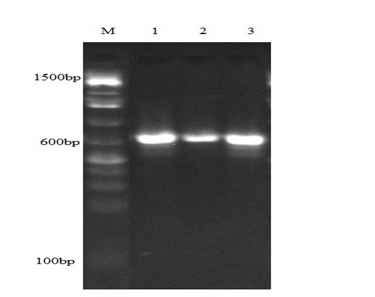
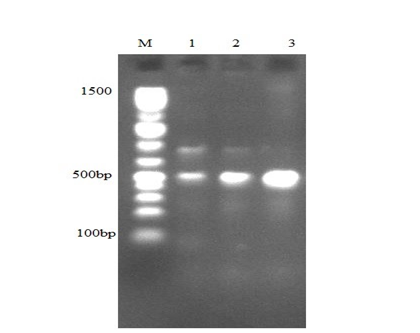
Table 3: Antibiotic Resistance Patterns and Multidrug Resistance of the Bacterial Isolates. Key: Aug=Augmentin; Ofl=Ofloxacin; Chl=Chloramphenicol; Amp=Ampicillin; Ery=Erythromycin; Gen=Gentamycin; Nit=Nitrofuratoin; Str= Streptomycin; Tet=Tetracyclin; + = Resistant; - = Susceptible Figure 1 showed blaTEM gene amplification at 600bp. Lane M contained the 1500bp molecular weight ladder while Lane 1 contained Staphylococcus spp sample 1. Lane 2 and 3 contained Staphylococcus spp sample 2 and Escherichia coli sample respectively. The primer generated the amplicon at 600bp. Figure 2 showed mecA gene amplification at 500bp. Lane M contained the 1500bp molecular weight ladder. Lane 1, 2 and 3 contained Staphylococcus spp sample 1, Staphylococcus spp sample 2 and Escherichia coli sample respectively. The primer generated the amplicon at 500bp.
Discussion
The pounded yam sold on the street in Yenagoa, Nigeria doesn’t need to be heated up before eating, hence its high risk. The pounded yam may act as a favorable substrate for the proliferation of bacteria, leading to food- borne disease outbreaks. Improper handling may also result in contaminations. The pounded yam sold by street vendors in Yenagoa, Nigeria, was found to have eight (8) different genera of bacteria, including Shigella spp., Bacillus spp., Staphylococcus spp., Pseudomonas spp., Salmonella spp., Klebsiella spp., and Proteus spp. Escherichia coli was confirmed by its characteristic green metallic sheen appearance. The street food vendors’ unsanitary food handling procedures may be responsible for the faecal contamination evident in the presence of Escherichia coli [18]. The antibacterial susceptibility of the bacterial isolates to the used antibiotics demonstrates widespread bacterial antibiotic resistance. For instance, every isolate described above, with the exception of Pseudomonas spp., was multidrug resistant, meaning that they were resistant to more than three different antibiotics (Table 3). Resistance to Erythromycin (6 isolates), Tetracyclin (5 isolates), Ampicillin (4 isolates), and Streptomycin (4 isolates) were the most common in our investigation, which is a deviation from the findings of Guo, et al. [19] that reported Ampicillin, Chloramphenicol and Tetracyclin as the most common. Bacillus spp was resistant to Chloramphenicol, Ampicillin, Erythromycin, Gentamycin, Nitrofuratoin, Streptomycin and Tetracyclin while Escherichia coli were resistant to Chloramphenicol, Ampicillin, Gentamycin, Streptomycin, and Tetracyclin. In contrast to our result where Escherichia coli recorded 34.4% resistance to Gentamycin, Kumar, et al. [20], Subedi, et al. [21], and Rimal, et al. [22] all reported 73.9% susceptibility to same antibiotic. The inappropriate use of third-generation cephalosporins may have contributed to this increase in resistance [23]. In contrast to Chakraborty, et al. [24] who listed Gentamycin as the most potent antibiotic, Augmentin and Ofloxacin were shown to be more effective in our investigation. Kabiru, et al. [25] discovered various food- borne infections to have a moderate resistance to antibiotics such Amoxicillin, Septrin, and Nitrofuratoin. Similar to our investigation, the isolates of Zhang, et al. [26] also show high resistance to Ampicillin, Trimethoprim, and Tetracyclin. It’s possible that excessive use, misuse, and abuse of antibiotics contributed to the general development of antibiotic resistance in these microorganisms.
The amplified blaTEM and mecA genes found in Escherichia coli and Staphylococcus spp. in this study are two examples of antibiotic resistant genes that are responsible for the high antibiotic resistance in these bacterial isolates (Figure 1 and Figure 2). Similar genes were found in Adefisoye and Okoh’s [27] investigation. According to Baraniak [28], these genes are an essential component of pathogen virulence because they let them adapt to various environments and increase their resistance to β-lactam antibiotics. Escherichia coli’s remarkable resistance to Ampicillin (37.5% resistance) is ascribed to the blaTEM gene [29]. Brinas, et al. [30] also reported that blaTEM genes are common in Ampicillin resistant Escherichia coli strains. The mecA gene is a common methicillin resistance gene found in Staphylococcus spp. It confers resistance to β -lactam antibiotics. According to Lerminiaux, et al. [10], horizontal gene transfer can be used to spread this gene among Staphylococcus strains, which may have happened with Staphylococcus samples 1 and 2 of this study. Escherichia coli samples also had the mecA gene. MecA was also found in Escherichia coli, Pseudomonas spp, Enterococcus spp, Proteus spp, Morgonella spp, and Salmonella spp, according to Hsu, et al. [31] and Seyedmonir, et al. [32]. Other isolates like Pseudomonas spp, Proteus spp, and Salmonella spp may have acquired the gene through horizontal gene transfer from either Escherichia coli or Staphylococcus spp, rendering them resistant to the tested antibiotics.
Conclusion
Even though our study only used eight bacterial isolates, the findings point to the presence of antibiotic-resistant pathogens in pounded yam sold on the streets of Yenagoa, Nigeria. The wide spread resistance of these food-borne pathogens is of extreme public health concern. This is because, as this study’s findings indicate, the majority of bacteria are resistant to commonly accessible antibiotics, making it challenging to control any disease epidemic caused by these pathogens. The report underlines the necessity of routinely training street food vendors and creating uniform food safety standards.
Conflicts of Interest
The authors declare no conflicting interest.
Acknowledgement
The laboratory team at the Central Research and Diagnostic Laboratory, Tanke, Ilorin, is highly acknowledged by the authors for their technical assistance.
References
-
WHO (2008) Foodborne disease outbreaks: guidelines for investigation and control. World Health Organization.
-
Jahan M, Rahman M, Rahman M, Sikder T, Lopez RAU, et al. (2018) Microbiological safety of street-vended foods in Bangladesh. J Consum Prot Food Saf 13: 257-269.
-
Anderson PH, Stone DM (1955) Staphylococcal food poisoning associated with spray-dried milk. J Hyg 53(4): 387-397.
-
Igbinosa EO, Beshiru A, Igbinosa IH, Ogofure AG, Uwhuba KE, et al. (2021) Prevalence and characterization of food- borne Vibrio parahaemolyticus from African salad in Southern Nigeria. Front Microbiol 12: 632266.
-
Beshiru A, Okareh OT, Okoh AI, Igbinosa EO (2020) Detection of antibiotic-resistant and putative virulence determinant genes of Vibrio strains isolated from ready- to-eat (RTE) shrimps in Delta and Edo States, Nigeria. J Appl Microbiol 129(1): 17-36.
-
Caniça M, Manageiro V, Abriouel H, Gilad JM, Franz CM, et al. (2019) Antibiotic resistance in foodborne bacteria. Trends in Food Science and Technology 84: 41- 44.
-
Thapa SP, Shrestha S, Anal AK (2020) Addressing the antibiotic resistance and improving the food safety in food supply chain (farm-to-fork) in Southeast Asia. Food Control 108: 106809.
-
Olorunfemi PO, Ngwuluka NC, Onaolapo JA, Ibrahim YK (2021) Susceptibility and molecular characterization of mec A-and mec B-positive community acquired methicillin-resistant Staphylococcus aureus isolates from students. Journal of Pharmacy and Bioresources 18(2): 155-171.
-
Cerqueira F, Matamoros V, Bayona JM, Berendonk TU, Elsinga G, et al. (2019) Antibiotic resistance gene distribution in agricultural fields and crops. A soil-to- food analysis. Environ Res 177: 108608.
-
Lerminiaux NA, Cameron ADS (2019) Horizontal transfer of antibiotic resistance genes in clinical environments. Can J Microbiol 65(1): 34-44.
-
Feglo P, Sakyi K (2012) Bacterial contamination of street vending food in Kumasi, Ghana. Journal of Medical and Biomedical Science 1(1): 1-8.
-
Womboh SB, Ajumobi VE, Ebute PA (2022) Assessments of contamination and susceptibility pattern of bacteria isolated from pounded yam sold along major roads in Makurdi metropolis, Benue State, Nigeria. UJMR Journal of Microbiology Research 7(2).
-
Cheesbrough M (2006) District laboratory practice in tropical countries. 2nd (Edn.), Cambridge University Press Publication, South Africa, pp: 1-434.
-
CLSI (2017) Performance standards for antimicrobial susceptibility testing. Clinical and Laboratory Standards Institute M100.
-
Onanuga A, Oyi AR, Olayinka BO, Onaolapo JA (2005) Prevalence of community-associated multi-resistant Staphylococcus aureus among healthy women in Abuja, Nigeria. African Journal of Biotechnology 4(9).
-
Santos TM, Caixeta LS, Machado VS, Rauf AK, Gilbert RO, et al. (2010) Antimicrobial resistance and presence of virulence factor genes in Arcanobacterium pyogenes isolated from the uterus of postpartum dairy cows. Vet Microbiol 145(1-2): 84-89.
-
Kaur M, Aggarwal A (2013) Occurrence of the CTX-M, SHV and the TEM genes among the extended spectrum β-lactamase producing isolates of Enterobacteriaceae in a tertiary care hospital of North India. J Clin Diagn Res 7(4): 642-645.
-
Giri S, Kudva V, Shetty K, Shetty V (2021) Prevalence and characterization of extended-spectrum β-lactamase- producing antibiotic-resistant Escherichia coli and Klebsiella pneumoniae in ready-to-eat street foods. Antibiotics 10(7): 850.
-
Guo S, Tay MYF, Aung KT, Seow KLG, Ng LC, et al. (2019) Phenotypic and genotypic characterization of antimicrobial resistant Escherichia coli isolated from ready-to-eat food in Singapore using disk diffusion, broth microdilution and whole genome sequencing methods. Food Cont 99: 89-97.
-
Kumar D, Singh AK, Ali MR, Chander Y (2014) Antimicrobial susceptibility profile of extended spectrum β-lactamase (ESBL) producing Escherichia coli from various clinical samples. Infect Dis 7: 1-8.
-
Subedi S, Chaudhary M, Shrestha B (2016) High MDR and ESBL Producing Escherichia coli and Klesbiella pneumoniae from Urine, Pus and Sputum Samples. Br J Med Med Res 13(10): 1-10.
-
Rimal U, Thapa S, Maharjan R (2017) Prevalence of extended spectrum beta-lactamase producing Escherichia coli and Klebsiella species from Urinary Specimens of Children attending Friendship International Children’s Hospital. Nepal Journal of Biotechnology 5(1): 32-38.
-
Shobha KL, Gowrish RS, Sugandhi R, Sreeja CK (2007) Prevalence of extended spectrum beta lactamases in urinary isolates of Escherichia, Klebsiella and Citrobacter species and their antimicrobial susceptibility pattern in tertiary care hospital. Ind J Pract Doct 3: 1-2.
-
Chakraborty S, Mohsina K, Sarker PK, Alam MZ, Karim MIA, et al. (2016) Prevalence, antibiotic susceptibility profiles and ESBL production in Klebsiella pneumonia and Klebsiella oxytoca among hospitalized patients. Periodicum biologorum 118(1).
-
Kabiru OA, Muibat OF, Nene H, Esther A (2013) Vended foods in Lagos, Nigeria: A potential reservoir for the spread of emerging strains of drug resistant bacteria. Journal of Health, pp: 675-680.
-
Zhang S, Wu Q, Zhang J, Lai Z, Zhu X, et al. (2016) Prevalence, genetic diversity, and antibiotic resistance of enterotoxigenic Escherichia coli in retail ready-to-eat foods in China. Food Control 68: 236-243.
-
Adefisoye MA, Okoh AI (2016) Identification and antimicrobial resistance prevalence of pathogenic Escherichia coli strains from treated waste water effluents in Eastern Cape, South Africa. Microbiologyopen 5(1): 143-151.
-
Baraniak A (2010) Molecular epidemiology and evolution of Enterobacteriaceae strains producing extended spectrum blactamases (ESBL) in Poland.
-
Ahmed MO, Clegg PD, Williams NJ, Baptiste KE, Bennett M, et al. (2010) Antimicrobial resistance in equine faecal Escherichia coli isolates from North West England. Ann Clin Microbiol Antimicrob 9: 12.
-
Briñas L, Zarazaga M, Sáenz Y, Larrea FR, Torres C, et al. (2002) β-Lactamases in ampicillin-resistant Escherichia coli isolates from foods, humans, and healthy animals. Antimicrob Agents Chemother 46(10): 3156- 3163.
-
Hsu CY, Hsu BM, Ji WT, Chen JS, Hsu TK, et al. (2015) Antibiotic resistance pattern and gene expression of non-typhoid Salmonella in riversheds. Environ Sci Pollut Res Int 22(10): 7843-7850.
-
Seyedmonir E, Yilmaz F, Icgen B (2016) Methicillin- Resistant Bacteria Inhabiting Surface Waters Monitored by mec A-Targeted Oligonucleotide Probes. Bull Environ Contam Toxicol 97(2): 261-71.
- Antifungal Activity of New Acetophenone Derivatives
- Interconnected Microbiomes Human Health Within an Environmental Framework
- Silkworm-Based Vaccine Production for H5N1: A One Health Approach to Pandemic Preparedness
- Microbial Diversity and Lipolytic Activity of Bacteria and Fungi from Oil-Contaminated Sites in Makurdi Metroplois
- Antibiotic Resistance Profile of Bacteria Isolated at the Central Laboratory of the National Hospital Center of Nouakchott
- Epidemiology and Sensitivity to Antibiotics of Germs Isolated from Blood Cultures in the Laboratory of the National Hospital Center of Nouakchott-Mauritania