A Case Report of Coenurus Cerebralis in a Goat at Dembecha District of Amhara Region, Ethiopia
This outbreak case with an unidentified etiological agent on caprine mortality was reported from Amhara Regional State, West Gojam zone, Dembecha district in February 2023 with the objective of investigating and determining the cause of the caprine death. During a clinical examination, the goat displayed seizures, lateral recumbency, and appetite loss. Additionally, paddling, convulsions, and unconsciousness were observed, which signs of a central nervous system disease. The case for coenurosis cerebralis has been established based on the clinical signs and the presence of certain hosts in the area. The brain was examined postmortem in order to confirm the diagnosis further. Cysts were found during necropsy in the left hemisphere's occipital lobe. The cysts were filled with a clear fluid, and the inner layer of the cysts had several clusters of scolice growing out of it. Larval form of Taenia multiceps, known as Coenurus cerebralis, is found in the small intestines of dogs and other carnivores in the wild. Worldwide, coenurosis is endemic, and it is particularly prevalent in Ethiopia's highlands, where there are many sheep. Commonly occurs in the life cycle of dogs and small ruminants. The larvae of this parasite are found in the brain and spinal cord of intermediate hosts, while the adult stage of the parasite lives in the small intestine of dogs, foxes, coyotes, and jackals. The definitive host becomes infected when it eats the brain or tissue become infected when the dog eats the brain containing the Coenurus cyst, which then develops into Taenia multiceps and begins to pass proglottids containing eggs on pasture. If a person accidentally consumes a parasite egg, they become infected with coenurosis. The primary method of controlling coenurosis is the regular administration of canine anthelmintics and the proper disposal of sheep and goat brain.
Introduction
Coenurosis cerebralis is important disease that affects sheep and goats and results in significant economic loss in their production [1]. Taenia multiceps are a taeniid cestode that inhabits the small intestines of dogs and other wild carnivores. The larval stage, Coenurus cerebralis, is typically found in the central nervous system. Gid, also known as sturdy, is a disease that primarily affects the central nervous systems (CNS) of sheep and goats and, to a lesser extent, cattle, buffalo, camels, pigs, deer, horses, yaks, and wild sheep, as well as humans [2]. In addition to being a serious health concern for sheep and goats everywhere, this disease could have important economic repercussions. The location, size, and extent of the cysts as well as the degree of brain compression all affect the symptoms [3].
When the cyst in Coenurus cerebralis grows, it causes CNS disorders that may be fatal. Coenurus cerebralis causes purulent meningoencephalitis [4]. Animals that have been infected display ataxia, blindness, circling, convulsions, teeth grinding, head tilting, lack of coordination, uncontrolled movements, salivation, paresis, and cerebral atrophy. Typically, 2–8 months after ingesting the pathogen, the majority of the clinical symptoms are seen [5]. Aside from the peritoneal and pelvic cavities, liver, intramuscular, tongue, parotid, lung, perineal fat, tunica adventitia of the aorta, and lungs, extracranial locations in goats have also been reported to harbor Coenurus cysts [6].
Radiology, ultrasonography, and computed tomography are imaging techniques that are rarely used to diagnose coenurosis instead of a clinical examination. Necropsy results are typically used to confirm the diagnosis [7].
Objective
The main goal of the investigation was to determine what caused an outbreak of caprine deaths and to gather samples and other data in the outbreak area.
Outbreak Areas
Dembecha is one of the words as in the Amhara Region’s West Gojjam Zone. Bure is to its west, Jabi Tehnan is to its northwest, Dega Damot is to its north, and the Misraq Gojjam Zone is to its east and south. The town is located 352 kilometers from Addis Abeba at latitude 10°33′N and longitude 37°29′E, with an elevation of 2083 meters above sea level. The outbreak took place on a private farm owned by Zangar Agricultural Development that is situated in lowland close to the Abay River in the Dembecha district’s Makar lega bedessa kebele. Farm of Zangar Agricultural Development is located 70 kilometers from woreda town.
Case Description
The owner of Zangar Agricultural Development was the first report the disease’s incidence to Animal Health Institute (AHI). The team of experts from institute traveled to Dembecha district Makar lega bedessa kebele to look into the origin of the outbreaks. Interviews with animal attendants and the farm manager for the Zangar Agricultural Development were done to find out the size and timing of the outbreak.
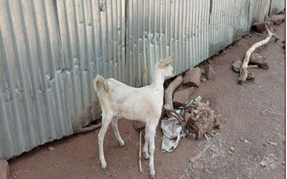
In the last year, the outbreak has resulted in the deaths the of 20 goats, the farm owner reported. The information provided by the animal attendant showed depression, circling to the left, altered head position, uncoordinated movement (ataxia), paralysis, inability to browse properly, and death within a week. All age groups were affected, but the young were more affected than the older generation. The team tentatively defined for coenurosis cerebralis and the confirmatory diagnosis for the disease was recommended by necropsy finding of the cyst. During a clinical examination, symptoms of a CNS disorder including paddling, convulsions, and unconsciousness were noted. Conjunctival hyperemia and keratitis were visible, and convulsion episodes were accompanied by opisthothonous, orthothonous, paddling, and oral foaming every 4-5 minutes. After the animal finally passed away from severe symptoms, a necropsy was carried out. We discussed a case of coenurosis in goats in this report, which was characterized by depression, circling to the left, altered head position, uncoordinated movement (ataxia), paralysis, inability to browse properly, and death within a week (Figure 1).
Results
Following the fieldwork, insightful conversations on the outbreak’s cause were had with every staff. Finally, the team came up with a case definition for coenurosis cerebralis, and after performing a necropsy, the disease was definitively diagnosed. Goats with coenurosis cerebralis cysts were found to have them in their brains (Figure 2A and 2B).
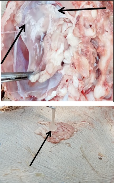
Figure 2A: A cyst settled in the left cerebral hemisphere.
Figure 2B: A cyst after protruded from the brain.
The necropsy finding of the cysts confirmed the case at the field level. Cysts were found during necropsy in the occipital lobe of the left cerebral hemisphere of the brain. The cysts had a translucent fluid inside of them, and the inner layer of the cysts had numerous scolices growing out of it. The cysts’ inner layer was distinguished by a thin transparent wall, while the outer layer had a thick fibrotic capsule.
Discussion
Globally, coenurosis is a serious issue with sheep and goats. According to the attendant’s information, villages where dogs and domestic animals coexist closely with domestic animals are much more likely to have the disease. Additionally, the majority of small ruminants are still slaughtered at home, and dogs in the village typically receive uncooked offal and carcass wastes. Numerous clinical symptoms, such as ataxia, blindness, circling, unsteadiness, drowsiness, head pressing, hind leg paralysis, and coma, have been reported in other studies Ozmen, et al. [8] and were also seen in the current case. According to some evidence, goats are less likely than sheep to develop coenurosis. This might be because goats spend less time grazing on the ground than sheep do. Since they tend to eat more specialized foods than sheep and typically eat the leaves that are on top of shrub and tree branches. Sheep are also effective hosts for the development of T. multiceps and coenurosis [9].
One of the cysts in the current report was located on the left hemisphere’s occipital lobe which similar with previous case [10]. Cysts interfere with CSF absorption as occupying masses and can raise intracranial pressure. Depending on the location, size, and level of pressure in the cerebral tissue, the clinical signs can vary [11]. Animals that are affected hardly ever exhibit clinical symptoms, and the cyst is typically only discovered after death or accidently being killed. Finally, we described the discovery of CNS in C. cerebralis in a goat.
Conclusion and Recommendation
Coenurosis disease occurs in brain and spinal cord of sheep and Goats caused by the intermediate stage of Taenia multiceps which inhabits the intestine of dogs, cats and wild carnivores. The best way of confirming the case was through clinical inspection and necropsy. The presence of free extending dogs and wild carnivores can worsen the spread of the parasite over a wide grazing land. Outbreak situation at about 20 goats were dead in the time outbreak, and daily goat deaths are occurring because there is no intervention. Coenurus cerebralis was identified in the outbreak case on the scene, and necropsies are typically used to confirm diagnoses.
The following recommendations were made in light of the aforementioned finding.
- Every residents should preventing dogs from accessing farms
- Not giving leftover meat/animal head from slaughtered animals to dogs
- Periodic administration of anti-helminthic medications to dogs and public awareness campaigns about the threats posed by this worm.
• Dead animals should be disposed of properly by pet owners.
Conflict of Interest
The authors declare that there is no conflict of interest.
References
-
Shiferaw A, Abdela N (2016) Public Health and Economic Significance Cerebral Coenurosis in Sheep and Goat : A Review. Acta Parasitologica Globalis 7(2): 54-65.
-
Diba S, Garoma A (2021) Epidemiology and economic loss of coenurosis in small Ruminants slaughtered at mojo halal export abattoir, Oromia reginal state, East Shoa Zon, Ethiopia. Int J Vet Sci Res 7: 127-137.
-
Varcasia A, Tamponi C, Ahmed F, Cappai MG, Porcu F, et al. (2022) Taenia multiceps coenurosis : a review. Parasites & Vectors 15(1): 84.
-
Evangelisti MA, Varcasia A, Deiana R, Zobba R, Passino ES, et al. (2016) Case Report Clinical evolution of cerebral coenurosis from invasive to chronic infection in sheep and a goat. J Infect Dev Ctries 10(10):1151-1155.
-
Soundararajan C, Sivakumar T, Balachandran C (2017) Coenurus cerebralis and its pathology in an organized farm of Tamil Nadu. Journal of Parasitic Diseases 41(2): 510-513.
-
Dezfouli MRM, Abbasi J, Nouri M, Golshahi H, Sureshjani MH (2019) A report on Coenuruses cerebralis infection in a wild goat ( Capra aegagrus). Vet Res Forum 10(1): 85-88.
-
Sykes JE (2022) Greene’s Infectious Diseases of the Dog and Cat. 5th(Edn.), 900 ill, Elsevier, pp:1172-1178.
-
Ozmen O, Sahinduran S, Halihur M, Sezer K (2005) Clinicopathologic observations on Coenurus cerebralis in naturally infected sheep. Schweiz Arch Tierheilkd 147(3): 129-134.
-
Gashe M, Sewalem M (2017) Review on Cerebral Coenurosis in Small Ruminants. Acta Parasitologica Globalis 8(3): 130-138.
-
Zewde D, Ashagrie T (2022) Coenurosis Cerebralis Case Investigation in Indigenous Goat at Womberema District of North Western Ethiopia. Austin Journal of Veterinary Science & Animal Husbandry 9(2).
-
Ajaj EA, Mohammad HA, Gharban HAJ (2021) First molecular confirmation of Coenurus cerebralis in sheep and goats with neurological behaviors in Iraq. Vet World 14(16): 1420-1425.
- Antifungal Activity of New Acetophenone Derivatives
- Interconnected Microbiomes Human Health Within an Environmental Framework
- Silkworm-Based Vaccine Production for H5N1: A One Health Approach to Pandemic Preparedness
- Microbial Diversity and Lipolytic Activity of Bacteria and Fungi from Oil-Contaminated Sites in Makurdi Metroplois
- Antibiotic Resistance Profile of Bacteria Isolated at the Central Laboratory of the National Hospital Center of Nouakchott
- Epidemiology and Sensitivity to Antibiotics of Germs Isolated from Blood Cultures in the Laboratory of the National Hospital Center of Nouakchott-Mauritania