Molecular Detection and Antimicrobial Susceptibility of Salmonella Enterica and Escherichia Coli Isolated From Honeybee Gut in Holeta Town, Western Shewa, Ethiopia
A cross-sectional study conducted in Ethiopia from December 2021 to June 2022 investigated the prevalence of Escherichia coli and Salmonella enterica in honeybee colonies in Holeta Town, West Shewa. Out of 200 honeybee samples analyzed using bacteriologic culture, biochemical, and PCR tests, 15 (7.5%) were positive for E. coli and 11 (5.5%) for S. enterica. Additionally, PCR targeting the invA gene detected S. enterica in 10 samples (5%). Notably, traditional hives showed higher rates of pathogen occurrence compared to modern colonies. None of the E. coli isolates exhibited virulence genes. The study found significant associations (P<0.05) between Salmonella isolation and factors such as feed supplement, water type, and colony collapse. Antimicrobial susceptibility testing revealed that all E. coli and 63.6% of S. enterica isolates were resistant to Ampicillin, Clindamycin, and Penicillin. Conversely, all E. coli isolates were susceptible to Streptomycin, while only Streptomycin (100%) and Trimethoprim (63.6%) showed effectiveness against S. enterica. The findings suggest that management practices play a crucial role in honeybee health and pathogen contamination. Implementing modern hives and adopting good management practices including inspection, feeding, sanitation, and disease control are recommended to mitigate the impact of pathogens on honeybee colonies in the study area.
Introduction
Beekeeping is the art of rearing honeybee colonies for economic benefit to exploit its products: honey, pollen, grain, propolis, and brood, and in practice for a long time [1]. The act of honeybee colonies was in cave art in Spain and Egypt in about 7000 B.C., for the first time [2]. It is one of the major agricultural activities that generate several opportunities for employment through its products [3]. Ethiopia has long been one of the leading producer countries of honey and beeswax [4]. Because of its excellent agro-climatic conditions and biodiversity, which have supported the establishment of a diverse honeybee flora and a large number of bee colonies, the country generates 98% of its honey and beeswax from traditional hives [5].
The health of honeybees has been one of the most critical areas of apiculture study in recent years. Globally, it deals with the mass of honeybee colony losses [6]. Colonies of honeybees are now declining in many regions of the world, which may be related to the negative impact of several pathogens that affect honeybees. In addition, one of the biggest challenges to managing bee colonies is the spread of parasites and diseases [7]. The exact cause of colony collapse disorder (the phenomenon that occurs when the majorities of worker bees in a colony disappears and leave behind a queen, plenty of food, and a few nurse bees to care for the remaining immature bees and the queen) is poorly understood. However, it commonly occurs because of several harmful circumstances [8]. Many pathogens can cause the collapse of a honeybee colony, including viruses, bacteria, and parasites [9]. In particular, several parasites attack honeybee colonies, resulting in significant harm. These pests include varroa mites, wax moths, small hive beetles, vespa hornets, and parasitic flies [10].
The gastrointestinal tracts of honeybees are home to diverse microorganisms, including bacteria [11]. As a result, numerous recent researches thus focus on the microbiota of their digestive tract [12, 13]. The virulence, adult host mortality, and transmission of honeybee disease were difficult to understand and poorly documented. Polluted water is a reservoir of pathogenic bacteria [6, 14], and access to it affects the health of insects, especially honeybees [15]. The use of reclaimed water sugar solution as drinking water has negative impacts on the deaths of honeybee colonies and can alter the shape of the midgut of honeybees [16].
The high incidence of bacteria present in the gut of honeybees is a public health risk, as the synanthropic behavior of bees may be conducive to dissemination through a wide variety of routes [17]. Worker honeybees collect food from sugar that is prepared, processed, and stored, and thus it may increase the likelihood of the risk of bacterial transmission. Beekeepers usually feed sugar solution during the lack of sufficient food, but the quality and diversity of sugar sources can affect the number of bees [18].
Much like the human gut microbiota, many bee gut bacteria are specific to the bee gut and can be directly transmitted between individuals through social interaction [19]. The main risk factor of pathogen transmission is water: apart from honeybee friendliness, water is highly contaminated with human pathogenic bacteria [20]. The usage of antibiotics for controlling infections affects other microbes, including the beneficial bacteria present in healthy hosts. The selection forces imposed by an antibiotic can result in an accumulation of resistance determinants, those often encoded on mobile genetic elements and readily transferred among community members [21]. Tetracycline and fumagillin are the two common antibiotics used to treat honeybee diseases nowadays [22]. Furthermore, disrupted gut microbiota due to antibiotic use, pesticide exposure, or dietary changes has been linked to higher pathogen loads and host mortality [23].
The frequent prevalence of bacteria found in the bee’s gut can result in a concern for public health, as the synanthropic behavior of bees may be conducive to dissemination through a broad range of routes [17]. The lack of sufficient food is partially a management issue in apiculture practices. Beekeepers usually feed sugar solution during starvation, but the quality and diversity of sugar sources can affect several bees [18]. The commonly isolated species of Enterobacteriaceae from honeybees include Enterobacter, Klebsiella, and Serratia [24]. Strains isolated from hives can cause mortality when administered to workers in the laboratory orally [25]. Potentially, these Enterobacteriaceae pathogens are under recognized as causes of mortality since infected worker honeybees usually leave the hive to die. S. enterica and E. coli is the usual pathogenic intestinal microflora of adult honeybees from Enterobacteriaceae [11, 24].
Low productivity and poor quality of honeybee products are the main economic impediments for honeybee apiculture and rural beekeepers. Most research on microbiomes in the intestine of honeybees has emphasized the lactic acid bacteria, which are known to have antimicrobial activity [26]. In Ethiopia, there are limited studies on honeybee health. Even if available, their main focus was on predators and pests visible to the human eye [27]. As the synanthropic behavior of bees may be conducive to dissemination through a wide variety of routes, the high incidence of bacteria present in the bee’s gut is a risk to public health [17]. There is a lack of studies on the pathogenic intestinal gut of microbial honeybees in Ethiopia. This study aims to isolate and identify E. coli and S. enterica from the honeybee gut, determine the antimicrobial susceptibility of the isolates, and identify potential risk factors associated with honeybee disease management in the study area.
Materials and Methods
Description of Study Area: The study was conducted from December 2021 to June 2022 in Holeta town in the central highlands of Ethiopia. Holeta, the capital of the Welmera district, is located in the Oromia special zone surrounding Finfine, 44 kilometers west of Addis Ababa on the highway to Ambo. The city is found at the latitude of 38° 30’E and 9° 3’N, as seen in Figure 1. It is about 2400 meters above sea level. With a bimodal distribution, the region receives a mean annual rainfall of 1100 mm, with 70% falling during the rainy season from June to September and the remaining 20% falling during the minor wet season from February to April. The annual temperature is 11 to 22°C with a relative humidity of 50.4%. The town has eight kebeles (the smallest administrative unit). Based on data from Holeta town administration, the total human population is about 36,705 [28].
![Figure 1: It is about 2400 meters above sea level. With a bimodal distribution, the region receives a mean annual rainfall of 1100 mm, with 70% falling during the rainy season from June to September and the remaining 20% falling during the minor wet season from February to April. The annual temperature is 11 to 22°C with a relative humidity of 50.4%. The town has eight kebeles (the smallest administrative unit). Based on data from Holeta town administration, the total human population is about 36,705 [28].](/fulltextimages/12769/fig_1.png)
In this study area, there are three types of beekeeping practices: traditional, transitional, and modern hives. The apiculture practice is profitable in this area, and the Holeta Bee Research Center encourages the community to participate in beekeeping activities and give bee colonies to the local community. Generally, Welmera district has 10876 traditional, 4512 transitional and 1856 modern hives: whereas, Holeta town has 42 Traditional, 40 Transitional, and 58 modern bees hives [29]. Study population: The study populations were adult worker honeybees managed in traditional and modern hives in Holeta town, Ethiopia. Study Design: A cross-sectional study was conducted to isolate and identify E. coli and S. enterica from honeybee guts and to determine the isolates’ antibiotic resistance profile.
Sample Size Determination and Sampling Strategy: Because honeybees are highly sociable insects, a colony is critical to their health. Unlike other animals, considering of a single honeybee is difficult. The proportion study, involving 40 honeybee colonies, was conducted using purposive sampling. Five honeybees were randomly selected from each colony, yielding 200 honeybees. The district’s agricultural bureau suggested four kebeles based on their potential for beekeeping (Table 1). Sample and Data Collection: Live honeybees were collected using screw-capped jars, assigned separate codes indicating colony number and date, transported by icebox, and then dissected aseptically at the Holeta Agricultural Biotechnology Research Center Laboratory [30]. Ethanol (70%) was used to sterilize the surface of honeybees. The dissected area and material were fixed with 70% and 97% ethanol, respectively. Honeybees were rinsed thrice with sterile water before using sterilized forceps to detach the stinger midgut, with the complete gut attached from the honeybee’s abdomen. Each gut was placed in a sterile petri dish. After carefully swabbing the stomach, each swab was placed in a test tube containing 9 mL sterilized buffered peptone water. The samples were incubated at 37C for 24 h under aerobic conditions. Finally, E. coli and S. enterica were detected in the samples.
| Site | Number of Colonies | Sample Collected Per Colony | Overall Number of Samples | ||
|---|---|---|---|---|---|
| Modern | Traditional | Total | |||
| Walmera | 4 | 1 | 5 | 5 | 25 |
| Sadamo | 3 | 2 | 5 | 5 | 25 |
| Meda Gudina | 2 | 3 | 5 | 5 | 25 |
| Gelgal Kuyu | 4 | 6 | 10 | 5 | 50 |
| HBRC | 15 | 0 | 15 | 5 | 75 |
| Total | 28 | 12 | 40 | 200 |
Table 1: Sample distribution among study kebeles/sites of Holeta town and hive type.
During the survey, information about honeybee disease management practices and potential risk factors were collected. Observational and questionnaire assessments were conducted to obtain information regarding the management conditions of beekeepers. The data collection format included details of professional training, feed and watering activities, and information on honeybee handling. Isolation and Identification of E. coli: The E. coli isolation was done according to the protocol of ISO-16654 2002 standard [31]. The pre-enriched gut swab samples were subsequently sub-cultured onto MacConkey agar for primary screening of E. coli and incubated at 37°C aerobically for 24 hours. Suspected colonies of E. coli (pinkish color appearance) were then subcultured on Eosin Methylene blue agar. The E. coli suspected colonies having a dark center and a greenish metallic sheen were sub-cultured onto nutrient agar for confirmation by biochemical tests such as triple sugar iron agar, catalase, indole, methyl red, Voges-Proskauer (VP), and citrate tests according to the standard procedure.
Isolation and Identification of Salmonella: Isolation and identification of Salmonella from the gut of honeybee swab samples were performed according to the procedure recommended by the International Organization for Isolation of Salmonella [32]. A loop full sample from the test tube containing 9 ml sterile buffered peptone water was transferred to 10 ml of Rappaport Vassiliadis-soy peptone broth (RV; Oxoid, UK) and incubated at 42 °C for 24 h. Since the international standard organization method specifies Xylose-Lysine Desoxycholate (XLD and Heckton Enteric agar (HE) agar are optional selective media for Salmonella species, plating onto agar media plates parallel on both XLD agar and HE agar were carried out after 24 h and 48 h of incubation. Typical Salmonella colonies were sub-cultured on nutrient agar at 42°C for 24 h and subjected to further biochemical confirmation [33].
Molecular detection of S. enterica (invA gene): To extract Salmonella genomic DNA, each isolate underwent culture on brain heart infusion (BHI) agar plates and was then incubated for 24 hours at 37°C, following the procedure detailed in the appendix. Subsequently, a pure colony was collected using a 10μl loop and suspended in nuclease-free water by gentle swirling in an Eppendorf tube, followed by vortexing for 30 seconds. The tube was heated in a thermal block at 95- 100°C for 10 minutes, allowed to cool for 2 minutes at room temperature, and then centrifuged at the highest speed in a mini-centrifuge for 5 minutes. After centrifugation, 50μl of the resulting supernatant was carefully transferred to a new tube, avoiding the pellet. This supernatant served as template DNA and was stored at -20°C until further use. Subsequently, up to 5μl of the collected supernatant per 50μl PCR reaction or up to 2.5μl per 25μl PCR reaction was utilized as a template for PCR amplification. Finally, the quality and quantity of the extracted DNA were assessed using a gel electrophoresis system and NanoDrop (spectrophotometer), as described by Bedassa, et al. [33].
The PCR reaction setup included the following components: Promega GoTaq Green Master Mix (12 μl), Primer Salm 3 (0.81 μl), Primer Salm 4 (0.87μl), Nuclease- free Water (8.82 μl), and 2.5 μl of DNA template (each). The DNA extracted from Salmonella isolates served as the template for amplifying the highly conserved region of the invA gene using primers Salm3 (5´-GCTG CGCG CGAA CGGC GAAG-3’) and Salm4 (5´-TCCC GGCA GAGT TCCC ATT-3´), which target a 389 base pair fragment of the conserved invA gene sequence specific to S. enterica [34].
Amplification was conducted in a thermocycler (BIO- RAD T100TM, Singapore) using the following cycling conditions: an initial incubation at 95°C for 5 min, followed by 35 cycles of amplification (denaturation at 95°C for 90 s, annealing at 60°C for 60 s, and elongation at 72°C for 90 s), ending with a final extension at 72°C for 7 min. Molecular detection of E. coli (virulence genes (stx1, stx2, and eaea): The DNA of E. coli was extracted using the boiling technique. Before DNA extraction, the isolates were cultured in LB broth at 37 °C for 18 h. Bacteria were pelleted from 1.5 ml LB broth, suspended in 200 μl of sterile deionized water, and incubated at 100 °C for 10 min. After centrifuging, the supernatant was used as template DNA and stored at −20 °C [35]. After extraction, DNA was subjected to PCR for the presence of virulence genes stx1, stx2, and eaea. The PCR reaction was set up in a 25μl mixture containing nuclease-free water (8 μl), both forward and reverse primers (2 μl), Gotaq master mix (Promega, USA ) (12μl), and template (3μl). The reaction mixture was amplified with an initial denaturation of 1 cycle for 3 min. at 950C; 35cycles, each consisting 40 s at 95°C, 40 s at 55°C, 30 s at 72°C; and a final extension of 1cycle for 8 min. at 72°C [36].
The amplified DNA products from Salmonella-specific PCR and virulence genes of E. coli were examined by electrophoresis on 1.2% agarose w/v gels (1.5 g of agarose was combined with 100 ml of 1 TAE buffer in a glass flask), stained with red gel, and visualized under UV light. Each gel received a current of 120 V. The PCR product was loaded onto an agarose gel (eight microliters along with 3 liters of the loading dye (B7025S, New England). A 100 bp DNA ladder was used as a marker for the PCR results [33]. Antimicrobial Susceptibility Test: Antimicrobial susceptibility tests were performed following the standard agar disk diffusion method, according to the Clinical Laboratory Standards Institute [37], using antimicrobial disks (Oxoid Basingstoke, England). The antimicrobials used in this study for both S. enterica and E. coli isolates were Ampicillin (AMP, 10μg), Chloramphenicol (CMP, 30μg), Ceftriaxone (CRO, 30μg), Streptomycin (S, 10μg), Tetracycline (TE, 30 μg), Oxy- tetracycline (OT, 30μg), Penicillin (P, 10μg), Clindamycin (CLN, 10μg) and Trimethoprim (TR, 5μg) (HIMEDIA, India). The isolates grown on nutrient agar were transferred to a test tube containing 5 ml tryptone soya broth (TSB) (Oxoid, England), and the broth culture was incubated at 37 °C for 24 h. The turbidity was then diluted with sterile normal saline for spectrophotometry at 620nM absorption between 0.08- 0.1ABS. A sterile cotton swab was dipped into the suspension and then swabbed uniformly in three directions over the surface of a Muller Hinton agar plate (Oxiod, England) and held at room temperature for 30 min to avoid excess moisture. Antibiotic disks were then placed on inoculated plates using sterile forceps. Antibiotic disks were gently pressed onto the agar to ensure firm contact and incubated at 37 °C for 24 h. After incubation for 24 h, the diameters of the zones of inhibition were measured and compared with the zone size interpretative guidelines for the family Enterobacteriaceae (Table 2) and determined to be sensitive, intermediate, and resistant [37].
Data Management and Analysis: The data collected through the questionnaire survey and laboratory results were entered into Microsoft Excel and analyzed using SPSS (SPSS version-20) statistical computer software. Descriptive statistics were used to describe the frequency and percentage of S. enterica and E. coli occurrences. Chi-square was used to see the association of bacterial occurrence with different risk factors. A p-value < 0.05 was considered indicative of a statistically significant association.
| Concentration (μg/disc) | Susceptible (mm) | Resistant (mm) | Intermediate (mm) | |
|---|---|---|---|---|
| AMP | 10 | ≥17 | ≤13 | 14-16 |
| CMP | 30 | ≥18 | ≤12 | 13-17 |
| CRO | 30 | ≥21 | ≤13 | 14-20 |
| CLN | 10 | ≥21 | ≤14 | 15-20 |
| S | 10 | ≥15 | ≤11 | 14-Dec |
| TE | 30 | ≥15 | ≤11 | 14-Dec |
| P | 10 unit | ≥17 | ≤14 | 15-6 |
| TR | 5 | ≥16 | ≤10 | 15-Nov |
| OT | 30 | ≥15 | ≤11 | 14-Dec |
Table 2: Zone diameter and Microbial inhibition concentration for Enterobacteriaceae. Key: AMP=ampicillin, CMP=chloramphenicol, C
Results
Among the bee samples examined, 5.5% and 7.5% were positive for S. enterica and E. coli, respectively, in culture. The highest number of isolates for S. enterica and E. coli were 2(8%) and 3(12%), respectively, from Meda Gudina kebele.
From the total modern hive sample examined, 2.9% had positive for S. enterica and 5.7% positive for E. coli. From the traditional hive samples examined, 11.7% and 11.7% were positive for S. enterica and E. coli, respectively, as described in Tables 3 and 4.
| Kebele | Hive Type | Number of Examined | Number of Positive | % | χ 2 | P-value |
|---|---|---|---|---|---|---|
| Meda Gudina | Traditional | 15 | 2 | 13.3 | 0.54 | 0.96 |
| Modern | 10 | 0 | 0 | |||
| Total | 25 | 2 | 8 | |||
| Sademo | Traditional | 10 | 1 | 10 | ||
| Modern | 15 | 0 | 0 | |||
| Total | 25 | 1 | 4 | |||
| Welmara | Traditional | 5 | 1 | 20 | ||
| Modern | 20 | 0 | 0 | |||
| Total | 25 | 1 | 4 | |||
| Galgel Kuyu | Traditional | 30 | 2 | 6.6 | ||
| Modern | 20 | 1 | 5 | |||
| Total | 50 | 3 | 6 | |||
| HBRC | Traditional | - | - | |||
| Modern | 75 | 4 | 5.3 | |||
| Total | 75 | 4 | 5.3 | |||
| Total | Traditional | 60 | 7 | 11.5 | 3.34 | 0.068 |
| Modern | 140 | 4 | 2.9 | |||
| Total | 200 | 11 | 5.5 | |||
| Kebele | Hive Type | Number of Examined | Number of Positive | % | χ 2 | P-value |
| Meda Gudina | Traditional | 15 | 2 | 13.3 | 2.14 | 0.71 |
| Modern | 10 | 1 | 10 | |||
| Total | 25 | 3 | 12 | |||
| Sademo | Traditional | 10 | 1 | 10 | ||
| Modern | 15 | 0 | 0 | |||
| Total | 25 | 1 | 4 | |||
| Welmara | Traditional | 5 | 1 | 20 | ||
| Modern | 20 | 1 | 5 | |||
| Total | 25 | 2 | 8 | |||
| Galgel Kuyu | Traditional | 30 | 3 | 10 | ||
| Modern | 20 | 2 | 10 | |||
| Total | 50 | 5 | 10 | |||
| HBRC | Traditional | - | ||||
| Modern | 75 | 4 | 5.3 | |||
| Total | 75 | 4 | 5.3 | |||
| Total | Traditional | 60 | 7 | 11.7 | 2.145 | 0.143 |
| Modern | 140 | 8 | 5.7 | |||
| Total | 200 | 15 | 7.5 |
Table 3: S. enterica isolated from the gut of a honeybee in HBRC and four kebeles of Holeta town.
Occurrence of E. coli and S. enterica at Colony Level: In this finding, from a total of 40 colonies of honeybees, 37.5% and 27.5% were positive for E. coli and S. enterica, respectively. The highest E. coli and S. enterica prevalence was observed in Meda Gudina, at 60% and 40%, respectively (Table 5). According to the evaluation of the prevalence of Salmonella and E. coli for the type of bee hive (from which the colony was sampled), the contamination was higher in the transitional than in the modern ones. Table 6 shows 58.3% prevalence for both pathogens in the transitional period, 28.6% for E. coli, and 14.3% for S. enterica in modern hives.
| Kebele | E.coli | Salmonella | |||||||
|---|---|---|---|---|---|---|---|---|---|
| N | n | % | ꭓ2 | P | n | % | ꭓ2 | P | |
| M/Gudina | 5 | 3 | 60 | 3.16 | 0.53 | 2 | 40 | 0.711 | 0.95 |
| Sedamo | 5 | 1 | 20 | 1 | 20 | ||||
| Welmera | 5 | 2 | 40 | 1 | 20 | ||||
| G/Kuyu | 10 | 5 | 50 | 3 | 30 | ||||
| HBRC | 15 | 4 | 26.7 | 4 | 26.7 | ||||
| Total | 40 | 15 | 37.5 | 11 | 27.5 |
Table 4: E. coli and S. enterica isolates at colony level in Holeta town.
| Hive Type | E. coli | S. enterica | |||||||
|---|---|---|---|---|---|---|---|---|---|
| N | n | % | ꭓ2 | P | n | % | ꭓ2 | P | |
| Traditional | 12 | 7 | 58.3 | 3.17 | 0.075 | 7 | 58.3 | 1.726 | 0.89 |
| Modern | 28 | 8 | 28.6 | 4 | 14.28 | ||||
| Total | 40 | 15 | 37.5 | 11 | 27.5 |
Table 5: E. coli and S. enterica isolates at colony level and type of hive.
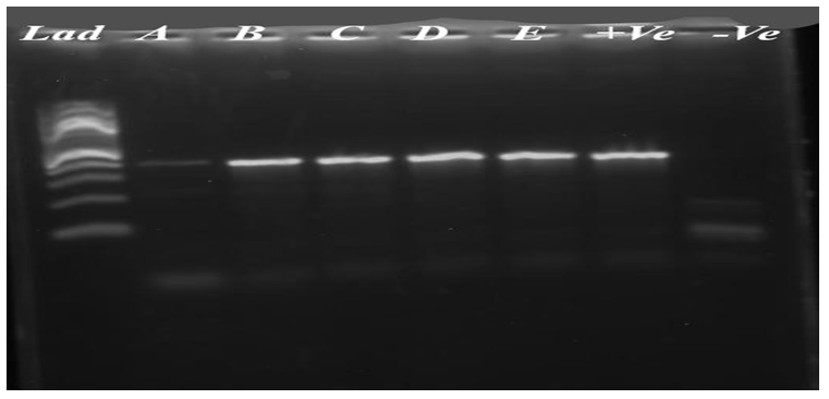
Overall prevalence of S. enterica by invA PCR: Presumptive S. enterica isolates that were Salmonella-positive and passed through the selective medium were confirmed by invA PCR. As a result, 5% (10/200) of the total prevalence was S. enterica (Figure 2). Occurrence of S. enterica and E. coli based on Risk Factors: Among beekeepers in the study area, 82.5% replied that they provided supplementary feed for honey during periods of feed scarcity. The most common locally available feed types used as colony supplements are shiro, sugar, and honey. Honeybees collect water from 40% of streams, 35% of rivers, and 25% of tap water. The respondents were asked whether they had received training in beekeeping and management techniques. Accordingly, 62.5% of the interviewed beekeepers received beekeeping training from the HBRC. Among the variables assessed, the type of supplementary feed (p=0.03), water source (p=0.009), and occurrence of colony collapse (p=0.001) were statistically significant (Table 7).
| Management | Categories | N | % | E. coli Positive | S. enterica Positive | ||||||
|---|---|---|---|---|---|---|---|---|---|---|---|
| N | % | X2 | P | N | % | ꭓ2 | P | ||||
| Do you Provide supplement feed? | Yes | 33 | 82.5 | 13 | 39.4 | 0.3 | 0.5 | 4 | 12.1 | 3.7 | 0.05 |
| No | 7 | 17.5 | 2 | 28.6 | 7 | 100 | |||||
| Type supplement | Sugar | 9 | 22.5 | 5 | 55.6 | 3.7 | 0.3 | 2 | 22.2 | 3.8 | 0.03 |
| Shiro | 13 | 32.5 | 6 | 46.2 | 3 | 23.1 | |||||
| Honey | 11 | 27.5 | 2 | 18.2 | 2 | 18.2 | |||||
| Nothing | 7 | 17.5 | 2 | 28.6 | 4 | 57.1 | |||||
| Type of water | Stream | 16 | 40 | 6 | 37.5 | 2.2 | 0.3 | 2 | 12.5 | 9.5 | 0.009 |
| River | 14 | 35 | 7 | 50 | 8 | 57.1 | |||||
| Tap | 10 | 25 | 2 | 20 | 1 | 10 | |||||
| Frequency of cleaning equipment | Every week | 15 | 37.5 | 4 | 26.7 | 1.3 | 0.3 | 3 | 20 | 1.8 | 0.41 |
| Every2 week | 13 | 32.5 | 6 | 46.2 | 3 | 23.1 | |||||
| Monthly | 12 | 30 | 5 | 41.7 | 5 | 41.7 | |||||
| Do you get beekeeping training? | Yes | 25 | 62.5 | 9 | 36 | 0.06 | 0.8 | 6 | 24 | 0.4 | 0.52 |
| No | 15 | 37.5 | 6 | 40 | 5 | 33.3 | |||||
| Have ever encountered colony collapse? | Yes | 29 | 72.5 | 9 | 31 | 1.9 | 0.2 | 2 | 6.9 | 22.4 | 0.001 |
| No | 11 | 27.5 | 6 | 54.6 | 9 | 81.9 | |||||
| Total | 40 | 100 | 15 | 37.5 | 11 | 27.5 |
Table 6: Cleaning, supplement feed, training, status of colony collapse, frequency of cleaning equipment of honeybee and S. enter
Antimicrobial Susceptibility Profiles of Isolates: The assessment of the antimicrobial susceptibility of all 15 E. coli and 11 S. enterica isolates from the honeybee samples collected from HBRC and four kebeles of Holeta town to the selected disks is shown in (Table 8). S. enterica isolates were highly susceptible to streptomycin (100%) and trimethoprim (63.6%), while 100% were resistant to Ampicillin, Clindamycin, and Penicillin. E. coli isolates were highly susceptible to Streptomycin (100%), Oxy-tetracycline, and Tetracycline (93.3% and Ceftriaxone (80%). All E. coli isolates were resistant to Penicillin and Clindamycin. Honeybee Management Practices: Of the 40 sampled owners and workers interviewed, about (80%) were male, and the rest (20%) were female. Workers of the most productive age, between 26-47 years, were actively participating in beekeeping. Among the households and workers interviewed, 22.5 %, 67.5%, and 10% were single, married, and divorced, respectively.
| Antimicrobial Agent | (µg/disc) | E. coli (n=15) | S. enterica (n=11) | ||||
|---|---|---|---|---|---|---|---|
| Susceptible N (%) | Resistant N (%) | Intermediate N (%) | Susceptible N (%) | Resistant N (%) | Intermediate N (%) | ||
| AMP | 10 | 0 (0) | 14 (93.3) | 1 (6.6) | 0 (0) | 11 (100) | 0 (0) |
| CMP | 30 | 10 (66.6) | 2 (13.3) | 3 (20) | 5 (45.4) | 1 (6.6) | 5 (45.4) |
| CRO | 30 | 12 (80) | 1 (6.6) | 2 (13.3) | 7 (63.6) | 2 (13.3) | 2 (13.3) |
| CLN | 10 | 0 (0%) | 15 (100) | 0 (0) | 0 (0) | 11 (100) | 0 (0) |
| S | 10 | 15 (100) | 0 (0) | 0 (0) | 11 (100) | 0 (0) | 0 (0) |
| TE | 30 | 14 (93.3) | 1 (6.6) | 0 (0) | 1 (9.09) | 5 (45.4) | 5 (45.4) |
| P | 10 U | 0 (0) | 15 (100) | 0 (0) | 0 (0) | 11 (100) | 0 (0) |
| TR | 5 | 11 (73.3) | 2 (13.3) | 2 (13.3) | 7 (63.6) | 2 (18.2) | 2 (18.2) |
| OT | 30 | 14 (93.3) | 1 (6.6) | 0 (0) | 5 (45.4) | 0 (0) | 6 (54.5) |
Table 7: Antimicrobial susceptibility profile of E. coli and S. enterica isolates. Key: AMP=ampicillin, CMP=chloramphenicol, CRO=
Regarding educational status, (32.5%) of the respondents had no formal education, (17.5%) attended primary education (25%) of them went to secondary and preparatory school (15%) attended a diploma, and (10%) were a degree or higher, as shown in Table 9. Consequently, both groups (literate and informal education) were practicing beekeeping. However, these findings indicate that honeybee farmers in Holeta Town have low levels of education. The respondents were interviewed for the variable to describe the frequency of inspecting their apiary and honeybee colonies. Among these, 45% replied that they look externally into the hives every week, 35% every three days, and 7.5% every day (Table 9).
| Demographic Variables | Categories | Frequency | Percentage (%) |
|---|---|---|---|
| Sex | Male | 32 | 80 |
| Female | 8 | 20 | |
| Education status | No formal education | 13 | 32.5 |
| Primary education | 7 | 17.5 | |
| Secondary/preparatory | 10 | 25 | |
| Diploma | 6 | 15 | |
| Degree | 4 | 10 | |
| Marital status | Single | 9 | 22.5 |
| Married | 27 | 67.5 | |
| Divorced | 4 | 10 | |
| Age | 15-25 years | 7 | 17.5 |
| 26-36 years | 14 | 35 | |
| 37-47 | 17 | 42.5 | |
| Above 47 | 2 | 5 | |
| Are you aware of bacterial honeybee disease? | Yes | 10 | 25 |
| No | 30 | 75 | |
| Frequency of observation | Every day | 3 | 7.5 |
| Every two day | 5 | 12.5 | |
| Every three day | 14 | 35 | |
| Every week | 18 | 45 |
Table 8: The proportion of sex, age, educational, marital, frequency of observation, and awareness of bacterial honeybee disease
Discussion
Honeybee colony losses have become a significant global issue, primarily due to the emergence of new honeybee diseases. Controlling these diseases is vital to protect honeybee populations. Traditionally, antibiotics like Tetracycline have been used to manage honeybee diseases. However, the use of antibiotics in apiculture is banned in many European countries due to potential health risks to both humans and bees. In our study, we investigated the prevalence of S. enterica and E. coli in honeybee colonies, focusing on different management practices and hive types in various kebeles (local administrative areas) in Ethiopia. In our study, similar to the findings reported by Zafar, et al. [38] and Diriba, et al. [39], we observed the highest prevalence of S. enterica at 8% in Meda Gudina kebele. Additionally, the overall occurrence of E. coli in honeybee gut samples was 7.5%, with the highest isolation rate of 12% also recorded in Meda Gudina kebele. Traditional hives had a higher prevalence of both pathogens compared to modern hives, indicating that management practices play a crucial role in disease prevalence.
Modern hive beekeepers were more likely to follow better management practices, including proper feeding, watering, and hygienic practices, as advised by HBRC researchers. Traditional hives are more challenging to manage and are more susceptible to pests and diseases, leading to higher pathogen prevalence [40]. Poor management practices, such as inadequate feeding and improper water sources, were associated with higher prevalence of S. enterica and E. coli. Feed supplements, water sources, and colony collapse were significantly associated with the presence of these pathogens [41]. A significant number of beekeepers (62.5%) received training from HBRC and livestock experts, leading to better production management and disease control. Proper training helps beekeepers improve their practices, reducing the prevalence of diseases [42]. S. enterica isolates showed high resistance to common antibiotics like ampicillin, clindamycin, and penicillin, with resistance levels ranging from 6.4% to 100%, while E. coli isolates also exhibited high resistance to the same antibiotics. This resistance is likely due to the overuse of these antibiotics in livestock, posing a challenge for both human and animal health [43].
Implementing a holistic approach to improve honeybee health and reduce disease prevalence involves several key strategies. Transitioning to modern hive designs can greatly facilitate better management practices, providing easier access for inspection, feeding, and sanitation. Alongside this, expanding beekeeping training programs is crucial, focusing on practical aspects like disease identification, prevention strategies, and effective control methods. Regular hive inspections, both externally and internally, are essential for early disease detection and prompt intervention. Providing honeybees with adequate feed supplements and clean water sources during feed scarcity periods helps boost their immune systems and reduces pathogen prevalence. Maintaining sanitary conditions in the apiary through equipment cleaning and hygienic practices minimizes disease risks. Furthermore, conducting additional research to identify pathogen sources and understand their effects on honeybee and human health is vital for implementing targeted control measures. By integrating these recommendations, beekeepers can work towards maintaining healthy colonies and sustaining honeybee populations [44, 45, 46, 47].
In this study, the overall Occurrence of S. enterica and E. coli in the gut of honeybees were 5.5% and 7.5%, respectively. In another way, the highest prevalence for these pathogens was Meda Gudina kebele. The occurrence of E. coli (20-60%) and S. enterica (20-40%) isolates among honeybee colonies in the study sites/kebeles signals the high distribution of S. enterica and E. coli present in the honeybee environment. Moreover, this shows that the honeybee gut is an alternative habitat for human pathogenic bacteria. The type of hive and management practice was suggested to be possible risk factors for the prevalence of S. enterica and E. coli in honeybees. The statistical difference observed in S. enterica isolates was in the feed supplement, water source, and colony collapse (P<0.05). It was evidence of a low level of public awareness about bacterial honeybee disease and associated risk factors in the study area. In another way, there is evidence of antibiotic resistance isolates from bees in the study area. In conclusion, the honeybee gut is an alternate habitat for human pathogenic bacteria as a high load of these bacteria recovered from the alimentary canal of honeybees. Our research underscores the significance of effective hive management techniques, hive design, and training in combating honeybee diseases. The adoption of modern hives and knowledgeable beekeepers plays a pivotal role in mitigating pathogen spread. Addressing antibiotic resistance and enhancing overall hive care are key factors in safeguarding honeybee populations and promoting the longevity of beekeeping practices. Further investigation is warranted to comprehensively grasp the implications of these pathogens and devise efficient disease control strategies.
Based on the above conclusion: the use of a modern hive instead of a traditional one is advisable for management practices like inspection, feeding, sanitation, and disease control easily; great emphasis should be given to training and extension programs for the community focusing on the practical aspects of general beekeeping, and more specifically on honeybee management; observation of bee colonies and cleaning feeding equipment are necessary for apiculture to improve honeybee health; further studies should be conducted to rule out the sources of targeted bacteria in the bee environment; the direct/indirect effect of the targeted bacteria to human and honeybee health should studied in detail.
References
-
Etxegarai-Legarreta O, Sanchez-Famoso V (2022) The Role of Beekeeping in the Generation of Goods and Services: The Interrelation between Environmental, Socioeconomic, and Sociocultural Utilities. Agriculture 12(4): 551.
-
Meikle WG, Mercadier G, Guermache F, Bon MC (2012) Pseudomonas contamination of a fungus-based bio pesticide: Implications for honeybee health and Varroa mite control. Biological Control 60(3): 312-320.
-
Kwadha CA, Ong’amo GO, Ndegwa PN, Raina SK, Fombong AT (2017) The biology and control of the greater wax moth, Galleria mellonella. Insects 8(2): 61.
-
Tsegaye A, Jenberie AW, Bihonegn AE, Lemma M (2014) Evaluation of different non-chemical wax moth prevention methods in the backyards of rural beekeepers in the North West dry land areas of Ethiopia. IOSR Journal of Agriculture and Veterinary Science 7(3): 29-36.
-
Belay FD, Oljirra AW (2016) The Significance of honey production for livelihood in Ethiopia. Jimma University, Ethiopia. Journal of Environment and Earth Science 6(4): 46-53.
-
Genersch E (2010) Honeybee pathology: Current threats to honeybees and beekeeping. Appl Microbiol Biotechnol 87(1): 87-97.
-
O’Neal ST, Anderson TD, Wu-Smart JY (2018) Interactions between pesticides and pathogen susceptibility in honeybees. Curr Opin Insect Sci 26: 57-62.
-
Maini S, Medrzycki P, Porrini C (2010) The puzzle of honeybee losses: A brief review. Bull Insectology 63(1): 153-160.
-
Evans JD, Schwarz RS (2011) Bees brought to their knees: microbes affecting honeybee health. Trends Microbiol 19(12): 614-620.
-
Ellis JD, Graham JR, Mortensen A (2013) Standard methods for wax moth research. In: Dietemann V, et al. (Eds.), The Coloss Bee Book 2: Standard methods for _Apis mellifera_ pest and pathogen research. Journal of Apicultural Research 52(1): 1-17.
-
Endo A, Salminen S (2013) Honeybees and bee hives are rich sources for fructophilic lactic acid bacteria. Syst Appl Microbiol 36(6): 444-448.
-
Mattila HR, Rios D, Walker-Sperling VE, Roeselers G, Newton ILG (2012) Characterization of the active microbiotas associated with honeybees reveals healthier and broader communities when colonies are genetically diverse. PLoS One 7(3): e32962.
-
Wu M, Sugimura Y, Iwata K, Takaya N, Takamatsu D, et al. (2014) Inhibitory effect of gut bacteria from the Japanese honeybee, Apiscerana japonica, against Melissococcus plutonium, the causal agent of European foulbrood disease. J Insect Sci 14: 129.
-
Khalil K, Lindblom GB, Mazhar K, Kaijser B (1994) Flies and water as reservoirs for bacterial entero-pathogens in urban and rural areas in and around Lahore, Pakistan. Epidemiol Infect 113(3): 435-444.
-
Staveley J, Law SA, Fairbrother A, Menzie C (2014) A causal analysis of observed declines in managed honeybees (_Apis mellifera_). Agricultural and Food Sciences, Biology, Environmental Science 20: 566-591.
-
Hananeh W, Al-Ghzawi AAM, Zaitoun S (2014) Does reclaimed water induce morphological changes in mid guts of honeybees (_Apis mellifer asyriaca_). Veterinary Science Development 4.
-
Menasria T, Moussa F, El-Hamza S, Tine S, Megri R, et al. (2014) Bacterial load of German cockroach found in hospital environment. Pathog Glob Health 108(3): 141- 147.
-
Pernal SF, Currie RW (2001) The influence of pollen quality on foraging behavior in honeybees (_Apis_ _mellifera_). Behavioral Ecology Sociobiology 51: 53-68.
-
Zheng H, Steele MI, Leonard SP, Motta EVS, Moran NA, et al. (2018) Honeybees as models for gut microbiota research. labanimal 47(11): 317-325.
-
Hussain T, Roohi A, Munir S, Ahmed I, Khan J, et al. (2013) Biochemical characterization and identification of bacterial strains isolated from drinking water sources of Kohat, Pakistan. African Journal of Microbiology Research 7(16): 1579-1590.
-
Sommer MOA, Dantas G, Church GM (2009) Functional characterization of the antibiotic resistance reservoir in the Human microflora. Science 325(5944): 1128 -1131.
-
Arbia A, Babbay B (2011) Management strategies of honeybee diseases. Journal of Entomology 8(1): 1-15.
-
Motta EVS, Raymann K, Moran NA (2018) Glyphosate perturbs the gut microbiota of honeybees. Proc Natl Acad Sci U SA 115(41): 10305-10310.
-
Burritt NL, Foss NJ, Neeno-Eckwall EC, Church JO, Hilger AM, et al. (2016) Sepsis and hemocyte loss in honeybees infected with Serratia marcescens strain sicaria. PLoS one 11(12): 0167752.
-
Raymann K, Shaffer Z, Moran NA (2017) Antibiotic exposure perturbs the gut microbiota and elevates mortality in honeybees. PLoS 15(3): 2001-2861.
-
Janashia I, Choiset Y, Robesona, Hwanhlem N, Bakuradze N, et al. (2016) Protection of honeybee _Apis mellifera_ by its endogenous and exogenous lactic flora against bacterial infections. Annual Agrarian Science 14(3): 177-181.
-
Shimelis S, Regassa F, Ballo S, Megersa S (2017) Survey of honey production system and honeybee disease and pests in Ejere district, West Shewa zone, Oromia regional state, Ethiopia. A Thesis submitted to the department of College of Veterinary Medicine and Agriculture of Addis Ababa University MScs. in Veterinary Epidemiology.
-
Holeta Town Administration (2011) Population Census Commission and housing census of Ethiopia 2010. Statistical report for Oromia region, Ethiopia.
-
Welmara Agricultural Office district (2022) Unpublished data from Welmara Agricultural Office district, Holeta, Ethiopia.
-
Engel P, James RR, Koga R, Kwong WK, Mc Frederick QS, et al. (2013) Standard methods for research on _Apis_ _mellifera_ gut symbionts. Journal of Apiculture Research 52(4): 1-24.
-
ISO (2002) Microbiology of food and animal feeding stuff-horizontal method for the detection of _Salmonella_ spices. Geneva, Switzerland, 6579: 511-525.
-
ISO (2007) Microbiology of food and animal feeding stuff. Horizontal method for detection of _Salmonella_ spp_._ in animal faeces and in environmental samples from the primary production stage.
-
Bedassa A, Nahusenay H, Asefa Z, Sisay T, Girmay G, et al. (2023) Prevalence and associated risk factors for _Salmonella enterica_ contamination of cow milk and cottage cheese in Ethiopia. Int J Food Contam 10(1): 2.
-
Gebeyehu A, Taye M, Abebe R (2022) Isolation, molecular detection and antimicrobial susceptibility profile of _Salmonella_ from raw cow milk collected from dairy farms and households in southern Ethiopia. BMC Microbiol 22(1): 84.
-
Firoozeh F, Saffari M, Neamati F, Zibaei M (2014) Detection of virulence genes in _Escherichia coli_ isolated from patients with cystitis and pyelonephritis. Int J Infect Dis 29: 219-222.
-
Abreham S, Teklu A, Cox E, Tessema TS (2019) _Escherichia_ _coli_ O157:H7: Distribution, molecular characterization, antimicrobial resistance patterns and source of contamination of sheep and goat carcasses at an export abattoir, Mojdo, Ethiopia. BMC Microbiol 19(1): 215.
-
CLSI (2018) Performance standards for antimicrobial susceptibility testing. 30th(Edn.). Clinical and Laboratory Standards Institute 38(3): 54-140.
-
Zafar H, Rahman SU, Ali S, Javed MT (2019) Evaluation of a _Salmonella_ Strain Isolated from Honeybee Gut as a Potential Live Oral Vaccine Against Lethal Infection of _Salmonella_ Typhimurium. Pol J Microbiol 68(2): 173- 183.
-
Diriba A, Fisaha M, Andualem D (2023) Causes of honeybee colony decline in south Ethiopia. Online J Anim Feed Res 13(4): 259-268.
-
Solomon S, Degu T, Fesseha H, Mathewos M (2021) Study on Major Parasitic Diseases of Adult Honeybees in Three Districts of Kaffa Zone, Southern Ethiopia. Vet Med Int 2021: 6346703.
-
Taulo S, Wetlesen A, Abrahamsen R, Mkakosya R, Kululanga G (2008) Microbiological quality of water, associated management practices and risks at source, transport and storage points in a rural community of Lungwena, Malawi. Afr J Microbiol Res 22: 131-137.
-
Bihonegn A, Begna D (2021) Beekeeping production system, challenges, and opportunities in selected districts of South Wollo Zone, Amhara, Ethiopia. Advances in Agriculture, pp: 1-10.
-
Pandey S, Doo H, Keum GB, Kim ES, Kwak J, et al. (2024) Antibiotic resistance in livestock, environment and humans: One Health perspective. J Anim Sci Technol 66(2): 266-278.
-
Barrasso R, Bonerba E, Savarino AE, Ceci E, Bozzo G, et al. (2018) Simultaneous Quantitative Detection of Six Families of Antibiotics in Honey Using A Biochip Multi- Array Technology. Vet Sci 6(1): 1.
-
Engel P, Kwong WK, Mc Frederick, Anderson KE, Barribeau SM, et al. (2016) The bee microbiome: impact on bee health and model for evolution and ecology of host-microbe interactions. mBio 7(2): e02164-e03115.
-
Gwimbi P, George M, Ramphalile M (2019) Bacterial contamination of drinking water sources in rural villages of Mohale Basin, Lesotho: exposures through neighbourhood sanitation and hygiene practices. Environ Health Prev Med 24(1): 33.
-
Moraes DMC, Almeida AMDS, Andrade MA, Nascente EDP, Duarte SC, et al. (2024) Antibiotic Resistance Profile of _Salmonella_ sp. Isolates from Commercial Laying Hen Farms in Central-Western Brazil. Microorganisms 12(4): 669.
- Antifungal Activity of New Acetophenone Derivatives
- Interconnected Microbiomes Human Health Within an Environmental Framework
- Silkworm-Based Vaccine Production for H5N1: A One Health Approach to Pandemic Preparedness
- Microbial Diversity and Lipolytic Activity of Bacteria and Fungi from Oil-Contaminated Sites in Makurdi Metroplois
- Antibiotic Resistance Profile of Bacteria Isolated at the Central Laboratory of the National Hospital Center of Nouakchott
- Epidemiology and Sensitivity to Antibiotics of Germs Isolated from Blood Cultures in the Laboratory of the National Hospital Center of Nouakchott-Mauritania