Occurrence of Third root (supernumerary / Radix Entomolaris / Radix Paramolaris) in the Primary Mandibular Molars - A Systematic Review
Purpose: The purpose of this systematic review is to evaluate the total prevalence of third roots seen in primary mandibular molars including both first and second in pediatric patients. Methods: An electronic search from three database was performed from inception till 2023 using different MeSH key words. Two independent authors carried out selection and analyses of the articles following PICO and PRISMA guidelines. To check the quality of included studies, modified New Ottawa scale was used. Results: Initial search following search of three electronic databases provided total 430 articles, in that only eight articles which fulfilled the inclusion criteria. The total prevalence of three rooted primary molars obtained ranged from 0.4% to 57% including only five different countries. There was no statistically significant difference found in gender wise comparison and also with right or left side occurrence. Its bilateral occurrence was shown only in two studies. Conclusion: There is insufficient data on overall prevalence of three rooted primary molars and also there are no quality studies undertaken across the globe. Future studies including more of cross-sectional and including advanced imaging techniques like CBCT is highly warranted in the arena of Paediatric Dentistry. This SR illuminate new pathways of knowledge about this root variation leading pediatric dentists to uncharted realms of understanding. Occurrence of lesion in Indian patient.
Abbreviations
SR: Systematic Review; PRISMA: Preferred Reporting Items for Systematic Reviews and Meta-Analyses; PICO: Population, Intervention, Comparison, Outcome; IOPAR: Intraoral Periapical Radiographs; CBCT: Cone-Beam Computed Tomography
Introduction
Roots in Primary molars show variation in number and canal anatomy enabling Pediatric dentist to encounter problems during pulpectomy or extraction procedure. Normally, the mandibular primary molars both including first and second molars have two roots, one placed mesially and other one distally. Very rarely an additional third root is found distally attached to the main distal root. This root is termed as “Radix Entomolaris” as evident in the available literature [1, 2, 3]. When this third extra root is seen buccally attached to the main mesial root then it is referred as “Radix Paramolaris” [4]. These accessory roots are usually found overlapped with the main roots and hence not visible on conventional radiographs. In order to locate their presence, authors have suggested to take radiographs with different angulations so that the outline of this root is visible on the radiographs.
The presence of extra roots in primary molars is associated with clinical significance and requires special consideration as it affects the treatment outcome in these teeth as this root is typically smaller than main distal root and usually curved. During pulpectomy procedure if the dentist is not aware of the existence of extra roots, may end up with failure in the treatment due to missing of the root. Even during extraction procedure, the angulated extra root may get entrapped while luxating the tooth resulting in root fracture of this extra root [5, 6]. Therefore, knowledge of occurrence of third root in primary mandibular molars is highly essential to obtain meticulous diagnosis which in turn help in enhancing quality treatment given to the young pediatric patients. Occurrence of third root is more common in permanent molars than in primary molars [7, 8, 9, 10]. Moreover, existence of third root is suggested as a racial characteristic of certain population including Mongoloid and Indians compared to Caucasian populations [11, 12, 13, 14, 15, 16, 17, 18, 19]. Although various case reports, review articles, case series and original studies have been undertaken providing greater evidence but there is no literature showing global level of prevalence including gender distribution, unilaterality or bilateral presentation of this third root in primary mandibular molars among different population [1, 2, 3, 4, 5, 6, 7, 8, 9, 10, 11, 12, 13, 14, 15, 16, 17, 18, 19, 20].
Therefore, the present systematic review was carried out keeping the research question as ‘what is the total prevalence of three rooted primary mandibular molars in Pediatric patients? By evaluating the total prevalence of an accessory third root seen in primary mandibular molars, the obtained data/findings may enable investigator to provide research evidence for making clinical guidelines, protocols and policies in the management of such teeth encountered with third roots during clinical practice in Pediatric Dentistry.
Materials and Methods
Protocol and Registration
The present SR was performed using the objective and transparent methods/guidelines of PRISMA (Preferred Reporting Items for Systematic Reviews and Meta-Analyses) statement given by Moher D, et al. in order to screen, evaluate and summarize all the relevant research output [21]. The protocol for this review was registered in PROSPERO registration database (International perspective register of systematic reviews) with the PROSPERO registration No. CRD42024521721. The research question of this SR was what is the prevalence of three rooted primary mandibular molars in pediatric patients across the globe?
Eligibility Criteria
PICO (Population, Intervention, Comparison, Outcome) Design schema was used to evaluate the prevalence of three rooted primary mandibular molars in pediatric patients as mentioned in Table 1.
| Population | Children of any gender, ethnicity and age ranging from 2 to 14 years |
|---|---|
| Intervention | Primary mandibular molars (first or second) diagnosed with extra roots (third root/RE) during examination or treatment |
| Comparison | Described in observational or clinical studies |
| Out come | Assessment of their prevalence in different population and usage of different diagnostic tools for their identification |
Table 1: PICO frame work for analysis
Inclusion Criteria
The inclusion criteria considered for selection of articles for analysis were publications showing cross-sectional studies, studies with primary or secondary objective including the term ‘variations in roots’ or ‘number of roots,’ prospective or retrospective studies, articles published in English language only, In Vivo studies and analytical or descriptive studies.
Exclusion Criteria
Articles on case reports, case series, conferences’ abstracts, summary articles, review articles, articles which are not in English language, author debates, commentaries and articles with no abstracts were not considered for SR analysis.
Search Strategy and Information Sources
The search strategy was performed by two independent authors (A and B). The relevant articles’ search was carried out using the well-known, most commonly used electronic databases such as PubMed, Embase and grey search using Google Scholar. Various MeSH key words were used in different combinations to obtain relevant articles from each electronic database as show in Table 2. Publications were searched from inception till December 2023 which are published only in English language without restriction to date and type of the study.
| Electronic Database | Search strategy |
|---|---|
| PubMed | three[All Fields] AND rooted[All Fields] AND primary[All Fields] AND (“molar”[MeSH Terms] OR “molar”[All Fields] OR “molars”[All Fields]) OR “radix”[All Fields]) AND entomolaris[All Fields] AND primary[All Fields] AND (“molar”[MeSH Terms] OR “molar”[All Fields] OR “molars”[All Fields]) OR “roots”[All Fields]) AND primary[All Fields] AND (“molar”[MeSH Terms] OR “molar”[All Fields] OR “molars”[All Fields]) three[All Fields] AND rooted[All Fields] AND primary[All Fields] AND (“mandible”[MeSH Terms] OR “mandible”[All Fields] OR “mandibular”[All Fields]) AND first[All Fields] AND (“molar”[MeSH Terms] OR “molar”[All Fields] OR “molars”[All Fields]) |
| Embase | Primary second AND (“mandible” OR “mandible” OR “mandibular” AND first AND (“molar”) OR Radix Entomolaris AND (“paramolaris”) |
| Google scholar | Three rooted primary mandibular molars or extra/accessory roots in primary molars/Radix Entomolaris/Radix Paramolaris in primary mandibular molars |
Table 2: Representation of search strategy in terms of MeSH terms and search words for each electronic database
Study Selection
Two independent reviewers searched the titles and abstracts considering the inclusion and exclusion criteria and identified relevant articles. Later, the same two authors independently reviewed the full text articles in depth which are unable to be excluded by title and abstract alone. Comparison of papers was performed between two authors with no disagreements regarding inclusion.
Data Extraction
Using data extraction form, data extraction was done from eight finally selected publications. The data extraction form consisted of the name of the first author, year of publication of the article, objectives of the study, design of the study, assessment tool used to obtain relevant information, results (prevalence of the third root seen) and conclusion of authors.
Assessment of Methodological Quality of the Included Studies
Two independent authors evaluated the methodological quality of the included studies following the assessment tool of modified Newcastle Ottawa Scale consisting of quality score based on criteria involving the categories like group selection, outcome and exposure. Based on this scale, for group selection and exposure, a maximum score of 5 points, for comparison group 2 points and maximum score of 3 points for outcome was considered as the highest methodological quality of the study which is represented in Table 3.
| Study with first author and year | Selection and Exposure | Outcome | ||||
|---|---|---|---|---|---|---|
| Representativeness of the sample | Sample size | Ascertainment of the exposure | Non-re- spondents | Assessment of outcome | Statistical test | |
| Hsu, et al. 2021 | 2 | 1 | 1 | 0 | 1 | 1 |
| Nagaveni, et al. 2018 | 2 | 1 | 1 | 0 | 1 | 1 |
| Jiang, et al. 2022 | 2 | 1 | 1 | 0 | 1 | 1 |
| Khosrozadeh, et al. 2021 | 1 | 2 | 1 | 0 | 1 | 1 |
| Nagaveni, et al. 2017 | 2 | 1 | 0 | 0 | 1 | 1 |
| Moyaho-Bernal, et al. 2021 | 2 | 1 | 1 | 0 | 1 | 1 |
| Tu, et al. 2010 | 2 | 1 | 1 | 0 | 1 | 1 |
| Liu, et al. 2010 | 2 | 1 | 1 | 0 | 1 | 1 |
Table 3: Study quality assessment using modified Newcastle Ottawa Scale (9 studies considered for assessment)
Results
Study selection and Flow diagram
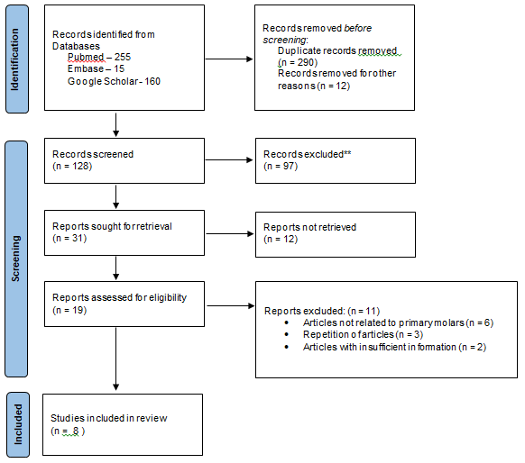
A total of 430 articles were identified in the initial search from three different electronic databases such as PubMed, Embase and Google Scholar. PubMed search resulted in 255 publications, Embase produced 15 articles and from Google Scholar 160 articles were identified initially. After removing the duplicates 128 publications were scrutinized. Following screening of titles and abstracts, 19 articles were selected for full text reading. From these 19 publications, 11 articles were excluded based on inclusion and exclusion criteria. Finally, only eight articles were selected for qualitative assessment. Eleven articles were excluded due to different reasons like, they were not related to primary molars, some articles showed repetition in different journals with same data and articles with insufficient information about assessment. The complete process of search strategy for selection of articles is shown in Table 4.
Results of Individual Studies
From PubMed source four articles were obtained, one from Embase and Google scholar produced three articles. Most of the studies evaluated for SR were retrospective studies and detail of these studies in elaborated in Table 5. Moyaho-Bernal MA, et al. evaluated the maximum study samples of 2284 pediatric patients followed by Hsu CL, et al. who evaluated 977 patient’s records (OPGs) [13, 14]. A wide range in the sample size ranging from 77 to 2284 was observed among the included studies. The population sample assessed for presence of extra roots were found from different countries like India, Taiwan, Mexico, Iran and China with maximum contribution from India, Taiwan and China (each country contributed two studies). In only one study, CBCT an advanced imaging technique was used as a diagnostic tool followed by intraoral periapical radiographs (IOPAR) in five studies and remaining two studies used vertical bitewing radiographs. Only one study used both OPG and CBCT scans (but they used these only in one case for assessment of third root) along with IOPAR. All studies have provided information on age of children considered for evaluation, gender predilection, unilateral or bilateral and right and left involvement with extra roots. Evaluation of both first and second primary molars for the presence of extra roots was shown in only two studies. From all these studies, the number of primary molars with extra third root identified was 733. This is the total prevalence obtained from five different countries across the globe.
Prevalence
The prevalence of mandibular primary molars with extra roots showed variation in different population (Table 5). The age of the children considered for evaluation in these eight studies ranged from 2 to 11.9 years old. Two studies showed prevalence of third root in both primary mandibular first and second molars. Four studies showed prevalence only in second molars (0.2% to 42%) and only two studies showed prevalence in first molars (0.4% to 57%). For gender wise distribution, the maximum occurrence was noted in males than females in many studies. When side involved by presence of extra roots was evaluated there was no much difference observed between right or left side. Compared to bilateral presentation, unilateral distribution of extra roots was found in maximum percentage as evident from these studies.
| Name of first author/Year of Publication/ Journal Name | Study Design/ Population sample | Screened patients/ Records/ Tooth evaluated | Patients found with extra roots | Age of the patients (years)/ Mean age | Gender N(%) | Side affected N(%) | Laterality N(%) | |||
|---|---|---|---|---|---|---|---|---|---|---|
| M | F | R | L | U | B | |||||
| Hsu CL, et al. [14] (2021) Journal of Dental Science | Retrospective Taiwan | 977 OPG PMSM | 591 (28.4) | 8.7 | 93 (29) | 75 (27) | 152 (25) | 113 (19.1) | 74 (12) | 97 (16.4) |
| Nagaveni NB, et al. [12] (2018) International Journal of Pedodontic Rehabilitation | Cross-sectional India | 77 patients IOPAR PMSM | 10 (6.5) | 3-10 | 1.5 | 1 | 5 (6.5) | 1 (1.3) | 4 | 2 |
| Jiang C, et al. [16] (2022) BMC Oral Health | Retrospective China | 247 CBCT Scans PMSM | 54 (24) | 7.3 | 35 (24.6) | 19 (22.9) | 55 (23) | 33 (14.2) | 88 (47.2) | 27 (49.1) |
| Khosrozadeh M, et al. [17] (2021) International Journal of Dentistry | Descriptive, Cross-sectional, Retrospective Iran | 300 patients IOPAR PMFM | 16 (57.1) | 3-10 | 6 (37.5) | 10 (62.5) | 4 (25) | 11 (68.8) | 15 (91) | 1 (6.2) |
| PMSM | 12 (42.8) | 7 (58.3) | 5 (41.7) | 9 (75) | 2 (16.7) | 11 (89) | 1 (8.3) | |||
| Nagaveni NB, et al. (2017) [11] CODS Journal of Dentistry | Cross-sectional, Descriptive India | 77 patients IOPAR PMFM | 2 (1.3) | 3-10 | 1 (1.2) | 1 (1.4) | 0 | 2 (2.6) | 2 (2.3) | 0 |
| Moyaho-Bernal MA, et al. [13] (2021) Acta Odontology Latinoamerica | Descriptive, Cross-sectional Mexico | 2284 patients IOPAR OPG (1 case) CBCT (1 case) PMFM | 10 (0.44) | Not mentioned | 4 (1.5) | 1 (1) | 5 (50) | 3 (30) | 8 (80) | 2 (20) |
| PMSM | 5 (0.22) | 3 (0.32) | 2 (0.15) | 1 (20) | 3 (60) | 4 (80) | 1 (20) | |||
| Tu MG, et al. [18] (2010) Journal of the Formosan Medical Association | Retrospective Taiwan | 227 children Vertical bitewing radiographs PMFM | 6(5) | 2.4-10.4 | 3(5.4) | 3(4.4) | 4 | 3 | 5 | 1 |
| Liu JF, et al. [19] (2010) Pediataric Dentistry | Retrospective China | 185 patient Vertical bitewing radiographs PMSM | 18(10) | 2.5-11.9 | 11 (12) | 7 (8) | 10 (11) | 13 (14) | 13 (73) | 5 (28) |
Table 4: Summary of the included studies representing the prevalence of third root in primary mandibular molars
(Data represented in N = Number, % = percentage) Table 5: Summary of the included studies representing the prevalence of third root in primary mandibular molars
Discussion
To achieve an answer for the focused research question, the current systematic review of the available literature was conducted to evaluate the prevalence of third root present in the primary mandibular molars including both first and second molars of children. From this SR, the prevalence of three rooted primary mandibular molars ranged from 2% to 57%. Only two studies evaluated the prevalence in both first and second primary molars, one from Iran and other from Mexico. These two studies showed the prevalence of three roots in primary mandibular first molars ranging from 0.4% to 57%, and in second molars it ranged from 0.2% to 42%. The wide range observed in these prevalences may be due to difference in sample size estimated, and racial predilection observed for this third root. Although retrospective, descriptive or cross-sectional studies are lacking among different population, most of the studies were reported from India, Taiwan and China. There are no studies showing its prevalence in other parts of the continent such as African, European and American countries. As a result, there is no research data depicting the global prevalence of extra roots. There was no gender wise predilection for the presence of extra roots, however, few studies showed male predominance compared to females. Pertaining to side affected, few studies showed more prevalence on right side and some on left side and regarding the laterality of occurrence, bilateral presentation is recorded as extremely rare.
The age of the patient evaluated ranged from minimum of 2.4 years to maximum of 11.9 years with mean age of 6.5 years because at this age roots of primary molars found with full length with still no evidence of root resorption found. For diagnosing extra roots, advanced imaging tools like CBCT scans were used in one study [16]. However, in a study performed by Moyaho-Bernal MA, et al, CBCT was used only in one patient [13]. The other diagnostic tool used were OPGs, bitewing and periapical radiographs. Recently, CBCT has revolutionized tremendously in the field of dentistry and has been used for various purposes [22, 23, 24, 25]. In pediatric endodontics, it is widely used to evaluate the root and canal anatomy in children due to its high resolution and non-invasive nature. Compared to conventional CT, the scan time and radiation dose of CBCT can be significantly reduced and also found superior to the combination of several 2D radiographic images regarding the radiation dose, cost and intrinsic information obtained. Therefore, it is slowly replacing conventional radiographs and has become an unavoidable trend and cost-benefits analysis. In a study performed [16], using CBCT, authors measured the geometric parameters of the disto-buccal and disto-lingual roots including the vertical root length, angle of root curvature using Schneider technique, level and angle of distal root furcation and the spreading angle and were then compared to the three-rooted first molars. Using CBCT evaluation, the length of the distolingual roots found was 7.4 mm, the curvature angle was 16.4 degrees, and spreading angle was of 34.8 degrees. This is the only study which enlightened the anatomical features of third root observed in primary second molars in Chinese children.
Two studies 18, 19 used vertical bitewing radiographs for diagnosis of extra third roots. In these studies authors have stated that, vertical bitewing film technique is more reliable and easier in detecting the primary molars with supernumerary roots compared to horizontal bitewing film technique. Compared to CBCT, the radiographic method is a non-invasive and practical method which reliably compare the results of studies pertaining to gender and bilateral presence of three rooted primary mandibular molars in different ethnic groups. Moreover, this technique also helps in detection of proximal caries and shows the condition of primary teeth root resorption in the one film itself. The only disadvantage associated with this technique is that it is difficult to differentiate distolingual (radix entomolaris) or distobuccal root (radix paramolaris) in primary molars.
Moyaho-Bernal MA, et al. [13] in their study along with evaluation of three rooted primary molars, have also assessed the crown morphological characteristics of primary molars based on clinical examination. In crown morphology, they found a triangular-shaped mandibular first molar, characterized by the presence of a distolingual lobe in combination with a cervical prominence. Other crown variations observed were paramolar cusp (tuberculum paramolare) which occurred in both mandibular first and second molars.
It is well known fact that prevalence of three roots is more common in permanent mandibular molars compared to primary molars [26, 27, 28]. Literature shows numerous publications including systematic reviews in permanent mandibular first molars compared to primary molars. A recent SR performed in permanent molars evaluated the global prevalence of three rooted permanent mandibular first molars. Pertaining to primary molars, there is insufficient precise data on this root variation in other geographic parts of the world. Although the exact cause is not known, but the present SR gives a pay for scope of future research to conduct more epidemiological cross-sectional studies. This will help in updating the literature on prevalence of extra roots in primary mandibular molars including detailed anatomy, diagnostic criteria and treatment protocol by encouraging researchers to utilize advanced imaging tools like CBCT. Studies showing the co-occurrence of extra roots both in primary mandibular second molars and permanent mandibular first molars are very rare. In the present analyses of SR, only one stud [14] evaluated the correlation of the occurrence between three-rooted primary mandibular second molars and three- rooted permanent mandibular first molars in children belonging to Taiwanese ethnicity. Authors found a prevalence of 28.4% and 27.6% in the primary mandibular second molars and permanent mandibular first molars, respectively. Bilateral presence of three-rooted mandibular molars was found almost similar (60%) in both primary and permanent molars. Therefore, it is evident from results of this study that the occurrence of 3-rooted primary mandibular second molar can strongly predict the possibility of three roots in permanent mandibular first molars. In addition to this, some recent research speculates that the primary mandibular second molars and permanent mandibular first molars originate from the same molar field because of their same time of development and similar crown morphology [29]. This was attributed to a theory proposed by a researcher, Butler in the year 1939. Moreover, a study performed by Song JS, et al. [30] also showed this association, and the probability of diagnosing extra third root in both primary second and permanent mandibular first molars was estimated as 94.3%.
From the present SR which evaluated eight original studies, the prevalence of three rooted primary mandibular molars in children estimated ranging from minimum of 0.2% to maximum prevalence of 57% from five different parts of the world. There was no much difference identified regarding gender wise comparison and side affected. Compared to unilateral presentation, bilateral occurrence was extremely rare. Due to insufficient and a smaller number of epidemiological and cross-sectional studies availability in the literature, future studies with quality research are highly recommended to enlighten the existing literature in the field of Pediatric Endo-Dentistry.
Strength of the Systematic Review
The strength and validity of the current SR is that it is the first SR which performed in evaluating the prevalence of three rooted mandibular primary molars in pediatric patients. All the reported cross-sectional and descriptive and retrospective studies have provided information till 2022. After that there is no enough evidence on literature showing the prevalence of extra roots in primary mandibular molars. Therefore, the novelty of this SR is that it provides an updated and overall research output on this domain which is highly essential in children in order to provide utmost treatment in the management of extra roots found during clinical practice.
Limitations
The limitation observed from this SR is that meta- analysis could not carry out as there were very few studies which were completely retrospective and also there was of insufficient homogenous and measurable data evident in these available studies. In addition to this, authors have referred only three electronic databases for search strategy which might have resulted in selection of few studies.
Why this paper is important to Pediatric dentist
- Primary mandibular molars including both first and second molars rarely found with supernumerary roots in some population.
- Management of extra third root which is also called Radix entomolaris is a great challenge to a pediatric dentist as it is more curved and shorter.
- An awareness about existence of this root is highly essential among all pediatric dentist to provide proper treatment.
References
-
Nagaven NB, Umashanakara KV (2012) Radix entomolaris and paramolaris in children. A review of the literature. J Indian Soc Pedod Prev Dent 30(2): 94-102.
-
Nagaveni NB, Umashankar KV (2009) Radix entomolaris in permanent mandibular first molars: Case reports and literature review. Gen Dent 57(3): e25-e29.
-
Gupta S, Nagaveni NB, Chandranee NJ (2012) Three- rooted mandibular first primary molar: Report of three cases. Contemp Clin Dent 3(Suppl1): S134-S136.
-
Nagaveni NB, Bajaj M, Shruthi AS, Poornima P (2014) Radix paramolaris (supernumerary third root) in primary mandibular second molar: Report of two cases. Niger J Exp Clin Biosci 2(2): 134-137.
-
Nagaveni NB, Umashankar KV, Radhika NB, Satisha TS (2011) Third root (Radix Entomolaris) in permanent mandibular first molars in Pediatric patients - An endodontic challenge. J Orla Health Community Dent 5(1): 49-51.
-
Nagaveni NB, Umashankara KV, Radhika NB (2012) A retrospective analysis of accessory roots in mandibular molars of Indian pediatric patients. Inter J Dental Anthropol 20: 38-46.
-
Aung NM, Myint KK (2022) Three-rooted permanent mandibular first molars: A Meta-Analysis of prevalence. Int J Dent 2022: 9411076.
-
Shekarian M, Majlesi M, Jahromi MZ (2023) Prevalence of C-shaped canals and three-rooted mandibular molars in the Iranian population by using cone-beam computed tomography. Clin Exp Dent Res 9(5): 906-912.
-
Gupta A, Duhan J, Wadhwa J (2017) Prevalence of three rooted permanent mandibular first molars in Haryana (North Indian) population. Contemp Clin Dent 8(1): 38- 41.
-
Khurayzi TA, Beleges EM, Dallak SA (2021) The prevalence of radix entomolaris (RE) in the mandibular permanent first molars among the Saudi arabian population - a systematic review. Saudi J Oral Dent Res 6(1): 22-30.
-
Nagaveni NB, Poornima P, Vilsan A, Mathew MG, Masroor S (2017) Prevalence of Three-rooted Primary Mandibular First Molars in Children of Davangere, Karnataka, India. CODS J Dent 9(1): 7-9.
-
Nagaveni NB, Poornima P, Valsan A, Mathew MG (2018) Prevalence of three-rooted primary mandibular second molars in Karnataka (South Indian) population. Int J Pedod Rehabil 3: 33-5.
-
Moyaho-Bernal MA, Carrasco-Gutierrez R, Jimenez- Flores R, Juarez-Luna G, Lopez-Del Pino GR, et al. (2021) Prevalence of three-rooted primary mandibular first and second molars: clinical and radiographic findings in a Mexican population. Acta Odontol Latinoam 34(2): 149-155.
-
Hsu CL, Huang JE, Chen HL, Tu MG, Liu JF (2021) The relationship of the occurrence between three-rooted deciduous mandibular second molars and three-rooted permanent mandibular first molars in children. J Dent Sci 16(2): 580-585.
-
Balaji K, Mani G (2021) Prevalence of three rooted primary mandibular molars in children visiting a university hospital in Chennai - A retrospective study. J Contemp Iss Busi Gove 27(2): 3030-3038.
-
Jiang C, Pei F, Wu Y, Shen Y, Tang Y, et al. (2022) Investigation of three-rooted deciduous mandibular second molars in a Chinese population using cone-beam computed tomography. BMC Oral Health 22(1): 329.
-
Khosrozadeh M, Mostafavi M, Hamrah MH, Niknejad E (2021) Prevalence of Three-Rooted Deciduous Mandibular Molars in the Children of Northwestern Iran. Int J Dent 2021: 5643668.
-
Tu MG, Liu TJ, Dai PW, Chen SY, Hsu JT, et al. (2010) Prevalence of Three-rooted Primary Mandibular First Molars in Taiwan. J Formos Med Assoc 109(1): 69-74.
-
Liu JF, Dai PW, Chen SY, Huang HL, Hsu JT, et al. (2010) Prevalence of 3-rooted primary mandibular second molars among Chinese patients. Pediatr Dent 32(2): 123-126.
-
Sirawut HU, Sunpatch B, Chidnarong A, Pornthip T, Pakjira T, et al. (2021) Prevalence of C-shaped canals and three-rooted mandibular molars using CBCT in a selected Thai population. Iran Endod J 16(2): 97-102.
-
Moher D, Liberati A, Tetzlaff J, Altman DG, PRISMA Group (2009) Preferred reporting items for systematic reviews and meta-analysis.: the PRISMA Statement. PLoS Med 6(7): e1000097.
-
Tafakhori Z, Rafiei M (2022) Frequency of radix molaris in mandibular first and second molars using cone-beam computed tomography images in a selected Iranian population. Caspian J Dent Res 11(2): 124-129.
-
Yang SE, Lee TY, Kim KJ (2021) Prevalence and morphology of C-shaped canals: A CBCT analysis in a Korean population. Scanning 2021: 9152004.
-
Javed MQ, Srivastava S, Alotaibi BBR, Bhatti UA, Abulhmael AM, et al. (2023) A Cone beam computed tomography-based investigation of the frequency and pattern of Radix Entomolaris in the Saudi Arabian population. Medicina (Kaunas) 59(11): 2025.
-
Sheth K, Banga KS, Pawar A, Wahjuningraum DA, Karobari MS (2024) Distolingual root prevalence in mandibular first molar and complex root canal morphology in incisors: a CBCT analysis in Indian population. Sci Rep 14(1): 443.
-
Talbani RM, Abdalrahman KO, Abdul RJ, Babarasul DO, Kazzaz SH (2022) Evaluation of Radix entomolaris and middle mesial canal in mandibular permanent first molars in an Iraqi subpopulation using cone-beam computed tomography. Biomed Res Int 2022: 7825948.
-
Mukhaimer R, Azizi Z (2014) Incidence of Radix entomolaris in mandibular first molars in Palestinian population: A clinical investigation. Int Sch Res Notices 2014: 405601.
-
Parakh H, Thosar NR, Khubchandani M (2023) An unusual case of bilateral Radix entomolaris treated with lesion sterilization and tissue repair in primary first molars. Cureus 15(12): e50837.
-
Butler PM (1939) Studies of the mammalian dentition. Differentiation of the post-canine dentition. Proceedings of the Zoological Society of London B109(1): 1-36.
-
Song JS, Kim SO, Choi BJ, Choi HJ, Son HK, et al. (2009) Incidence and relationship of an additional root in the mandibular first permanent molar and primary molars. Oral Surg Oral Med Oral Pathol Oral Radiol Endod 107(1): e56-e60.
- Understanding Pediatric Multiple Sclerosis: Clinical Presentation, Diagnostic Criteria, Therapeutic Advances, and Supportive Care Approaches
- Hemophilia in Children
- Xia-Gibbs Syndrome- A Case Report
- A Study to Assess Effectiveness of Play Therapy in Reducing Post-Operative Pain among Children Age 2 To 5 Year who have Undergone General Surgeries in Selected Pediatric Hospitals of Vadodara
- Preterm Birth: Scope of the Problem, Cost of Care, Potential Complications and Current Guidelines for Management
- Noradrenaline: Can we Use it to Manage Hemodynamic Instability among Neonatal Septic Shock at the NICU?