A Review of Mosquitoes (Diptera: Culicidae) and Their Biodiversity, Medical and Veterinary Importance
Background: Insects, especially mosquitoes, make up a large part of the creatures on the planet, and almost all humans and animals encounter various insects throughout the day and night. Most of the infectious diseases that are registered annually in the world by the World Health Organization (WHO) are caused by insects, especially mosquitoes of the Culicidae family. The main purpose of this study is to investigate the mosquitoes of the Culicidae family, the diseases transmitted by them and the ways of disease transmission. Methods: This is a general review study to increase the knowledge of researchers in the field of diseases of insects and especially mosquitoes of the Culicidae family, which examines the most important parasitic and viral diseases in the field of medicine and veterinary medicine. Results: Parasitic diseases of Malaria and Dirofilariasis, viral diseases of Dengue Fever, Zika virus, West Nile virus, Chikungunya virus and is one of the most important diseases transmitted by mosquitoes of the Culicidae family, which are mostly transmitted by different species of Aedes and Culex. Conclusion: Today, with the changes in the global climate, in some countries, we are facing an increase in the number of mosquitoes that transmit diseases. Parasitic and viral diseases are the main diseases transmitted by this family of mosquitoes. One of the best ways to deal with important diseases such as Malaria, Yellow fever and Dengue fever, which make millions of people sick and even die every year, is to control the growth and reproduction of Culicidae mosquitoes.
Ghorbani A¹*, Dekkiche K², Ghorbani A³ and Garedaghi Y⁴
Keywords: Mosquitoes; Culicidae; Biodiversity; Transmission; Pathogenic Agents
Introduction
Arthropods are the main one of the most important organisms on the planet, which make up more than 80% of all organisms, and many life cycles and nature depend on this group of organisms, and in the absence of these organisms, food cycles Humans and animals suffer many problems. But the species of these organisms, besides their positive characteristics, can be considered as a parasitic agent due to their blood sucking from other organisms, or they can also play a role in the transmission of various diseases and cause the occurrence of many infectious diseases for humans and animals. From this point of view, the study of these organisms is of high value in medical and veterinary science. The most important class of arthropods is Class Insecata, which includes mosquitoes and flies [1]. Insects have been living on earth for about 350 million years. They are one of the first creatures on the planet that have survived to this day. Most insects have the ability to fly. Flying has given mosquitoes and flies the ability to try to find better places and better food conditions. that this factor played a very important role in the spread and dispersion of these organisms [2].
More than 3500 species of mosquitoes have been identified, but still many species of mosquitoes remain unknown due to their dispersion and high number [3]. Culicidae family is the main family of insects that has two sub families named Anophelinae and Culicinae, which have 482 species and 3119 species respectively [4]. Various parasitic, viral and bacterial diseases are transmitted by the Culicidae family. Familiarity with the classification and morphology of the Culicidae family is very important in the investigation and control of diseases transmitted by these insects.
Classification and Morphology
The Classification of Culicidae
Kingdom: Animalia Sub-kingdom: Metazoa Branch Line: Arthropoda Sub-branch: Hexapoda Class: Insecta Subclass: Pteregota Order: Diptera Sub-order: Nematocera Family: Culicidae Subfamily (1): Anophelinae, genus Anopheles Subfamily (2): Culicinae, five genus:
- Culex
- Aedes
- Culiseta
- Orthopodomyia
- Uranotaenia
The Morphology of Culicidae
Mosquitoes are holometabolous insects; they have a complete metamorphosis, so that the life stages (adult, larvae and nymphs) have very different morphologies, which make it possible to identify subfamilies, genera and species [5].
Eggs
After absorbing blood, the female, exclusively hematophageal, lands in a place to shelter and digest her hematophagic meal, a few days later, she lays eggs. The eggs are fusiform, measuring about 1 millimetre in length: whitish at the time of laying and quickly take, by oxidation of certain chemical components of the theque; a brown or black color [6, 7]. Classically the Culicidae egg includes from the inside to the outside: the embryo, the pellucid vitelline membrane, the thick endochorion, the more or less pigmented, embossed or areola [8]. Eggs can be laid in isolation (Stegomya, Anopheles, Orthopodomyia) or in bulk in the form of a pod (Culex, Culiseta) [9, 10]. Anopheles eggs are laid in isolation on the surface of the water. Their shape is more or less ovoid and equipped with side floats allowing them to maintain a horizontal position. Aedes eggs are elongated narrowed and show a network of fine depressions. They float horizontally on the surface of the water, Culex eggs grouped in pods are cylindro-conical and stand vertically [10, 11].
The Larvae
Mosquito larvae are distinguished from other aquatic insects by the absence of legs (apodes) and the presence of a bulbous thorax that is both wider than the head and abdomen [12]. It is usually 1 to 2 mm long, but can reach 4 mm in larger species. The fourth larval stage (L4) is approximately 5 mm long [13]. The body of the larva is divided into three parts: the head, chest and abdomen.
Head
Consists of three chitinous plates, one dorsal odd and median, the fronto-clypeus, the other two lateral and symmetrical. The head included in a sclerotinized capsule. The mouthpieces are grinding, and mainly composed by thick mandibles with sharp tips, and a triangular and serrated chin plate called mentum [14, 15]. It also has many bristles that can be used for species identification, including internal and external preclypelal bristles [16].
Thorax
In Culicidae larvae, the thorax does not appear segmented, but consists of three coalescing segments [8, 17]. Each has many bristles, the shape and size of which vary according to their implantation (the ventral bristles are different from the dorsal ones) and according to the species. These bristles have been numbered chetotaxia and can be used for specific diagnosis [16].
Abdomen
Culicidae larval abdomen is elongated sub-cylindrical, composed of nine segments, the first 7 segments are morphologically comparable, but the 8th and 9th segments are very different [18]. At the dorsal part of the eighth segment are located the stigmatic orifices: sessiles in the Anophelini, they open at the end of a chitinous tube or siphon in the Culicini. The ninth segment gives insertion to the complex system of anal bristles as well as to two hyaline appendages, the papillae themselves framing the anal orifice. The respiratory siphon carries spines in the form of a siphon comb and bristles, the shape and position of which are used for specific diagnosis [16].
Nymph
The Culicidae nymph, or pupa in English, is characterized by a head and thorax united in a single globular mass, the cephalothorax, and a tapered and curved posterior part constituting the abdomen. The general shape of the nymph is shaped like a comma. The mosquito nymph is mobile through sudden contractions of the abdomen, allowing it to move efficiently and escape predators [16]. The nymph does not feed, but during this stage, the mosquito undergoes profound transformations.
The Adults
The adult is of medium size about 9 mm, generally light brown, with clear anterior bands on the abdominal tergites, is easily distinguished from other families of nematodes, especially by the scales from which their body is covered by the trunk (or proboscis) which is very elongated [12]. Adults are thin and relatively small insects, usually about 3-6 mm long. Females are distinguished from males by glabrous antennae. Males have feathery antennae and more tapered morphology [19]. Females live longer than males who die shortly after mating. The three fundamental parts of the mosquito’s body are very distinct: the head, thorax and abdomen [20].
Head
Usually globose, it carries two faceted eyes, voluminous and almost joined, often blue or metallic green; a pair of antennae with fifteen segments, feathery in the male, almost glabrous in the female. It also has prickly-sucking mouth appendages. Females have long, vulnerable, rigid tube- shaped mouthparts of the prickly-sucking type [14]. In Chapter 1 Bibliography of Culicidae 16 males; the two pairs of mandibular and maxillary stylus are reduced or missing, making them unable to prick [17].
Torex
Is a fusion of three rigid segments: prothorax, mesothorax and metathorax? Each of these segments has a pair of long, thin, scaly legs. The legs are long and thin, each formed of nine successive parts articulated: the hip, the trochanter, the femur, the tibia and the 5 articles of the tarsus, the last of which has two lateral claws, allowing the mosquito to cling to the substrate [16]. The wings have ribs covered by dark or light scales [13].
Abdomen
The abdomen of an adult mosquito has 10 segments, eight of which are visible, are comparable and each consists of a rigid dorsal (tergite) and ventral (sternite) chitinous plate [18]. It also has scales, often used for identification [16]. In the female, the last segment carries the cerques. The anal orifice is dorsal compared to the vaginal orifice. In males, there is a 180° rotation of segments VIII. IX and X. This phenomenon, described by Christopher in 1915(8). Appears between the 12th and 24th hour, after hatching. Thus, the anal orifice becomes ventral, while the genital orifice becomes dorsal. The tip of the abdomen is tapered or truncated and used for gender identification [21].
Life Cycle
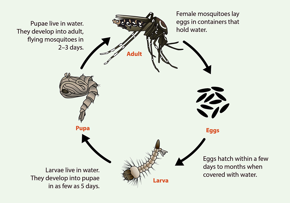
Culicidae have four life stages: eggs, larvae, nymphs and adults. The larval stage passes through three successive larval stages. Which are characterized by a noticeable increase in size, which can be of the order of 10 times from the first to the fourth stage [16]. This life cycle goes through a pre- imaginal phase that takes place in the water, it concerns the egg, the larvae, the nymph, and an aerial phase that concerns the winged adult, during this latter phase, the breeding and dispersal period occurs (Figure 1) [22].
Communicable Diseases
Culicidae family is one of the most important family of insects that cause many diseases for humans and animals. Countries with hot and humid climates are more susceptible to the spread of diseases caused by these insects due to the climatic conditions and the suitable conditions for the growth and reproduction of insects of the Culicidae family. Most of the diseases transmitted by the Culicidae family occur in the countries of East Asia, Central and South America, the Indian Peninsula and Africa. Parasitic and viral diseases are the main diseases that are transmitted by these insects, of course, bacterial diseases can also be transmitted by this group of insects, but bacteria can cause secondary infections due to blood feeding and insect bites. Here we mention the most important diseases of the region from mosquitoes of the Culicidae family.
Parasitic diseases
Malaria
Malaria is the most important disease transmitted by the Culicidae family, according to statistics published by the World Health Organization (WHO) in 2020, 241 million people in the world were infected with this disease, and 627,000 people died. Among these, the most age group involved with this parasite are children under 5 years of age, which include 77% of the total population of infected people [23]. Seven species of Anopheles from the Culicidae family are involved in the transmission and spread of four types of malaria (Plasmodium falciparum, Plasmodium vivax, Plasmodium malariae and Plasmodium ovale), which are A. Meigen, A. fuviatilis, A. culicifacies, A. dthali, A. stephensi, A. maculipenn A. superpictus [23, 24, 25]. The highest incidence of malaria is related to hot and humid areas in East Asia, Indian Peninsula and Africa, where the most common type of malaria in East Asia is P. vivax and the most common type of malaria in Africa is P. falciparum [26]. In the past years, many control programs have been carried out by the World Health Organization (WHO) to control malaria, but research between 2013 and 2018 in Burkina Faso shows that the average number of people under 5 years of age who refer to health centers for malaria is It has been increasing that this factor indicates a more detailed investigation and better control of insects [1].
Dirofilariasis
Dirofilariasis is a helminthic parasitic disease common between humans and animals, which is now considered as an emerging tropical disease. Global climate changes, especially the increase in temperature in the summer season, have increased the activity and better reproduction of this parasite and accelerated its spread. Dirofilaria spp parasites cannot reach full maturity in humans, but they have the ability to cause eye and skin lesions in humans [27]. The two main species causing filariasis that are important in medicine and veterinary medicine are Dirofilaria immitis and Dirofilaria repena [28]. Culicidae family mosquitoes such as Aedes, Culex and Anopheles are the main carriers of this parasite, but the species Aedes albopictus is considered as the most important mosquito species that transmits this worm parasitic infection in Europe and other parts of the world [29]. Dogs are the main reservoirs of this parasite, but cats can also be considered as reservoirs of this disease. Dirofilaria immitis, which is known as dog heartworm, is of great importance in veterinary medicine and infects mostly old dogs [30]. Canine heartworm disease is one of the most important insect-borne parasitic diseases of canines in the United States, and is prevalent in all southern states of the country. Worm diseases, unlike viral, bacterial and even parasitic protozoan diseases, cause longer infections for their hosts, and the best way to prevent such diseases is to conduct annual tests, especially for pets such as dogs and cats [31].
Viral Diseases
Dengue Fever
Viruses are much different and more dangerous than parasites and bacteria because they are small organisms whose laboratory tests are much different than other pathogens. Millions of people die every year due to viral diseases. Mosquitoes of the Culicidae family, which are considered as one of the most important disease vectors in the world, also play a role in the transmission of viral diseases. Dengue fever is one of the most common viral infectious diseases in the world, transmitted by Culicidae mosquitoes [32]. By 2022, 4,110,465 cases of dengue fever have been registered in the world, every year about 50 to 100 million new cases will be added to the number of people affected by this viral disease, and about 20,000 cases of this disease will lead to death [33, 34].
This virus belongs to the genus Flavivirus and has four serotypes named DEN-1, DEN-2, DEN-3 and DEN-4. Dengue fever exists in more than 100 countries of the world, including the Eastern Mediterranean, Eastern Asia, the American continent, and Africa, which have hot and humid climates, but the most important focus of this disease is the Western Pacific countries, especially Malaysia, which has about They account for 75% of all the infected people in the world. They are related to the countries of the western Pacific Ocean, it is a virus of East and Southeast Asia, especially Malaysia [35]. A. Aegypti and A. Albopictus are the most important species that transmit Dengue Fever, which causes symptoms such as fever, headache, muscle and joint pain after blood feeding and disease transmission, and in severe cases, causes bleeding in humans. Hemorrhagic fever is more common in children. Mosquitoes that transmit dengue fever are very interested in hiding in the resting place of people, especially between the walls of houses, and the best way to deal with it is to use insecticides in the seam between the walls and the rest rooms of houses [36].
Zika Virus
Zika virus is a newly emerging single-stranded RNA virus that was first isolated from a rhesus monkey in 1947 in the Zika Forest of Uganda [37]. The first human report of this virus dates back to 1953 in Nigeria. Several small epidemics of this virus have occurred throughout history, such as the 2007 epidemic in the Federated States of Micronesia, which infected 5,000 people, or the 3,200 people infected in French Polynesia in 2013 and 2014 [38]. A. albopictus, A. luteocephalus, A. furcifer and A. aegypti species play a role in the transmission of Zika virus, but A. Aegypti is the most important vector of this virus that transmits Zika virus from one human to another [39]. But there are also evidences of transmission of this virus through sex, blood transfusion, and mother-to-fetus transmission [40]. This disease has symptoms such as digestive disorders, sputum, headache, itching, conjunctivitis, which are basically latent symptoms and disappear between 3 and 12 days.
West Nile Virus
West Nile virus (WNV) is a virus from the Flavivirus family that infects most birds and its main host is birds, but humans and other animals can also be infected with this disease. It was first discovered in the 1950s in the African continent [41, 42]. In 2020, EU countries reported 316 local cases of WNV infection, most of which were reported by Greece, 143 cases, Spain 77 cases and Italy 66 cases. Eight provinces in Bulgaria, Spain, the Netherlands, and Germany have reported local human cases for the first time, indicating further spread of WNV in Europe and requiring careful surveillance and vector control [43]. Culex spp mosquitoes such as Cx. pipiens and Cx.restuans are the main carriers of this virus [44]. The clinical manifestations of this disease in humans are very rare and only children or the elderly may have flu-like symptoms that resolve on their own after several days.
Chikunguny Virus
Chikunguny virus is a virus of the Togaviridae family and was first discovered in Tanzania [45]. During the last 20 years, more than 10 million cases have occurred in more than 125 countries of the world [46]. In tropical countries with hot and humid climates, this virus causes a self-limiting fever and the patient recovers with almost absolute rest, but in cases that have happened mostly in India, it can cause severe nervous disorders and purging. It is also clinical [47]. The main carriers of this virus are Aedes mosquitoes and A. albopictus, A. Aegypti and A. polynesiensis species [48].
La Crosse Virus
La Crosse virus was first discovered in 1960 in the body of a 4-year-old girl in the state of Minnesota, USA. A. triseriatus is the main carrier of this mild virus. In general, the symptoms of this disease are similar to the flu, and in very few cases, it may cause severe disorders in humans. The United States of America is the most important center of this disease [49].
Japanese Encephalitis Virus
Japanese encephalitis virus is a virus of the Flavivirus genus and the Flaviridae family that exists in Asia, India, eastern Australia and the Pacific Ocean region [50]. The main hosts of this virus are pigs and water birds, but humans or horses may also be infected with this virus. According to the World Health Organization, in 2011, there were about 68,000 cases of this virus and about 13,000 deaths .Cx. tritaeniorhynchus is the main mosquito that transmits this virus [51].
St. Louis Encephalitis Virus
St. Louis Encephalitis Virus was first discovered in 1933. This virus, which is a virus transmitted by insects, is present in the entire American continent from Canada to Argentina, but human cases of this disease have only been discovered in the United States. Cx. pipiens, Cx. quinquefasciatus and Cx. nigripalpus are the main carriers of this disease [52].
Bacterial Diseases
Bacteria are one of the most important microorganisms in the world that have positive effects on the human life cycle, but also cause major diseases. In most cases, bacterial diseases are caused by direct contact or breathing in contaminated environments, but Culicidae mosquitoes do not play a direct role in the transmission of bacteria to humans or animals. Culicidae mosquitoes can transmit secondary bacterial infections in addition to viruses and parasites to their hosts when they feed on blood from their hosts. Staphylococcus spp, Bacillus spp, Streptococcus spp bacteria transfer to their hosts [53].
Results
We examined the most important diseases transmitted by mosquitoes of the Culicidae family, and malaria, yellow fever and dengue fever were among the most important diseases transmitted by this group of insects. Most of the diseases caused are related to hot and humid tropical regions such as Africa, Southeast Asia, Indian Peninsula, Central and South America. Due to the power of flying, Culicidae
mosquitoes have the possibility of spreading more diseases, and nowadays due to climate changes and increasing air temperature, the conditions for the growth and reproduction of these mosquitoes have improved, and these factors help the spread of diseases.
Other diseases such as Eastern equine encephalomyelitis, Venezuelan equine encephalomyelitis, Ross River, Germiston, Oriboca, etc. are also transmitted by mosquitoes of the Culicidae family, but the most important regional diseases are listed in the Table 1.
| Type of Disease (Reference) | The Name of the Disease | The Host | Family | Genus | Important Species | The Center of the Disease |
|---|---|---|---|---|---|---|
| Parasitic [24] | Malaria | Human | Culicidae | Aedes | A. superpictus - A. maculipenn - A. stephensi - A. dthali - A. culicifacies - A. fuviatilis - A. Meigen | East Asia, Indian Peninsula, South and Central America, Africa |
| Parasitic [29] | Dirofilariasis | Human and animal | Culicidae | Aedes and Culex | A. Albopictus - A. vexans - C. molestus - C. pipiens sensu lato - C. pipiens | Mediterranean countries, America, Australia, Japan |
| Viral [36] | Dengue Fever | Human and animal | Culicidae | Aedes | A. Aegypti - A. Albopictus | Eastern Mediterranean, Eastern Asia, America and Africa |
| Viral [39] | Zika virus | Human and animal | Culicidae | Aedes | A. albopictus - A. luteocephalus - A. furcifer - A. aegypti | Africa, Asia, South Pacific, South and Central America, Caribbean, southern North America |
| Viral [44] | West Nile virus | Human and animal | Culicidae | Culex | C. pipiens - C. restuans | Africa, Europe, North America |
| Viral [48] | Chikunguny virus | Human | Culicidae | Aedes | A. albopictus - A. Aegypti - A. polynesiensis | Africa, East Asia, South, Central, and North America |
| Viral [49] | La Crosse virus | Human and animal | Culicidae | Aedes | A. triseriatus | United States (eastern) |
| Viral [51] | Japanese Encephalitis Virus | Human and animal | Culicidae | Culex | C. tritaeniorhynchus | Asia, India, eastern Australia and the Pacific region |
| Viral [52] | St. Louis Encephalitis Virus | Human and animal | Culicidae | Culex | Culex pipiens, Culex quinquefasciatus, and Culex nigripalpus | South, Central, and North America |
Table 1: Important diseases transmitted by mosquitoes of the Culicidae family.
Discussion
All humans and animals are in contact with different types of arthropods during the day and night, among which insects have the most contact with different creatures. And they create major problems for the medical and veterinary community. Many researches have been conducted in different countries to investigate and control mosquitoes of the Culicidae family. In 2007, Moosa-Kazemi conducted investigations to identify different mosquitoes in the southeastern region of Iran (Sistan and Baluchistan), which is a hot and humid region on the coast of the Oman Sea, which indicates good and suitable conditions for the growth and reproduction of species [54]. Different from Culicidae family such as Cx. arbieeni, Cx. bitaeniorhynchus, Cx. deserticola, Cx. hortensis, Cx. perexiguus, Cx. pipiens, Cx. pseudovishnui, Cx. pusillus, Cx. quinquefasciatus, Cx. sinaiticus, Cx. theileri, Cx. tritaeniorhynchus, Culiseta longiareolata, Ochlerotatus caballus, Oc. caspius, and Uranotaenia unguiculata, A. vexans. It was also found that as we move towards the coast of the Oman Sea and Chabahar city, due to the increase in air humidity, the species of mosquitoes also increases [54]. Marco E. Metzger in the state of California discovered foci of Aedes aegypti and Aedes albopictus mosquitoes in 2011, and then research showed that yellow fever spread in the region in 2013, which indicates a direct connection between foci of this mosquito and it is the spread of disease. The California region in the United States of America is a region with a high population as well as a hot and humid climate on the edge of the Pacific Ocean. Create for humans and animals [55, 56, 57, 58]. Today, control methods are one of the most effective ways to reduce the number of diseases in medicine and veterinary medicine, and should be done with methods such as vaccination, ways of transmission and spread of diseases, and especially important diseases of the mosquito family Culicidae, such as Malaria, Yellow fever. The important point in controlling insects is that environmental conditions and biological pollution should be taken into account, because many health organizations use chemical poisons to control mosquitoes, which requires further investigations and taking into account the health of the human society and Animals are in that area.
Conclusion
The study of arthropods, especially mosquitoes, is of great importance and value. Our research team has also tried to point out only the important diseases in this study. Our main goal is to familiarize students of veterinary, medical, zoology, and biology departments with the basic principles and important diseases transmitted by mosquitoes, especially the Colchicine family. Given climate change and the increase in the mosquito population, it is important to be familiar with the species and diseases transmitted by them in simple and concise language. In this short study, an attempt has been made to describe all aspects of the important diseases and the species of mosquitoes that transmit the disease, but there may be other diseases and other species of mosquitoes of the Colchicine family that are less important. Therefore, other researchers can expand this study to examine other aspects of the diseases and their transmission routes.
References
-
Ghorbani A (2023) An Overview of the Science of Parasitology Simply for the General Public. International Journal of Medical Parasitology & Epidemiology Sciences 4(1): 12-18.
-
Matile L (1993) Morphology of Diptera.
-
Boulares M, Rehimi N, Houhamdi I, Baaloudj A, Soltani N, et al. (2023) Systematic and ecological study of mosquitoes (Diptera: Culicidae) at lake Fetzara (Annaba, Northeast Algeria). Ukrainian Journal of Ecology 13(1).
-
Wilkerson RC, Linton YM, Fonseca DM, Schultz TR, Price DC, et al. (2015) Making mosquito taxonomy useful: a stable classification of tribe Aedini that balances utility with current knowledge of evolutionary relationships. PloS one 10(7): e0133602.
-
Carnevale P, Robert V (2017) Anopheles: biology, transmission of Plasmodium and vector control. IRD Editions.
-
Séguy E (1955) Introduction to the biological and morphological study of dipteran insects. Introduction to the biological and morphological study of dipteran insects, pp: 260-260.
-
Menakh M (2000) The Impact of ecological factors on the degree of infestation by mosquitoes of the Culicinae subfamily in the Oum-El-Bouaghi region (Doctoral dissertation, Oum-El-Bouaghi).
-
Rioux JA (1958) Culicides of the Mediterranean South: systematic and ecological study. Lechevalier.
-
Brumpt E (1949) A Precis of Parasitology.
-
Gubler DJ (1998) Dengue and dengue hemorrhagic fever. Clinical microbiology reviews 11(3): 480-496.
-
Lounaci Z (2003) Biosystematics and bioecology of Culicidae (Diptera, Nematocera) in rural and agricultural environments. Doctoral dissertation, Master’s Thesis, National Agronomic Institute, El-Harrach, Algeria.
-
Harbach RE (2007) The Culicidae (Diptera): a review of taxonomy, classification and phylogeny. Zootaxa 1668(1): 591-638.
-
Wood DM, Dang PT, Ellis RA (1979) The insects and arachnids of Canada. Part 6. The mosquitoes of Canada. Diptera: Culicidae. Canadian Government Publishing Centre. Canada
-
Rodhain F, Perez C (1985) Precis of medical and veterinary entomology; notions of epidemiology of vector diseases. Agris FAO.
-
Owen WB (1985) Morphology of the head skeleton and muscles of the mosquito, Culiseta inornata (Williston) (Diptera: Culicidae). Journal of Morphology 183(1): 51- 85.
-
Carnevale P, Robert V, Manguin S, Corbel V, Fontenille D, et al. (2009) Anopheles: biology, transmission of. Plasmodium and vector control.
-
Becker N, Huber K, Pluskota B, Kaiser A (2011) Ochlerotatus japonicus japonicus–a newly established neozoan in Germany and a revised list of the German mosquito fauna. Eur Mosq Bull 29(88): 102.
-
Berchi S (2000) Resistance of some populations of Culex pipiens pipiens L. to malathion in Constantine (Algeria) (Diptera, Culicidae). Bulletin of the French Entomological Society 105(2): 125-129.
-
Balenghien T (2006) From the identification of West Nile virus vectors to modeling the risk of infection in the south of France.
-
Bendali F (2006) Bioecological, systematic and biochemical study of Culicidae (Diptera: Nematocera) from the Annaba region. Biological anticulicide control. Doctoral Thesis in Biology.
-
Darnis G, Robert D, Pomerleau C, Link H, Archambault P, et al. (2012) Current state and trends in Canadian Arctic marine ecosystems: II. Heterotrophic food web, pelagic- benthic coupling, and biodiversity. Climatic Change 115: 179-205.
-
Darriet F (2017) Mosquitoes and men: Chronicle of an announced outbreak. IRD Editions.
-
Wombo JBL, Ibinga E, Oyegue-Liabagui SL, Limoukou RKI, Okouga AP, et al. (2023) Severe malaria in children and adolescents in Southeast Gabon. BMC Infectious Diseases 23(1): 1-8.
-
Bafghi AF, Sepehr MM, Mozayan MR, Bagheri P, Dehghani A, et al. (2023) Passive Case Findings on Malaria in Yazd as a Central Province of Iran During 2011-2020. Iranian Journal of Medical Microbiology 17(1): 117-122.
-
Ghorbani A, Sadeghi-Nasab A, Ghaderi E, Moradi G, Nouri B, et al. (2024) Investigating the prevalence of Malaria between 2014 and 2021 in Fars province, Iran. 5th Research congress of hormozgan medical sciences student.
-
Yang Y, Liu Y, Xie Z, Wu S, Yang L, et al. (2018) Epidemiology of malaria in Yulin, South China 1999–2016: Imported Malaria threatens zero local case status. Vector-Borne and Zoonotic Diseases 18(10): 533-538.
-
Alsarraf M, Dwużnik-Szarek D, Hildebrand J, Mierzejewska EJ, Kloch A, et al. (2023) Occurrence of Dirofilaria repens in wild carnivores in Poland. Parasitology Research, pp: 1-9.
-
Șuleșco T, Thien HV, Toderaș L, Toderaș I, Lühken R, et al. (2016) Circulation of Dirofilaria repens and Dirofilaria immitis in Moldova. Parasit Vectors 9(1): 627.
-
Genchi C, Rinaldi L, Mortarino M, Genchi M, Cringoli G (2009) Climate and Dirofilaria infection in Europe. Veterinary parasitology 163(4): 286-292.
-
Raoof P, Garedaghi Y (2017) Investigation of infection with Dirofilaria immitis parasite in stray dogs in Tabriz city of Iran. Livest Sci 8: 38-42.
-
Upton KE, Budke CM, Verocai GG (2023) Heartworm (Dirofilaria immitis) in carnivores kept in zoos in Texas, USA: risk perception, practices, and antigen detection. Parasites Vectors 16(1): 150.
-
Abidemi A, Aziz NAB (2020) Optimal control strategies for dengue fever spread in Johor, Malaysia. Computer methods and programs in biomedicine 196: 105585.
-
Majeed MA, Shafri HZ, Zulkafli Z, Wayayok A (2023) A Deep Learning Approach for Dengue Fever Prediction in Malaysia Using LSTM with Spatial Attention. International journal of environmental research and public health 20(5): 4130.
-
Jamil Y, Al-Azab AM, Al-Azab F, Selwi NA (2023) Larvicidal Effects of New Organophosphorus Schiff base compounds against Dengue Fever Vector Aedes aegypti (Diptera; Culicidae). Sana’a University Journal of Applied Sciences and Technology 1(1).
-
Taib NM, Atil A (2023) Tackling Dengue Issue Need Proactive Measures: A Narrative Review of Current Level of Prevention and Control of Dengue in Malaysia. Archives of Epidemiology & Public Health Research 2(1): 148-153.
-
Seang-Arwut C, Hanboonsong Y, Muenworn V, Rocklöv J, Haque U, et al. (2023) Indoor resting behavior of Aedes aegypti (Diptera: Culicidae) in northeastern Thailand. Parasites & Vectors 16(1): 1-4.
-
Zika V (2015) Zika virus: a review to clinicians. Acta Med Port 28(6): 760-765.
-
Prasad R, Kumar K, Dohare R (2023) Caputo fractional order derivative model of Zika virus transmission dynamics. J Math Comput Sci 28(2): 145-157.
-
Hennessey M, Fischer M, Staples JE (2016) Zika virus spreads to new areas—region of the Americas, May 2015– January 2016. American Journal of Transplantation 16(3): 1031-1034.
-
Yeasmin M, Molla MMA, Masud HMAA, Saif‐Ur‐Rahman KM (2023) Safety and immunogenicity of Zika virus vaccine: A systematic review of clinical trials. Reviews in Medical Virology 33(1): e2385.
-
Calistri P, Giovannini A, Savini G, Monaco F, Bonfanti L, et al. (2010) West Nile virus transmission in 2008 in north‐eastern Italy. Zoonoses and Public Health 57(3): 211-219.
-
Aguilera-Sepúlveda P, Napp S, Llorente F, Solano- Manrique C, Molina-López R, et al. (2022) West Nile virus lineage 2 spreads westwards in Europe and overwinters in north-eastern Spain (2017–2020). Viruses 14(3): 569.
-
European Centre for Disease Prevention and Control. Epidemiological update: West Nile virus transmission season in Europe.
-
Landesman WJ, Allan BF, Langerhans RB, Knight TM, Chase JM (2007) Inter-annual associations between precipitation and human incidence of West Nile virus in the United States. Vector-Borne and Zoonotic Diseases 7(3): 337-343.
-
Soumahoro MK, Boelle PY, Gaüzere BA, Atsou K, Pelat C, et al. (2011) The Chikungunya epidemic on La Reunion Island in 2005–2006: a cost-of-illness study. PLoS neglected tropical diseases 5(6): e1197.
-
Souza WMD, Lima STD, Mello LM, Candido DS, Buss L, et al. (2023) Spatiotemporal dynamics and recurrence of chikungunya virus in Brazil: an epidemiological study. Lancet Microbe 4(5): e319-e329.
-
Tandale BV, Sathe PS, Arankalle VA, Wadia RS, Kulkarni R, et al. (2009) Systemic involvements and fatalities during Chikungunya epidemic in India, 2006. Journal of Clinical Virology 46(2): 145-149.
-
Bartholomeeusen K, Utt A, Coppens S, Rausalu K, Vereecken K, et al. (2018) A chikungunya virus trans- replicase system reveals the importance of delayed nonstructural polyprotein processing for efficient replication complex formation in mosquito cells. Journal of Virology 92(14): 10-128.
-
Bennett RS, Cress CM, Ward JM, Firestone CY, Murphy BR, et al. (2008) La Crosse virus infectivity, pathogenesis, and immunogenicity in mice and monkeys. Virology journal 5: 1-5.
-
Zhu Y, Chen S, Lurong Q, Qi Z (2023) Recent Advances in Antivirals for Japanese Encephalitis Virus. Viruses 15(5): 1033.
-
Su CL, Yang CF, Teng HJ, Lu LC, Lin C, et al. (2014) Molecular epidemiology of Japanese encephalitis virus in mosquitoes in Taiwan during 2005–2012. PLoS neglected tropical diseases 8(10): e3122.
-
Curren EJ, Lindsey NP, Fischer M, Hills SL (2018) St. Louis encephalitis virus disease in the United States, 2003–2017. The American journal of tropical medicine and hygiene 99(4): 1074.
-
Aljoboory RKI, Lafta SM (2022) Diagnosing Different Types of Bacteria on Culex Mosquito Spp.(Diptera: Culicidae) in Baghdad. Cureus 14(12).
-
Moosa-Kazemi SH, Vatandoost H, Nikookar H, Fathian M (2009) Culicinae (Diptera: culicidae) mosquitoes in chabahar county, sistan and baluchistan province, southeastern iran. Iranian Journal of Arthropod-Borne Diseases 3(1): 29.
-
Metzger ME, Yoshimizu MH, Padgett KA, Hu R, Kramer VL (2017) Detection and establishment of Aedes aegypti and Aedes albopictus (Diptera: Culicidae) mosquitoes in California, 2011–2015. Journal of Medical Entomology 54(3): 533-543.
-
Garedaghi Y, Branch T (2011) Flea infestation in farm animals and its zoonotic importance in East-Azerbaijan province. American Journal of Animal and Veterinary Sciences 6(4): 192-195.
-
Raoof P, Garedaghi Y (2017) Investigation of infection with Dirofilaria immitis parasite in stray dogs in Tabriz city of Iran. Journal of Livestock Science 8: 38-42.
-
Garedaghi Y, Khaki A (2014) Evaluation of the Effectiveness of Ethanolic Extract of Solanum Surattense against Plasmodium Berghei in Comparison with Chloroquine in Sourian Mice Using in Vivo Tests. Crescent Journal of Medical and Biological Sciences 1(3): 76-79.
- hMPV: Is It Another Covid-19 Like Situation?
- Streptomyces: Sources of Novel Discoveries in Antibiotic Research to Combat Antimicrobial Resistance
- Past and Current Immunotherapy in Cancer
- Hematological Cancer and Viral Infection
- The Growing Threat of Antimicrobial Resistance in India: Challenges and Solutions
- The Role of NEU1 in Coronavirus Infection and Pathogenesis