
Citation: Wong TH. Single Stage Instead of Two-Stage Bilateral Total Knee Arthroplasty to Avoid Leg Length Discrepancy in Severe Varus Knee Deformity- Propose a Mathematical Model to Support the Hypothesis. J Ortho Bone Disord 2017, 1(2): 000108.
*Corresponding author: Tze Hong Wong, Department of Orthopedics, National Taiwan University Hospital Hsin-Chu Branch, Hsin Chu, Taiwan, ROC Taiwan, Tel: +886-972654018; Email: tzehongwong@gmail.com
Single total knee arthroplasty (TKA) may be beneficial for its one admission, one anesthesia, cost reduction and faster recovery including shorter rehabilitation. However, controversial opinion against the idea for its higher complication rate, such as deep vein thrombosis and more blood loss. Limited discussion upon the point of leg length discrepancy (LLD) as a result of one side TKA in severe varus knee deformity. In this entity, single stage TKA may gain its significant beneficial effect to avoid LLD. A mathematical model is proposed to calculate the LLD severity in a standardized condition and provide the formula to predict LLD before a two stage bilateral TKA is going to be performed.
Method and MaterialBefore to set up the formula, some assumptions are to be made as following:
The original leg length 2H will be changed to 2P due to the correction of the varus deformity angle ϴ to 6 degree valgus.
2H = R x Cos ϴ/2 + R x Cos ϴ/2………1
2P = R x Cos 3° + R x Cos 3°…………..2
LLD = 2P -2H = 2R x ( Cos 3° - Cos ϴ/2 )……….3
If R = 50 cm (for standard patient )
LLD = 100 x ( Cos 3° - Cos ϴ/2 )
According to above table, for a patient with 50cm femur and tibial length, LLD up to 6.6 cm may be consequently happened in a severe 30 degree bilateral varus deformity (Figures 2-7).
In above example post Op LLD was noted due to correction from 16 degree varus to 6 degree normal Valgus.
This patient received single stage bilateral TKA and no LLD noted as correction to bilateral normal valgus 6 degree at the same surgery.
DiscussionLLD = 2R x (Cos 3° - Cos ϴ/2 )
The formula provided can be a predictive tool to calculate the possible LLD before a two stage TKA for severe angular deformity knee.
According to Figure 2, less than 10 degree deformity may be considered as ignorable as only up to 0.4 cm LLD will be resulted. On the other hand, more than 16 degree deformity seems to be unacceptable because of 1.6 cm LLD noted
For the patient whose Femur and tibial length R’ can also make use the table of Figure just simply multiply the LLD by R’/R ratio.
The formula above is a reasonable hypothesis to obtain helpful and useful data before difficulty TKA cases are going to be performed. More accurate measurement will be performed in the future for more practical and clinical purposes.
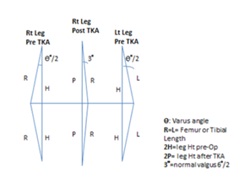
Figure 1
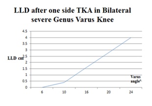
Figure 2: LLD after one side TKA in Bilateral Severe Genus Varus Knee.
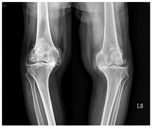
Figure 3: severe varus pre-Op.
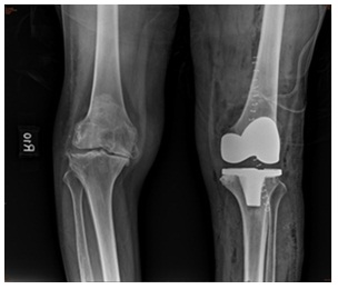
Figure 4: Lt TKA correction of varus to 6 degree Valgus but LLD noted.
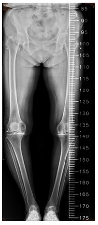
Figure 5: Pro-Op Scanography.
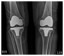
Figure 6: Post-Op Standing View.
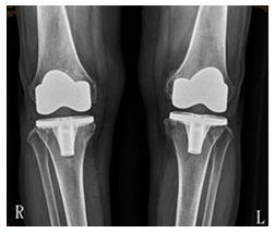
Figure 7: Three Months Post Op.
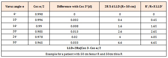
Table 1


