Citation: Jagema T and Jarso D. Study on Epidemiology and Socioeconomic Impact of Epizootic Lymphangitis in Carthorses in Southwestern Shoa. J Vet Sci Res 2016, 1(3): 000114.
*Corresponding author: Jarso D, Addis Ababa University, College of Veterinary Medicine and Agriculture, PO Box: 34, Bishoftu, Ethiopia, Email:djmojo74.ethio@mail.com
A study was conducted between December 2008 and April 2009 on 705 carthorses in three towns of Southwestern Ethiopia namely Sebeta, Asgori and Woliso to investigate the Epidemiology and Socioeconomic impact of Epizootic lymphangitis (EL). The study has revealed an overall prevalence of 25.1% (177/705). There was no statistically significant (X2=3.88, P>0.05) difference on the occurrence of the disease in the three districts. The highest prevalence being observed at Woliso with 27.9% and the lowest was at Sebeta with 20.2%. The result of histopathological and mycological examinations has revealed characteristic features of HCF. Result of differential leukocyte count had shown a statistical significant difference across the severity of the disease and neutrophil count (r=0.87, F=6.08, P<0.005) while lymphocyte count were inversely related to the severity stage of the disease (r=0.94.F=23.28, P<0.001). The yeast forms of Histoplasma capsulatum var. farciminosum were isolated on the sabouraud’s dextrose agar. The result of questionnaire survey from 35 carthorse owners indicates that EL was the first and the major important disease of carthorses by creating a negative impact on the economy of the carters. Despite its impact, awareness on the transmission and control mechanism of the disease was not uniformly known by the carthorse owners. Therefore, further study on the extent of the disease and educating the owners both on the disease and its way of transmission was recommended.
Keywords: Carthorse; Epizootic lymphangitis; Prevalence; Socioeconomic impact
According to FAO (1999) [1] the world horse population is 60 million of which 8% is found in Africa, of this Ethiopia possesses 2.75 million horses, 5.02 millions donkeys and 0.63 million mules. In northwestern part of Ethiopia, equids have been used not only for transportation of agricultural products but also for ploughing activities showing their contributing in crop production [2]. Furthermore, as the topography of the rural part of the country is not convenient for the modern transport technology. Thus, the major means of transportation of both goods and man are equines. Despite all these uses, they are suffering from several diseases such as Epizootic lymphangitis (EL), African horse sickness, ulcerative lymphangitis, dourine, rabies, anthrax, horse mange and babesiosis [3]. Among these, Epizootic lymphangitis seems the first in Ethiopia [4].
Epizootic lymphangitis, grouped as a list B disease of the Office International des Epizootics, was first described in 1873 by Rivolta as a chronic infection of horses, mules and donkeys caused by the thermally dimorphic fungus, Histoplasma capsulatum var. farciminosum, and is characterized clinically by a spreading, supurative, ulcerative pyogranulomatous dermatitis and lymphangitis. The organism has also been classified by the genus name Zymonema, Cryptococcus, saccharomyces, or Blastomyces [5]. The parasitic phase is yeast-like, and the soil saprophytic phase is mycelia, being characterized by septated and branched hyphae [6,7] which until recently was known as Histoplasma farciminosum [8]. HCF causes the induction of both humeral and cellular immunity [9,10] because the tissue form of organism can reside both intracellular and extracellular [11,12].
Three varieties of dimorphic ascomycete H.Capsulatum were identified: (1) H.capsulatum var. Capsulatum, the cause of histoplamosis in animals and humans; (2) H.Capsulatum var. duboisii, the cause of a distinct variant form of disease in humans, African histoplasmosis; (3) H.Capsulatum var. farciminosum (HCF), the agent causing epizootic lymphangitis in horses in Africa, the Middle East, and parts of Asia. Only H. Capsulatum var. capsulatum is found in North America [13].
The disease occurs as outbreaks in horses, donkeys and mules in parts of Iran, Asia, India, Northern Africa, and the Mediterranean littoral. Most outbreaks occur in autumn and winter or when large numbers of horses are gathered together for military or other purposes [5]. Rare cases of human infection have also been reported [14,15]. After gaining entry through wounds, the fungus invades subcutaneous tissue, sets up a local granuloma or ulcer and spreads along the lymphatic vessels. It has three forms, namely the cutaneous, the ocular and pulmonary depending on the route of entry [14]. The ocular form of the disease results from inoculation of the organism in to the eye, likely by biting flies [5]. The skin form of the disease occurs when contaminated soil contacts traumatized skin; the conjunctiva form is believed to be spread by flies while, the pulmonary form occurs after inhalation of organism [16].
The disease is primarily an ulcerating, suppurative, pyogranulomatous dermatitis and in most cases lymphangitis [14]. Thickening of the skin in the area and general swelling of the whole limb are common signs and the lesions are quite painless. The lesions usually develop on the limbs, particularly about the hocks, but may also be present on the back, sides, neck, vulva and scrotum. Occasionally, lesions appear on the nasal mucosa just inside the nostril and do not involve the nasal septum. Ocular involvement is manifested by keratitis and conjunctivitis, sinusitis and pneumonia occur in the pulmonary form of the disease [5].
Histopathological, a characteristics pyogranulomatous reaction with fibroplasias and with a predominance of macrophages and giant cells is observed. Numerous ovoid to round yeasts 3-5 x 2.5-3.5µm in diameter are found both extracellular and intracellular in macrophages and giant cells that are usually surrounded by a ‘halo’ when stained with gram stain or Haematoxyline and Eosin [16].
Lesions are usually confined to the skins subcutaneous tissue, lymph vessels and lymph nodes most commonly in the extremities, chest wall, and the neck. It can also be present as ulcerative conjunctivitis of the palpebral conjunctiva, or as multi focal pneumonia. The diagnosis of EL is based on the clinical examination of the lesions, microscopic examination of the yeast form of HCF in pus, serological tests, and skin hypersensitivity testing [14].
Histoplasma farciminosum is usually cultured at 25○C SDA enriched with 2.5 percent glycerol, or pleuropneumonia–like organism (PPLO) nutrient agar enriched with 2 percent dextrose and 2.5 per cent glycerol and a PH -8 at 25°C [17]. Addition of antimicrobial to the medium is recommended cycloheximide (0.5g/l) or chloramphenicol (0.5g/l). After two to six weeks of culturing, dry, yellow to dark brown, granular and wrinkled colonies appear. Dimorphism is tested by growing the fungus at 37○C in its yeast phase on BHIA enriched with 10 % horse blood in an atmosphere of 20 % carbon dioxide complete conversion to the yeast phase may, however, be achieved only after four to five repeated serial transfer on to fresh media every eight days. Yeast colonies are flat or raised slightly or deeply wrinkled, and white to light or grayish to brow, and have a pasty consistency [17].
The result of the socioeconomic study conducted by Ameni and Siyoum (2002) [18] indicated that an estimated loss of US $ 3000 annually due to morbidity and mortality of carthorses. In general, the results of a few studies conducted in the country have shown the rampant occurrence of the disease and to some degree its socioeconomic implications and thus warrant for appropriate control strategies so that its impact is minimized [4].
Many treatments have been tried largely without success. Parental Iodides and Amphotericin B have been reported as treatment options. Sodium iodide is administered as a 10% solution at a dose of 1ml per 5kg intravenously once weekly for 4 weeks [12]. Amphotericin B is administered at a dose of 0.2mg per kg every 48 hours for three treatments [5]. A combination of Sodium iodide and penstrip was found to have higher therapeutic values on EL cases as compared to the other drugs [4]. Out breaks in uninfected areas are best controlled by slaughter of affected animals. In enzootic areas severe case should be destroyed and less severe cases kept in strict quarantine while undergoing treatment. All infected bedding, harness and utensils should be destroyed or vigorously disinfected. Formularized aluminum hydroxide adsorbed, and heat attenuated vaccines have been widely used apparently with success [5].
Despite the rampant occurrence of the disease, very few studies have been conducted in Sebeta, Asgori and Woliso towns. Therefore, the present study was conducted to show the impact of the disease and its distribution across those towns. The specific objectives of this study were:
Description of Study Area
The field study was conducted in three towns namely Sebeta, Asgori and Woliso. In these towns, carthorses are used for the transportation of people and goods. These towns were selected because of the availability of epidemiological information on EL and their proximity to the main laboratory.
Sebeta town: It is situated within Alemgena district, about 25 km Southwest of Addis Ababa, on Jimma road. It has on altitude of 2260 m a.s.l. and an average annual rainfall of 95.1 mm, temperature 23.94○C and relatively humidity of 49.3 %. There are 368 formally registered horses drawn carts in the town [19].
Asgori Town: It is located 60 km Southwest of Addis Ababa, on Jimma road. It has on altitude of 2060 m a.s.l and mid low land topography with hot and humid weather condition. It has an annual rainfall ranging from 150-650 mm and a temperature of 15-25○C. There are 1100 formally registered horses drawn carts in the town [19].
Woliso Town: It is located 114 km Southwest of Addis Ababa, on Jimma road. It has an altitude of 2800 m a.s.l. The topography is mid low land. The weather condition is hot and humid. There are 230 formally registered horse drawn carts in the town. It has an annual rainfall ranging from 950-1200 mm and a temperature of 10.5-25○C [19]. The laboratory was carried out at the Aklilu Lemma Institute of Pathobiology a Biomedical research Institute of the Addis Ababa University, located in the southern campus of the Technology faculty of the same University and National Animal Health Diagnostic and Investigation Center /NAHDIC/ located in the Sebeta town.
Sampling of Study Animals and Study Design
The study involves cross-sectional and purposive methods to estimate the prevalence of the disease and to evaluate the profiles of leukocytes across different stage of the disease respectively [20]. Classification of case in to four severity stages (mild 1 (01), mild 2(02), severe 1 (03), severe 2 (04), was done on the basis of clinical examination as previously described by Endebu (1996) [21] and Hadush et al. (2008) [4].
Mild stage 1: cases with a cutaneous nodule or nodules on any parts of the body, detectable either visually or via palpation.
Mild stage 2: case with clinical lesions characterized by the formation of line of infection along the lymphatic consisting of 2-3 nodules and /or ulcers oozing serous fluid.
Severe stage 1: cases with extensive abs cessation and ulceration of the superficial lymphatic vessels, coalescence of adjacent ulcers.
Severe sage 2: nodules and “cording” of the lymphatic vessels with involvement of any two or more parts of body.
Survey of EL in carthorses
A total of 705 carthorses were visited and recorded on parking stations and on marketing days. The market days were chosen because carthorses were used as beast of burden to carry crop products and other goods to the markets and therefore, were seen in relatively large numbers on these days. The limbs, sterna region, scrotal region, oral region, head region, and cervical regions of each carthorse were visualized and examined for clinical lesions and animals which had suggestive nodular and ulcerating lesions along the lymphatic vessels were recorded as positive for EL. Those carthorses which have healed lesions or scars were recorded also as positive for EL. For further confirmation from clinically positive animals, pus samples were taken from lesions and brought to the laboratory for direct microscopic examinations and culture.
Microscopic and Mycological Examination
Direct Microscopic Examination of pus: Samples for microscopic examination were collected from closed nodules with the aid of a sterile syringe on a clean microscopic slide. Fresh smears were prepared and subjected to methanol fixation on the field and brought to the laboratory for gram staining [22] and Giemsa staining [23] and examined under microscope.
Cultivation of Histoplasma Capsulatum var. Farciminosum: The contents of unruptured nodules were aseptically aspirated using sterile needle and syringe, and inoculated aseptically in to screwed universal bottles containing slants of Sabourauds Dextrose Agar (SDA, Oxide) enriched with 2.5% glycerol containing chloramphenicol (0.5 gm /liter). For the isolation of the mycelial form, incubation was made at 26○C while isolation of the yeast form was made at 37○C [14]. The media were checked periodically for growth of HCF.
Gross and Histopathological Examinations
For post mortem and histopathological examinations, one stray horse was brought from town and post mortem examination was done after killing the animal with intravenous administration of simulate, with a recommended dose of 1ml per 10kg body weight. Spleen, liver, kidney, lungs, heart and lymph nodes were examined one by one and samples were taken from these organs for histopathology in 10% formalin. Small tissue was placed in a tissue basket and washed for 24 hours. Tissues were dehydrated in the 70%, 80%, 90%, and absolute ethyl alcohol for 4, 4, 2 and 1 hours in each preparation respectively. The volume of alcohol was made to be 50% of the volume tissues. Then the tissues were inserted in cedar wood oil for clearing. After the specimens kept overnight in the cedar wood oil, the tissues were put in to the xyline for few seconds [24]. Xyline treated tissue then allowed to be changed in well melted paraffin wax for three changes of which the tissues expected to be embedded (impregnated). The paraffin wax was melted in 62○C for 24 hours. In the paraffin wax the tissues were kept for 1 hour of which three changes are made. For impregnation the amount of temperature used was 65○C. Immediately after the last change tissues were blocked with a paraffin wax. Then the tissues were allowed to cool and dry within the paraffin block. And then the tissues were prepared for sectioning. The feed mechanism on the microtome was turned back almost as far as gone its last limit. Then the appropriate knife inserted in holder and screwed tightly in position and checked that the tilt of the knife is set at the correct angle. The block holder was moved to forward, upward, downward and backward until the block came almost touching the knife. After insuring the edge of block is parallel to the edge of the knife, the surfaces of block were trimmed by making the section thickness gauge to 15µm and the microtome operated until the sections the tissues were cut. The thickness gauge was adjusted at 5µm and the microtome operated gently until good sections were coming [24]. During cutting the tissue ribbons were compressed and creased. For this reason before they became attached to the slides, the creases removed and flattened with warmed water at a temperature of 50○C. The tissue ribbons which were good sections were allowed to float on warm water in the flotation water bath. As the sections came flattened they were lifted on albumenized slide (smeared with Mayer’s egg albumin which was prepared from equal amounts of egg white and glycerin) by brought in contact with the section. Then the slides allowed drying on an oven. The sections were deparafinized with three changes of xyline for 5 minutes each. Then placed in absolute alcohol, 90% alcohol, 80% alcohol, 70% alcohol, for 2 minutes each. Then the slides hydrated with tap water. The hydrated slide then stained with Haematoxyline for 15 minutes and washed with running water for 15 minutes. Then after it counter stained in the eosin for 2 minutes, the section became dehydrated by dipping once only in ascending grades (70%, 80%, 90%, absolute) of alcohols. Then three changes in the xyline made and after waiting for 2 minutes in each the slides mounted with DPX. The stained slide was examined for the presence of pathological changes in the 10x, 40x and 100x microscopes [24].
Differential Leukocytes Count
Blood sample for differential leucocytes count were collected from the jugular vein of eight cases of EL with different severity stages as grouped by Hadush et al. [4]. Then smears were subjected for methanol fixation in the field. The smears were then stained with Giemsa stain in the Laboratory. Differential leucocytes count was performed using oil immersion microscope [4].
Questionnaire Survey
The socioeconomic impact of the disease was assessed through prepared questionnaire format (Annex-1). From this study a total of 35 carthorse owners were asked.
Data Analysis
Data entry and analysis was made by SPSS Ver.10 statistical Software. Percentage was used to evaluate the socioeconomic impact and prevalence of the disease. Continuous variables were summarized using averages. Correlation coefficient was used to assess degree of linear relationship between severities of EL and differential leukocyte count. All results were reported as significant if P-value is less than 0.05.
ResultsPrevalence of the Disease
EL was diagnosed based on clinical examination of characteristic lesions and confirmation was made through identification of the yeast from pus or swab smears through gram stain. Table 1 shows the overall prevalence of EL recorded by the present study at the three towns. The overall prevalence of EL in carthorses at three towns were 25.1% (177/705) and 27.9% (57/204) at Woliso, 20.2% (41/203) at Sebeta and 26.5% (79/298) at Asgori. The difference in prevalence between the three towns was association but not significant (X2 =3.88, at df =2, P >0.05). However there was significant (severity: X2=139.5, P<0.001; clinical positive: X2=17.4, P<0.001 and site: X2=17.4, P<0.001) association between those variables and EL lesion in carthorse.
Characteristic and Distribution of Lesions
In the study area, the cutaneous form of the disease was most frequently seen. The local lymphatic was involved. Almost all of the cases seen had one or more nodular lesions on different body parts most commonly on sterna region, extremities, scrotal region, head region and cervical region. Recently erupted nodules up on palpation were firm and freely movable over the sub cutis, in some cases they were observed to occur in a line following lymphatic vessels appearing as a rigid knotted rope. Aged and un ruptured nodules were usually flabby with no hair and smooth tip indicating the site of rupture, in severe cases ruptured nodules were observed to be arranged in a line and discharging white to yellow pus. A few case of pulmonary EL with bilateral nasal discharge and cutaneous lesion were found. In advanced cases the nodules and ulcers were found to involve almost all body parts with unpleasant odor.
Post mortem and Histopathological Examinations
Post mortem Examination: Post mortem examination of the observed case (Plate 1) revealed cutaneous multifocal suppurative lymphangitis, ulcerative suppurative dermatitis, in the involved areas. All internal organs looked normal but multifocal pneumonia was seen on lungs.
Histopathological Examinations: Histopathological examination of tissue collected from liver when stained with Haematoxyline and Eosin; there was excessive thickening of capsule (cirrhosis) due to fibrous tissue proliferation as well as infiltration of mononuclear cells. There was haemosidirosis in the portal triad areas as well as sinusoids. Hepatic cells showed different degenerative changes that is cloudy swelling and changes. The cells were enlarged having large nucleus and eosinophilic granular cytoplasm. Besides, the lesions consist of pyogranulomatous inflammation with fibroplasias. The presence of numerous yeasts in tissue sections stained with H and E were seen.
Differential Leukocyte Count
Figure 1 shows the association between severities of EL and neutrophil count. The association between severity of EL and neurophil count was statistically significant (r=0.87. F= 6.08, P< 0.005. On the other hand also shows statistically significant (r=0.94, F=23.28, P< 0.001) inverse relationship was observed between severity of EL and lymphocyte count.
Mycological Investigation
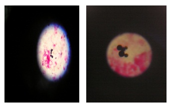
Direct Microscopic Examination:Gram stain smears from pus revealed Gram-positive yeast forms with a halo (unstained capsule like) structure around them. The yeast forms were lemon-shaped with one edge wider and the other bluntly pointed. They could occur individually or in groups either free or intracellular phagocytes with in macrophages. Yeast forms with whole unstained transparent lemon shaped cytoplasm space and some of these forms were seen in a state of budding from one end.
Isolation of the Yeast and Mycelia forms: The colonies of the yeast form appeared flat, raised, wrinkled and white to grayish white in color and pasty in consistency (plate 5) but the mycelia form not grown.
Questionnaire Survey
The result of an interview indicated that the EL was increased from time to time and cause significant impact on the carthorse owners. It indicates that among the disease that affects carthorses, EL is considered as the first and major important disease that kills their horses there by subjecting them to poverty. The result of questionnaire survey from the carthorse owners indicates that an estimated loss of 1, 600.00 Ethiopian birr because of death of affected horse. According to this study, it was observed that there was an average loss of 40 ETB per day per carthorse owner due to decreased working capacity (morbidity) and unproductive feed expense. As the disease progresses, the working power of affected horse decreases gradually and, the cost of feed for horse exceeds than the daily income. The final fates of severely sick horses were left outdoors for scavengers.
DiscussionThe present study has tried to determine the prevalence of the EL, the profile of leukocytes count on different severity stages of the disease, characteristics of histopathological lesions and its socioeconomic impact. The observed constraint was that the numbers of study cases per the different severity stages were low due to the unwillingness of the carthorse owners for giving blood sample. Regardless, of the above indicated constraints, the result of this study indicated that EL was endemic to the study areas with an average prevalence of 25.1%. This might be due to that the affected and susceptible horses were stabled together at marketing place and carthorse parking stations, the presence of stray horses, sharing contaminated harnesses between horses and lack of awareness of the carthorse owners for transmission of the disease. Similarly, damage of the skin because of improper harnessing system and carry on abrasive materials favor the entry of pathogen. A similar study conducted by Endebu [21] indicated that histoplasmosis farciminosi is the first most important disease of carthorses at Debre-zeit and Akaka towns and an average prevalence of 21% was recorded from the data collected from 28 towns that use carthorses for the transportation of man and goods [18]. The highest prevalence was recorded at Mojo (39%) followed by Ejaji (36.5%), Bati (36%), Woliso (33%) and Debre-zeit (29%).
As indicated by previous workers [4,8-10,18,21,] EL was observed to affect any parts of the body with frequent exposure to injury are highly affected. In carthorses, harness that pass across the chest cause frequent wounding of the chest, which facilitates the entrance of the organism [18,21,23].
The Results of differential leukocytes count revealed that neutrophilia in advanced cases which was in agreement to Al-Ani and Al-Delaimi (1986) [11] and Hadush (2004) [4] who reported high neutrophil count in advanced cases of EL. This was because as the disease progresses the type and number of bacterial contaminants increase thereby causing proliferation and infiltration of neutrophils [25]. On the other hand, lymphocyte had statistical significant inverse relationship with severity of cases. This was due to HCF is dominantly intracellular organism and stimulates proliferation of lymphocyte [4]. Histopathological liver showing mixed population of neutrophils, lymphocytes, macrophages and multinucleate giant cells were seen when stained with Haematoxyline-Eosin but spleen, lungs, lymph nodes, kidney and heart were not stained with H and E.
The result of an interview conducted on 35 carters showed that EL was increased from time to time except at Woliso. The relative decreased prevalence of the disease from previous reports of [18] in Woliso could be attributed to season of the year. According to them EL locally is called “Sarare” or “Nidft” and is termed as “Horse’s AIDS’ as infected horses do not recover; is the first and major important disease of carthorses that kills their horses thereby subjecting them to poverty. The economic losses which were now being caused by the disease because of reduced performance of the horse and death of affected animal was a very serious problem. The infected animals were left outdoors as the disease attains severe stage since they act as a source of disease transmission.
Although, the survey result indicated that EL was considered to be the first and major disease of carthorses, the method of transmission of the disease and its control mechanism were not uniformly known throughout of all the study areas but some carters seriously complain the problem after the disease have been occurred. This indicates that there is a need of awareness creation in the society on the extent and transmission of the disease.
The result of the socioeconomic study conducted by Ameni and siyoum [18] indicated that there is an estimated loss of US $ 3000 annually due to morbidity and mortality of carthorses in Ethiopia. EL is endemic to Ethiopia and has been recorded almost in all veterinary clinics across the country and its distribution covers humid and hot areas with altitude arranging from 1,500 to 2,300 meter above sea level [26]. Study conducted so far shown that it is the first most important disease of carthorses in several towns. The affected skin and subcutaneous tissue is thickened, fibrous, and firm lymphatic vessels are distended. Transmission generally involves infection of wounds by flies and contaminated by feeding on the open wound of infected animal [12,10]. The organism has been isolated from the gastrointestinal tracts of flies [12]. Because of all the study areas have hot and humid climates promote the survival of the environmental form of the HCF. Further, such environmental conditions favour the breeding of flies, which play a significant role in the transmission of EL. Sick horses which have lesions (nodules or/and ulcers) became restless because of infestation of the lesions with flies.
There is progressive loss of appetite and condition as the severity of the disease increases. In this study, it was observed that the owners handled affected animals inhumanely in that they provided no veterinary care, no nursed them, and no provided adequate feed and water. The reason for this appears to be that most (probably > 95%) of cases do not recover and owners consider that looking after such horses in simply a waste of money and energy. Owners exploit the power of sick horse until its health deteriorates whereupon the animal is left outdoors for scavengers after it becomes unable to pull the cart and the disease reached it severest stage.
So far, the veterinary services of the country have given little attention to EL although the present study suggests that EL is an important endemic disease of equids in the southwest parts of Ethiopia and causing great economic losses and social crisis. The disease does not have effective treatment so far and the final fate of sick horses is to be left outdoors for scavengers. The disease is rarely responding to treatment. One of the reasons could be due to the complication of the lesions with different bacteria in addition to its chronic nature. In general,Histoplasma Capsulatum var. Farciminosum is less responsive to antibiotics. Nevertheless, its response to drugs like Amphotericin B, sodium iodide and Clotrimazole has been reported from other parts of the world [4]. When it comes to the Ethiopian situation, there is no drug prescribed to Epizootic lymphagitis in any of the veterinary clinics across the country. As a result of absence of treatment, sick animals are left out doors for scavengers after their working power is exhausted.
Conclusion and RecommendationsThe result of the study has shown that EL was prevalent and endemic to the three towns and the distribution of the disease across the towns was increasing from time to time. The prevalence of the disease increases when horses stabled together in marketing places and parking stations, sharing contaminated harnesses and use of in appropriate harnessing material. The economic losses associated with a high prevalence, reduced work performance of a horse and death of affected animals was a very serious problem for carthorse owners. From the result of differential leucocytes count, there was neutrophilia in advanced cases and one can imagine that the death for EL would be bacteremia in addition to starvation and the effect of HCF. On the other hand, lymphocytes decreased as the disease severity increases. On the basis of this conclusion the following recommendations are forwarded:

Plate1: Horse: circumscribed nodular lesions on the skin of left limb.
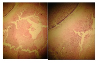
Plates 2 and 3: Microscopic appearance of liver tissues, showing mixed population of neutrophils, lymphocytes, macrophages and multinucleate giant cells (Haematoxyline-eosin stain, 100X).
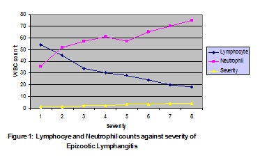
Pigure 1: Lymphocye and Neutrophil counts against severity of Epizootic Lymphangitis
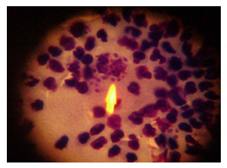
Plate 4: Giemsa stain of pus from un raptured nodules showing yeasts.
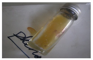
Plate 5: Colony of yeast form on SDA
Plates 6 and 7: The yeast observed under microscope from culture of HCF.
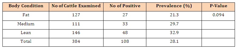
Table 1: Prevalence of Epizootic lymphangitis in carthorses in three towns of Southwest Ethiopia.


