Alien Trace Metal Elements in the Body: Changes May Explain Recent Epidemics and Diseases
It is now well-known that many elements of the periodic table find a way into our bodies and have the potential for disturbing the delicate homeostasis (balance) of body chemistry. Nevertheless, in spite of the volume of research published in this area, only vague suppositions result concerning any involvements and little progress has been made concerning human health or addressing the preponderance of the innumerable epidemics that now are evident. This results from the inability for statistical analyses to be sufficiently precise to pinpoint specific causes. As a result, a simpler analysis is presented herein based on the premise that environmental chemical epidemics can only arise from an increased change. As a result, global diets and lifestyles have been examined in detail to establish items of change. Such a needed criteria in the recent decades poses strict requirements eliminating most candidates. Two areas of rapid growth in life-style changes clearly are evident and involve the diet of fish (sushi) (high methyl mercury) and the increase in medical vaccines (aluminum hydroxide). It is apparent that people now live never knowing whether they are at risk from these due to genetic susceptibilities. This is especially important for women of child-bearing years, where a fetus is always at high toxic risk levels. As a result, the availability of general population testing now is desperately needed especially for the neurotoxins, Hg, Al, As, and Pb, all alien species to the body together with Se that appears to be the body’s natural healthy chelator.
Introduction
The human body is a remarkable example of excellence in design. It displays automatic self-sufficiency, survival maintenance and repair. Unfortunately, we humans casually accept this, generally think very little of it, until it malfunctions. Then we more fully appreciate these built- in capabilities, but soon continue our lives fully hoping that this finely tuned mechanism will continue unchecked for an extended longevity. Various epidemics of illnesses have always arisen over the centuries, occur naturally, are often unexpected and in the past have been caused by some modified viral form. Historically, this has always been a factor of life and accepted as a hazard. However, times now appear to differ, and some current concerns that are at epidemic levels are no longer only viral but appear to center more on environmental causes, and for which the body has no response. This has initiated necessary new conjectures by the medical community that previously often had known treatments, either a vaccine or a medication to control or cure such an event. Now, with no understanding in many cases, the introduction of environmental aspects in the modern age poses problems with impossible long lists of potential causes. It would appear to be a very difficult if not an impossible task to unravel any single item or group for explanations. This is partially true, but if the cause produces an epidemic this introduces innumerable required conditions that are necessary and begins to eliminate many potential suggestions. The medical literature is now ripe with such exercises and has considerably reduced the options. Additionally with neurological epidemics, the possibilities are even more restrictive, as the brain itself has to be affected. Such epidemics if not common in the past also need the basic factor of a needed change. As a result, although we now live in the realm of genetic explanations, our genes are not apt to suddenly change drastically. As a result, genetic explanations are necessarily not a primary but have to be a secondary component if involved, possibly due to epigenetic changes that are now being suggested as moderating genetic susceptibilities [1]. This paper concerns environmentally induced epidemics that have arisen, are global, and have become apparent in recent decades. A generally accepted consensus now is that these appear connected to changes in life-style that have occurred in modern-day living. As a result, partly based on such concerns, the field of medicine has turned more aggressively to a better understanding of body chemistry, its balances and the nature of possible changes. This has been facilitated by advances in analytical ability such that in recent years it has become possible to monitor numerous components of the human body through analysis of its measurable components. This has resulted in an overwhelming wealth of publications that for medicine introduces the further difficult task of analysis. If anything, it has illustrated more clearly how every human is different [2]. As a result, even with extensive statistical aid, analyses become limited in value due to the array of variable parameters and the necessary variations introduced in any collected data. The dilemma is that although it would seem obvious that if a large sample of a control group are compared to a group that has a certain illness, the differences should clearly imply a role from any apparent observed correlations. The problem is that the control sample may not be a truly rigid constant representative group in that its body chemistry can constantly vary depending on behavior and many other factors. This is true for the test case group also. It is hoped that large sampling will dilute and buffer such differences but this does not appear to be the case. The data needs to have a more significant degree of precision for a rigorous solution [3]. As a result, although hundreds of such analyses have been published relating to toxic substances and enumerable illnesses, although certain groups of correlations have appeared possible, analyses are always left with non-specific conclusions and suppositions. Even so, such analyses are continuing today further examining multiple illnesses to no avail [4, 5, 6, 7, 8, 9]. Moreover, it is interesting that none of these studies appear to be repeated to gain any level of validation. No scientific rigorous singular solutions are possible under such circumstances. I know this from personal experience in science when one of my apparently correct statistical fit to a large body of combustion data was finally shown to be in error due to such minor variations in the input data [10, 11, 12]. In other words, medical analysis if to be meaningful has to turn to a different approach. The continuing collection of data with little hope of meaningful analysis may be of some interest but is not worthwhile. A most recent analysis of blood plasma in Tunisian children with autism monitored 33 chemical elements and although an impressive effort only shows all to be present at some level [13]. An alternate common-sense aspect not yet in extensive practice is to simply look for changes that have occurred in the world. Are there geographic or life-style factors that fit into this required time slot and occur globally? This paper emphasizes such an approach concerning such an area that has been quite extensively studied but previously not been open to such a simple concept. A more scientific approach now is presented. What has emerged so far in previous research is a new interest concerning the inorganic chemistry of the body. Initial analyses of the generally used biomarkers were surprised to find a large array of elemental compounds in the body, especially many that had no biological roles. These now have become suspect due to their natures and ready presence in everyone. If nothing else, the many studies with humans and animals now have indicated a potentially important role for the inorganic elemental materials. Also, that the delicate homeostasis (balance) of the elements in the body can be easily disrupted by some illness. The question addressed in the present study is whether these elements can be aggravators capable of playing major roles as inflammatory sources. Out of the myriad of medical publications concerning humans or animals is their now sufficient evidence to highlight any single element or group that has changed significantly? Moreover, are such consequences in the body quite general and form a basis not only for being a cause in a single but for several disorders?.
The Significant Elements Found in the Blood and those Alien to the Human Body: The Basic Value of Large General Surveys
Partly as a result of the ICP-MS (Inductively Coupled Plasma Mass Spectrometer), most of the metal elements in the periodic table can be readily monitored to very minimal levels. Blood is one useful biomonitor for humans and analyses of all the metals present down to low levels are available in a vast array of published surveys from around the world. Some appearing in recent years that are studies solely sampling general populations to observe their distributions and averages have widened the number of elements measured now up to 45 or even 52 elements [14, 15]. Since the 1960’s, the US in its National NHANES program also has monitored about 5000 people/year and included coverage of about 14 of the trace elements in their blood or urine samples [16]. Human biology now is well studied and the important elements required as essential nutrients are well defined having either major or minor roles [17]. As listed in Table 1, 8 elements are basic to the body’s requirements and are present in blood at significant levels on a gm or mg/L level.
| Required Elements | Alien Elements | ||
|---|---|---|---|
| Major (mg/L) | Minor (µg/L) | (µg/L) | (µg/L) |
| Ca 55 | CrIII < 2 | Ag < 1 | Hg ≤ 9 |
| Cu 1.5 | Co < 1 | Al ≤ 6? | Pb ≤ 50 |
| Fe 1 | Mn ≤ 15 | As ≤ 12 | Rb ≤ 2.5 mg/L |
| K 170 | Mo < 3 | Ba < 7 * | Sb ≤ 3 |
| Li 7 | Ni < 1.5 | Be < 1 | Sc ≤ 1 |
| Mg 50 | Se 240 | Bi < 1* | Sn ≤ 5 |
| Na 3300 | Cd ≤ 2 | Sr < 35* | |
| Zn 10 | Ce ≤ 2 | Ti ≤ 1* | |
| Cs ≤ 3 | Tl ≤ 1 | ||
| Ga < 1 | V ≤ 0.4 | ||
| Ge < 10* |
Table 1: Approximate suggested reference values (RV95) for health of trace elements monitored in human adult blood samples. a Pre
Table 1: Approximate suggested reference values (RV95) for health of trace elements monitored in human adult blood samples. a Preliminary set of values drawn from several references [18, 19, 20, 21, 22] and numerous other surveys. They will vary country to country and are still being established. *Values can be variable and larger at times depending on location/population, and other factors such as dialysis, vaccines, prosthetic devices (Al), therapeutics (Bi), industry (Ge), and prosthetics (Ti). Sr is generally significant due to its chemical similarity and association to Ca. Value uncertain at present due to the introduction of Al as the major adjuvant in vaccines.
Al, As, Hg, Mn, Pb, Se and Tl are the known major neurotoxins. From an evolutionary point of view this Table is quite remarkable. It appears as a necessary measured recipe for life.
The ASTDR US Agency for toxic substances and disease registry [23] now has accepted responsibility for releasing detailed toxicological and environmental reports that are freely available on the Web. They now include 26 metal elements and generally are the most current reviews. They generally supersede earlier reports available in the World Health Organization’s EHC Monograph Reports (Environmental Health Criteria), or their similar IPCS, INCHEM, and CICAD reports on chemical safety [24], which do still include valuable extensions to include Pt, Pd, and Ti elements and do actually have more recent dated reports for Hg, Ag, Sr and Tl. Six other elements are listed in Table 1 as being essential, playing various minor roles in the body. Although in several cases these can be toxic (Cr if in its hexavalent form), they are minor and are required only in µg/L amounts. Two neurotoxins, Mn and Se, important to the body do fall in this category. As will be seen, Se is now becoming established as a very essential element from this list. It is interesting that the body utilizes only elements from the initial part of the periodic table and the seven later elements of the first row of transition elements. Any possible minor roles for As and V still remains unclear. Mo at atomic number 42 is the heaviest element required. Ones lighter than this, that seem to have been overlooked in the body’s design are namely Be, Al, Sc, Ti, V, Ga, Ge, Rb, Sr, Y, Zr, and Nb that play no roles. Some of these are toxins but only at more elevated levels. Together with Hg, Pb and Tl, these are listed in Table 1 as Alien elements, all found in the body but serving no purpose. Due to their neurotoxic access to the brain, and also having basic body toxicity Al, As, Hg, Mn, Pb, Se and Tl gain in importance. With the exception of thallium, their concentrations generally are a little larger than the other non-essential alien elements. Thallium, generally still rare in most environments is not widely encountered, existing mainly in specific geographical locations. Surprisingly, as noted in the surveys, most of these non-essential elements of the periodic table do creep into most diets. What is more surprising is that the chemicals in the body are in a delicate balance (homeostasis). The introduction of only traces of a toxin, a biological organism or even stress can disturb this balance and induce serious illnesses. Several elements, such as Ba, Rb, Sr, Sb, and Sn can be present at low levels and are tolerated by the body. The possible elevated levels for Rb, Sr and Ba generally arise from their chemical connection and similarity to the other alkali or alkaline earths. A recent inventory of global anthropogenic atmospheric emissions listed 12 typical hazardous trace elements, all listed in Table 1 and all found present in the human body [25].
Several countries have published blood survey data indicating their approximate general baseline values observed for their citizens. There are many other such studies but these often limit the measures to selected combinations of elements such as Pb, Cd and Hg, due to their ever presence and considered importance. Such survey results are generally presented in statistical form, the frequency distribution often being portrayed by listing values that encompass the 10, 25, 50, 75, 90 and 95% of the population tested. From this, an average value normally is acquired and occasionally a maximum value quoted. They are rarely shown pictorially. However, if so produced, as here in Figures 1-4, the nature of their distributions is more readily apparent and the variations and magnitudes more obvious. The cases illustrated, namely for As, Cd, Cu, Hg, Mn, Pb, Se, and Zn, represent data from various countries. Magnitudes span a range of mg/L (zinc and copper), selenium (< 300 µg/L), whereas the other 5 for Cd to Pb and are on lower scales of 0-60 µg/L. These magnitudes are interesting in themselves and show several generalities immediately. Firstly, differences and similarities can exist between nations, distributions can have similar shapes and average values can vary globally. Levels of Cu, Zn and Se can have similar distribution shapes with differing averages. Mn appears quite tightly controlled by the body. The other 4 minor species, As, Cd, Pb and Hg show differing averages, similar shapes but obviously are geographically affected. Consequently, although humans are all genetically different, there is an obvious underlying similarity of body chemistry even for the alien elements Figure 1.
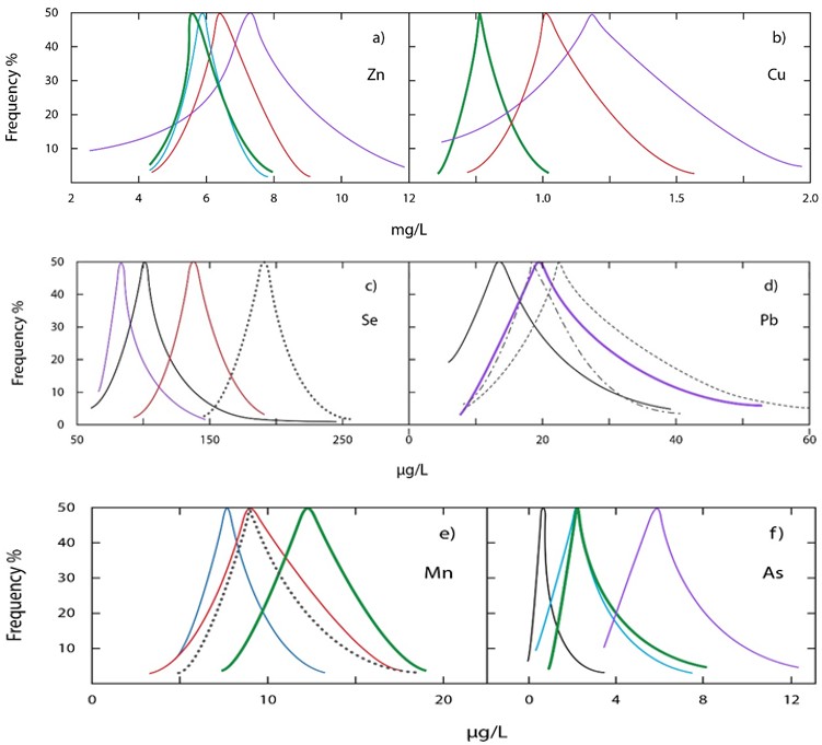
Figure 1a, b: Survey distributions of blood testing for Zn and Cu. [26] red, Italy; [27] blue, France; [28] green, China; [20] purple, Brazil.
Figure 2c, d: Survey distributions of blood testing for Se and Pb. [26] red, Italy; [20] purple, Brazil, [29] black, Finland; [30] …. USA, [31], heavy dash-dot Norway; [32] light dash line, Korea.
Figure 3e, f: Survey distributions of blood testing for Mn and As. [26] red, Italy; [27] blue, France; [28] green, China; [20] purple, Brazil, [29] black, Finland; [30] …... USA.
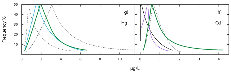
Figure 4g, h: Survey distributions of blood testing for Hg and Cd. [27] blue, France; [28] green, China; [20] purple, Brazil, [29] black, Finland; [32] … Korea, [33] dash-dot, USA.
Elements that May be Introducing Risk: Survey Implications
As a result of many such global surveys it is possible to begin to assess which elements may possibly be posing risks to health. It is not unreasonable to assume that those elements essential for life are managed effectively and automatically by the body. Any fault or disruption with them will not be long in becoming evident and generally be effectively diagnosed and corrected. Over the years, toxicity limits have been established for most of the elements from human and animal studies and values obtained that correspond to levels where symptoms affecting the body become apparent, the so-called NOAEL and LOAEL, no or lowest observed adverse effects levels. These have formed the basis for establishing minimum risk levels (MRL), Biomonitoring values that can be suggested by taking a built-in safety buffer of possibly 10- to 100-fold lower margin than the NOAEL value if possible, and this has become a useful medical guide. However, on several distributions it has become noted that the fall–off region can vary to different extents and readily stretch even beyond a 95% cut off of the sample and that an MRL value did not adequately reflect risk in such slow fall-off distributions. This was becoming commonly noted particularly for mercury where a large part of current survey distributions can extend well beyond the US MRL suggestion [34].
As a result, the actual value encompassing 95% of the survey now is being accepted as a better risk assessment magnitude, the so-called Reference Value (RV95) and is becoming accepted as a better diagnostic value [18, 19, 20, 21, 22, 35]. Such values are listed already in Table 1. However, this is still based on the accepted premise that if levels are sufficiently low, they can be accepted. This of course results from the fact that we have been living with nature quite well for many centuries indicating that the human body appears to tolerate many elements at levels that occur naturally. None the less, such a set of benchmark values remains invaluable and a basic guide for doctors. Surveys arise by sampling normally labeled healthy people. As a result, the field of toxicity is not an exact science and some of its assumptions now are being questioned, especially as humans are living longer and some elements found in the body may not have been in common use for long. However, in a basic manner this remains irrelevant in the present discussion. Changes are required and if a noted risk factor is relevant to an epidemic it has to satisfy various conditions such as having global availability. This and other requirements become very restrictive and extensively minimize considerations of periodic table options. Additionally, metals heavier than Mo do become suspect automatically as alien species in the body, but are also less encountered in most diets. Other than the aspect of slow accumulation, which may be relevant to diseases of the elderly, the question of higher exposures is obvious. However, without symptoms there is always an assumption of health. Levels falling within the RV95 values of these figures are automatically acceptable. This is apparent in the distributions illustrated in Figs.1 -4. For any of the toxic elements other than Mn and Se needed by the body, all distributions are characterized by a slow fall-off to outlier cases occurring well beyond even the 98% of the distribution, often in some cases to higher values. Until recently there has been little interest in such extended values that can be creeping towards levels of toxicity. For elements required by the body the risk assessment for health differs in needing to examine extremes, essentiality and toxicity [36]. There are now models relating dose-response aspects to assess risk [37]. The problem becomes different though for alien elements when only the upper limit is of interest. As a result, for most of these, the important data mainly come from survey levels, human observations and animal studies. Lead which appears to be the most toxic alien element has been very extensively studied. Human and animal studies note cognitive decline at even low levels, leading to the conclusion that a zero tolerance is most appropriate implying no model is needed [38, 39, 40]. Tests on rats now confirm oxidative stress and neurodegeneration also with low doses of methyl mercury [41] and arsenic exposure also indicates neural impairment at low levels [42]. Surprisingly, even strontium thought to be benign in the body now has been reported to produce oxidative stress in pregnant women [43]. Low concentrations of these trace toxic alien species in mixtures (As, Cd, Hg, Pb) are reported to have deleterious effects on mice and rats and moderate their bodily distributions [44, 45, 46]. Pregnancies are of particular concern as safe levels still need establishing [47]. Consequently, even though supposedly safe blood levels are being suggested, these are only a useful rough guide for medical consultations and remain the best realistic supposition. It is the nature of variation between humans that remains complex and additionally the variation of genetic susceptibilities that is apparent in the data. Nevertheless, the simple conclusion that can be drawn from all toxicity studies is that less is recommended.
Assessment of Diets and Which Alien Elements are More Commonly Encountered?
The assessment of what foods humans need to eat for good health arose from the advance of analytical analysis only about 50 years ago. Before that our diet was fixed by habit, culture and experience. A large body of data has been analyzed since, assessing the needed components and recommendations derived for bodily daily intake levels [48]. With such a baseline set of values, countries have been able to better gauge the adequacy of diets, and manage and control their general food supplies now in a more meaningful way. The detailed analysis of the elements in food examines not only sufficiency in the diet but can highlight dangerous possible health aspects. As a result, basic diets are now analyzed in chemical detail in many countries of the world. One outcome of this, is also the monitoring of the alien elements that we unknowingly ingest. The major importance of course is to ensure adequacy in required elements and bring this into balance by managing their sources. Such dietary reports are important and available, ones are for the US [49], UK [50], France [51], Germany [52], China [53], Cambodia [54], Sub-Sahara Africa [55, 56], Brazil [57], and Norway [58]. Their ranges of values are seen to vary to some degree on comparing different countries. However general magnitudes are consistent. The UK study had the important conclusion that, over the recent period of 30 years, dietary changes were minor in the UK. In fact, in solely examining most foods, there appears to be little significant global change in eating in these surveys consistent with explaining any epidemic. However, this assessment now is in error due to life-style changes that have occurred and have not yet been fully included. One significant addition to diet, overlooked in all these previous reports, is the eating of fish, Japanese sushi style. Although around for several decades, sushi-style of eating fish began in the US as a rather limited exotic food. Then as people changed to consider a healthier lifestyle that included dieting, it rapidly became accepted due to its high nutrition, low-fat and calorie character. It began to flourish in the 90’s and particularly in the US began to experience exponential growth now witnessed globally [59, 60].
The Japanese government recently estimated that outside of Japan there are more than 20,000 Japanese restaurants globally. The US has at least 4000 and now almost every grocery food store carries a sushi display. Fish are now a major source of alien mercury in its most toxic organic form, methyl mercury. It has also re-introduced unexpected mercury poisoning cases for the medical profession to diagnose among the general public consuming high fish diets. The World Health Organization had long realized the potential risk factor of mercury in fish consumption having completed a risk analysis for this in 2006 [61]. They established a recommended daily intake, however noting then that this left 6% of women of child-bearing age above the then accepted EPA blood level recommendation of ≤ 5.8 µg/L. This value has been accepted ever since but now is clearly regarded as too high a level extensively exceeded and a missing factor in people’s diets. Normal diets generally are sufficient to satisfy the body needs for the major and minor elements in Table 1, and it is rare to ingest these to levels of excess, the body normally has automatic controls on its major elemental needs. This is true both for food and water supplies even though some may show toxicity especially from highly contaminated water supplies. Toxicity also can be complex in that it depends on the chemical form of an element, particularly its water solubility, and also its valence state. For example, the ore of mercury is cinnabar, HgS, totally insoluble in water and as a result non-toxic. Similarly, chromium has two major valences, trivalent (III, non-toxic and required by the body) and hexavalent (VI), for example chromate compounds that are soluble and highly toxic. Water sources become a consideration due to solubility and the nature of their source. Surface supplies appear to have less problems for potable water than from water wells, but major impurities of As, Mn, Pb or U tend to be a regional geographic problem now quite widespread. The World Health Organization (WHO) realizes this problem and currently is reevaluating guidelines for drinking water [62]. Domestic filtering of drinking water is helping to reduce this component of diet in more advanced cultures. However, certain countries, such as parts of India and Bangladesh, are geographically unfortunate in having water wells now very rich in arsenic to the point that drinking water alone causes many cancer deaths per year [63, 64, 65, 66]. The US has less of such a problem, now being able to suggest lowering its arsenic National water standard to <10 µg/L [67]. Even so, there do remain sections of the South Western United States where many wells remain in excess [68]. Modern day industrialization and urbanization can also lead to groundwater contamination certainly of the heavy metals as witnessed in the Pearl River Delta region of China [69]. The Flint Michigan region of the US accidently raised Pb levels in its drinking water to dangerous levels, but it also remains a problem in other areas of the US and certainly in Bangladesh where spice adulteration with lead also is a problem of concern [70, 71]. As a result, water problems are ever present and non-negligible regarding illnesses. For example, currently Mo and Pb are raising concerns in various US counties [72, 73] and MN in water is suspected as having effects on children [74, 75, 76]. Even strontium levels have been addressed recently in the water of some Indian villages [77]. Consequently, although water cannot be casually disregarded, its quality is very variable. Also, although the alien elements are ingested to varying levels, global sources will generally remain more from food than from water. An interesting observation is that food and water supplies in normal diets are fortunately not often both rich in the same element. Also, it has to be remembered that some elements can be important even in minor quantities such as the need for vitamin B12 with its molecule having simply a single necessary Co atom at the center of a very complex structure. This raises the additional question of whether quantity can also be an important aspect in some disease assessments. Most humans live most of their lives eating their conventional diet and survive quite adequately. Nevertheless, some documented increase would appear to be necessary. Consequently, if the trace metals are to be held responsible for any epidemic a change in behavior has to be evident as discussed above with the example of enhanced fish intake sushi-style. Looking at the list in Table 1 of alien elements generally found in the body, it is soon apparent that most of these will be of little concern, mainly due to their very minor concentrations if presence at all in the body. The list can be broken down into several groups.
- Elements that appear harmless or have low toxicity at low levels (Ag, Al, Ba, Ce, Cs, Ga, Ge, Rb, Sn, Sr)
- Non-carcinogenic (Bi, Cd)
- Toxic and carcinogenic (As, Be, Hg, Pb, Sb, Sc, Tl)
- Neurotoxins (Al, As, Hg, Pb, Tl) The major basic consideration as outlined above is the requirement of life-style changes, either in human diets or other aspects. Smoking has been one custom, but now is in decline in many countries, but medical advances with vaccines is another that has significantly expanded. Such an examination of the low ingestion of numerous of these elements coupled with food, water, life-style and medical services now can further reduce the above list of potential hazardous alien elements to Al, As, Ba, Cd, methylHg and Pb as having possible deleterious effects on the body that are worthy of further consideration. The question of whether quantity is important remains uncertain, but as outlined may be irrelevant. Nevertheless, many experiments are now emerging from animal studies that do indicate effects from even low levels, well below the reference or safe minimum reference levels or from co-exposures [46]. As already indicated, the safe level for lead now is recommended as zero that clearly illustrates this may be part of any problem. Nevertheless, in order to explain global epidemics, change is required. Before the epidemics, people lived with contaminated food and water irrespective of their safe reference levels and quantities. The question now is what is different. They were living adequately before but why not now.
What Ingestion of Alien Elements has Really Changed?
As suggested, the question of quantity may be irrelevant unless it has increased significantly. Nevertheless, one relevant aspect of such research is that it has illustrated not only effects of trace quantities but has highlighted possible synergistic toxic effects between elements in some cases, which could also be a contributing factor. Although considered in the literature now for almost a decade, very little research has involved such mixed trace metal feeding. Studies with rats and mice do indicate low level effects and a disturbance of the body homeostasis balances with relative differences in organ absorptions [78, 79, 80, 81]. One study already has examined possible human brain mechanisms for metal mixtures [82]. Meanwhile, for some of these trace elements, environmental public pressure has in fact resulted in their actual demise in society. Cd ingested partly from smoking has decreased in some countries, and a very significant reduction of lead and elemental Hg has occurred in recent years as most of their previous commercial applications now have become widely banned. Surveys are beginning to note the decreases particularly for Cd and also Pb [16, 83]. From the list above of potentially important alien elements in the body Ba has been used medically for many years with no apparent adverse effects even though it is actually a heavy- metal. It does not readily accumulate in the body, is non- carcinogenic and is not a neurotoxin, and was once used commonly, added as a soot suppressant in oil combustion but now appears to raise no health concerns in any regard. In normal situations its toxicity is of no concern and its level in the body is rarely even tested. As mentioned above, cadmium is highly toxic and carcinogenic. It retains its inorganic nature, not forming organometallic compounds in the body. It generally is a low trace level in blood samples as seen globally in Figure 4h. It can be ingested as deposition from leafy vegetables especially from Cd rich-soil areas in China [84, 85, 86], from rice [87, 88], fish and seafood [89, 90, 91], free- living game [92], and also from tobacco smoking [93]. It has a high retention rate by the body and can affect the kidneys. A recent review of cadmium’s toxicity suggests its induced oxidative stress on the liver and kidneys can cause pathogenic risks for a variety of cancer forms [94]. It is slowly chelated naturally by the body’s selenium via its many protective protein metallothionein forms [95, 96, 97, 98, 99]. However, due to its long body half-life, aspects of it managing to pass into the brain still remain uncertain as also its possible synergism with other toxins. However, a major point of interest here stems from the reduced levels of tobacco smoking in the world, which is reflected as decreases in surveys of cadmium blood levels. Aluminum enters this discussion by recently having become the major adjuvant in most US and other vaccines, generally in the form of its hydroxide. Generally regarded as being medically safe, this is based stressing mainly oral data. However, because the intestines have a very low absorption rate of only about 1 to 2% for such orally ingested aluminum, this assumption now is becoming questioned in that inoculations increase this retention value closer to 100% [100]. As a result of the great success and value of vaccines, this now reflects into a significant change and re-assessment for this element in its human-lifestyle consumption and also modifies many prior assessments from studies with both humans and animals. It is particularly important for children in the US, who receive about 35 inoculations by the age of five, a much-enhanced rate from earlier years. As a result, when considered with other neurotoxins, this has concerned many major groups [101, 102, 103]. Depending on body weight, it can readily introduce a high risk of toxicity, as suggested even from animal studies [104, 105, 106]. Significant levels also have been noted in brain autopsies [107, 108], and in cases of death from autism with ages from 15-50 years old [109]. This latter study, finding one case at age 15 is particularly disturbing. Although selenium has been shown to counteract the aluminum in the brains of mice and rats, uncertainties remain and glutathione peroxidase may even become depleted possibly suggesting a slow rate of its replenishing formation capability [110, 111, 112, 113, 114, 115]. Additionally, a study with human brain cells has noted a synergistic collaboration between Al and Hg as neurotoxins [116]. As a result, the addition of a new ingested source of Al to the already normal dietary intake is significant particularly for young people [117]. The case with arsenic is particularly interesting as it is already an epidemic of major proportions in several parts of the world. The extreme case is in Bangladesh and has resulted in recent decades through changing the water supply from surface sources to ground water wells. It is now affecting millions of people and as a result about 20,000 deaths occur per year in Bangladesh [118]. This has facilitated many studies in various countries concerning arsenic toxicity. It obviously leads to a myriad of medical illnesses affecting the skin, lungs, kidneys, a series of cancers, cardiovascular risk, and is thought to induce fetal loss, premature births and low birth weights [119, 120, 121]. There are indications of neural effects in some cases, but are evident as cases of dementia and memory loss effects. Studies with rats do clearly show possible enhanced oxidative stresses and neural changes [122, 123]. The Western countries such as the US and Canada have low arsenic and few if any cases of such arsenic poisoning [124]. Arsenic is unavoidable and often present in both food and water. Dietary intake also is complicated by arsenic’s tendency to methylate and be in an organic form. However, some of these forms become less toxic, as is the case in fish entailing a full speciation analysis for an exact arsenic toxicity level [125, 126, 127]. Rice is another general food source, affected by soil and pollution conditions, sometimes becoming methylated [128, 129, 130, 131]. Whether Se protects the brain from more serious illnesses such as Alzheimer’s Disease remains uncertain but the eating of lentils rich in selenium has been proposed in Bangladesh [132], and selenium appears protective in arsenic induced impairments in mice and rat studies [133, 134]. The geographic comparisons between countries tends to minimize a critical overall role for arsenic in the major epidemical illnesses in the West, but arsenic is ubiquitous in the world as a potential toxic collaborator and a source of some illnesses. Even though lead has been a health risk factor from at least Roman days, it has remained an important commercial element until recent decades. However, through extensive environmental public pressure it has gradually been banned from most commercial products. Nevertheless, even today I recently heard of a child being poisoned in the US from a newly purchased lead-glazed bathtub. It also remains as a glaze for clay pottery in Mexico [135], and is still a major concern due to its continuing presence. It is with us in all older homes, present as old paint, it is on copper water pipes joints in any Pb/Ag solder and may even result from old lead connecting water pipes, besides being possibly in a drink served from a cut crystal glass decanter. Lead is detected in all humans showing a significant level in blood. Physicians now are content if it is below 50 µg/L in adults or children [136], even though it is known that the safe healthy level for lead is zero [38, 39]. It remains a significant concern in pregnancies [47], especially as lead and most of the other metal elements have unrestricted flow in blood through the placenta [137]. It has a toxic potential regarding human reproduction even at low levels [138]. Effects on children are more significant than adults and Pb exposure at low levels continues to be a major public concern [139, 140]. Young children are particularly prone to paint dust gathering on floors. One analysis of data from an earlier drinking water “lead crisis” in Washington DC (2000-2004) concluded that fetal death rates and reduced birth rates were also a consequence [141]. Consequently, even though lead-based paints remain in use in many countries [142, 143, 144, 145], lead blood levels are noted to be in decline globally in all developing societies [146, 147, 148]. As a result, lead does not appear to be a primary element inducing recent epidemic growths in illnesses but may be an epigenetic modifier as noted in animal studies [149]. Its levels in water and food though remain a medical concern and it may continue to play a major role triggering or contributing to certain illnesses. A role for selenium as a chelator remains uncertain but in areas of lead pollution, residents have been noted to have a selenium deficiency [150]. Numerous studies with mice and rats clearly document the potential oxidative stress that arises from lead exposure and the possible beneficial role of selenium [151, 152, 153, 154, 155].
Mercury now is regarded as the neurotoxin of most concern to health. Although it has played a major role in commerce for millennia, its health risk became more seriously apparent 70 years ago through the neurological birth defects originating from the environmental disaster in the Minamata village in Japan. Until then, neural illnesses were not really at a significant level to be of major concern. The phrase “Mad as a hatter” arose from the felt-hat industry where mercuric nitrate was used in the process. Mercury’s main use centered on its elemental ability to amalgamate with other metals, most importantly gold and silver. From even Egyptian times this has been a major role and although now no longer commercially mined, artisanal mining for gold is still an important occupation in numerous parts of the world ultimately utilizing mercury amalgamation and separation. Its unusual chemical symbol, Hg, originates from the Greek “hydrargyrum” (liquid silver). It found use in innumerable processes in industry and medicine. These ranged from switches, thermometers to the chloralkaline process and it has been important in dentistry being used as a silver/tin-mercury amalgam in teeth. It is also common in certain medications and still as a skin lightener [156, 157, 158]. Although generally banned, this latter use remains common particularly in India and Saudi Arabia. Until recently, one of mercury’s main medical uses for decades has been as the primary adjuvant and preserver in vaccines as the water-soluble organic form of thimerosal (sodium 2-ethylmercurithiosalicylate). In the US it has now been replaced in recent years by aluminum hydroxide in most vaccines, but remains present in many of those exported and can be reintroduced into US vaccines during pandemic times. Mercury now has been extensively banned globally and only remains in the illicit mining for gold, in the largely still uncontrolled gaseous emission from certain combustion processes (primarily coal and cement industries), in several US vaccines and still is used by some dentists, although banned and replaced now in numerous countries [159]. It is no longer mined from its ore cinnabar and not easily purchased having lost its intrinsic value. Human ingestion can be in the elemental form as its vapor, possibly from its leaching and vaporization from dental fillings before being absorbed and then oxidized to its inorganic divalent ion in the blood. It is also in most fish diets as the more toxic organic form, methyl mercury, having been extensively bio accumulated in nature and the fish as this methylated structure. Pollution of most water expanses now has arisen and remains from historical colonial days when silver and gold mining were extensive [160].
Although now coupled to possibly decreasing anthropogenic additions to the environment, the still large evasive, recycling of these historical excesses in the earth’s waters and land retains an uncertainty that will remain into the foreseeable future before it may one-day return to natural pre-industrial levels. Mercury is the most researched element in the periodic table with about 5000 publications/ year in the areas of medical and environmental interests. Nevertheless, even with the reduction of mercury in vaccines, the growth of consuming sushi from the 1980’s in the US diet to now significant levels, and also throughout the world has elevated methyl mercury to a major human toxin, especially due to its neurotoxic organic nature and ready bio accessibility. Studies of heavily fish-eating communities have been used to establish the NOAEL and LOAEL, no or lowest observed adverse effects levels. Through such studies of the Faroe Islands community (whale consumers), and that of the Seychelles (Ocean fish-eaters) differences have highlighted a missing factor. This appears to be the selenium fish component, now realized to be the body’s natural chelator for mercury through its many seleno-enzymes and also for many of the other alien elements consumed in diets. From the current wealth of research several conclusions now can be concluded. One is that mercury’s toxicity depends on its form, elemental, ionic or organic. From the gold miners, chloralkali and dental employees many of whom have been subject to prolonged mercury elemental vapor inhalation, they show mainly the symptoms of kidney function difficulties (prime target organ), with some lesser secondary neural effects [161, 162, 163].
Obviously, over time elemental mercury does appear to probably pass across the blood/brain barrier. Once in the blood the elemental form is oxidized to the divalent ion [164]. Peru still has artisanal gold miner’s shops located in its towns. However, there are no reports of severe neural consequences in their neighborhoods from released inorganic mercury vapor [165]. However, miners are diagnosed with oxidative stress and DNA methylation that may have some effect on their health [166]. Elemental mercury’s half-life in the body is measured in days or several weeks. It is excreted mainly in urine and feces. Any related neural effects in fact appear reversible with time [167] but in some cases of dental fillings, dementia appears to persist [168]. Radioactively labeled tooth fillings of sheep were reported to be leached and retained by the body [169], however low doses of inorganic mercury with rats did show signs of slight entry into the brain causing oxidative stress and cell death [170, 171]. Organic forms of mercury, namely the thimerosal in vaccines and the methyl mercury in fish are very neurotoxic and have been extensively examined in humans and numerous animals, from mice and rats to rabbits, hamsters and monkeys, some studies with labeled mercury. They are readily absorbed and have the brain as a major target, passing freely through the blood/brain barrier. One study with monkeys was very detailed [172]. Thimerosal, readily breaks down in the body to ethyl mercury and thiosalicylate. In monkeys, the ethylHg has a shortened blood half-life of about seven-fold compared to the 22 days for methylHg, and similarly a shorter brain half-life of 24 compared to 60 days. What was noteworthy in studies was a process of de-alkylation in the brain that was significantly more with ethylHg, about 70% becoming inorganic compared to 10%. Both in vitro and in vivo studies now have been extended and reviewed confirming these behaviors [173]. For human diets, from fish-eating studies the half-life in the blood is quite variable reflecting human susceptibilities, data varying from <30 to >120 days [174, 175].
Brain half-lives for inorganic mercury also are uncertain but surmised to be years in magnitude [176]. It became evident that seafood exposure to methyl could be neurotoxic even at low levels [177, 178, 179, 180]. Also, it has been noted in 25 countries around the world that women of child-bearing age have high dietary mercury levels [181]. The two large cohort studies on humans utilizing the peoples of the Faroe and Seychelles Islands now have spanned several decades. Both groups have predominantly seafood diets [182, 183, 184]. Through these, from their contradictory results it was finally accepted that the differing levels of selenium in the fish (whale meat or ocean fish) was a major missing factor that was modifying their data. It was providing a countermeasure of protection from the mercury [185, 186, 187]. This was clearly confirmed by selenium’s neural protection from methylHg now reported in rats and mice [188, 189]. The research has highlighted the health benefit value of selenium in the body and how it differs from the other elements [190, 191, 192, 193]. The fact that the body is programmed to produce 25 seleno- proteins appears purposeful [194], as well as its magnitude over all the alien elements found in the blood. It is clearly apparent that selenium is the body’s natural chelator, not only for mercury, but for the alien elements. It has also been suggested as a protector against manganese, possibly not necessary in a healthy person [195]. However, this aspect of selenium now introduces a need for a reassessment of fish in diets [196, 197, 198]. As long as selenium is present in excess of the mercury on an atomic level, it has the potential to neutralize or sequester mercury’s deleterious effects [199]. The fact that selenium has an atomic weight of 79 compared to mercury’s 201 introduces an additional factor of 2.5 advantage for selenium on an atomic basis. This is important as many fish analyses are generally analyzed by weight and now require conversion to molar quantities to test for this ratio [200, 201, 202, 203, 204]. In addition, the nature of the specific selenoprotein appears to be involved and complicates this further [205, 206]. Also, it has also been suggested that high levels of organic mercury together with other alien elements may lead to a dangerous deficiency of required selenium enzymes [207]. Selenium is a unique element being necessary for life but is also a neurotoxin. It displays a narrow dosage range between being therapeutic or toxic. An acceptable dietary range is 40-250 µg/day, optimum being an intake of 100-200 µg/day [208], but a consistent dose of 300 µg/day for 5 years now has shown the possibility of increased mortality [209]. Many soils around the world are deficient in selenium and are of great concern for agriculture and diets [210, 211, 212, 213]. Since 1984, Finland has been using Se- enriched fertilizers to enrich crops [214], while others tend to enrich feed stocks. Nevertheless, it has now been realized that selenium plays a very important role in the body in the form of its glutathione peroxidase enzymes, which regulate body inflammation [215], and have a role also in brain signaling [216]. Nevertheless, some doctors presently do not recommend selenium use during pregnancy [217] due to the observed overall uncertainties [206].
Are Epidemics Due to Increased Levels of Alien Elements that Deplete Selenium’s Protection?
The medical profession has realized for some time that the heavy metals are a major health concern, their mechanism being one of oxidation [218]. As indicated above, their mitigation in the body can be by a large regimen of selenoenzymes that chelate and sequester them before elimination [219]. Canada has recognized this importance, monitored their total population for blood selenium levels, and finding it fully in the desirable 100-400 µg/L range with a mean close to 200 µg/L [220]. Nature has endowed most people with body selenium levels in excess over any alien metals present [221], an interesting feature in itself. Currently the literature is very rich with research on selenium surmising that its deficiency may be associated with a whole string of many illnesses that involve oxidative stress, namely organ failure in critically ill children [222], thyroid problems [223, 224, 225], strokes, atherosclerosis, oxidized LDL (low density lipids), osteoporosis, and all cardiovascular illnesses [226, 227, 228], cancers [229, 230, 231, 232], Huntington’s disease [233], Parkinson’s [234, 235, 236], Alzheimer’s [235, 237, 238, 239, 240, 241], pregnancy and adverse outcomes [206, 242, 243, 244, 245, 246], autism [247, 248], amyotrophic lateral sclerosis (ALS) [249, 250], schizophrenia [251], and others. Its importance in pregnancy currently is being emphasized, recommending maternal blood levels of at least 100 µg/L. Recent research is important in possibly explaining how a fetus survives the high toxicity of its environment [252]. In two noteworthy studies it was reported that the selenium blood level in the umbilical cord was much higher than that of methyl-mercury. Converting
these to molar or atomic quantities it was found that the Se to Hg ratios were 62-fold [253] in one study, and in a range of 5 to 626 [254] in another, indicating possibly the overlooked required protection needed for the low weight fetus during pregnancy. Missing data now needed are the potential rates of depletion and formation of these selenoenzymes and whether selenium can become depleted [255]. Such research remains primitive and selenium’s biology of selenoprotein degradation and metabolism remains uncertain [256].
Conclusion
On analyzing the necessary components of human diet, it can safely be concluded that few pronounced changes have occurred in recent decades. However, it is quite extraordinary that diets do contain a significant number of elements that serve no purpose and are regarded as alien species, do nothing other than increase the body’s burden for required elimination. Many of these are toxins and neurotoxins but fortunately generally at low levels in the blood. A detailed examination for the changes necessary to trigger modern epidemics concludes that the only elements of pronounced interest are Al, As, Hg, Pb, all in fact neurotoxins and hazardous. From these Al and Hg have gained more importance due to their global growth through life-style changes. Arsenic poses severe problems in drinking water around the world but is largely controlled in the Western developed countries. Pb is a major toxin that certainly still remains dangerous particularly for young children but is in a continuing state of decline in diets, particularly in adults, as it gets more heavily controlled and removed from commerce. This tends to leave Hg and Al as the prime suspects at present for most medical illnesses. Animal research also is noting that these can also be accentuated by association with other trace elements. They are necessarily in all peoples who are vaccinated and/or eat a fish diet. The latter is most common around the world and although Al now is being reported in many brain autopsies, Hg is most probably the more toxic, occurring also more frequently now in individuals as mercury poisoning from fish diets. The medical profession emphasizes the nutritional benefits of fish, which is true, and as a safeguard smaller class of fish now should be eaten for which Se > Hg rather than the larger fish categories that remain rich in methylHg. As a result, it can be quite positively surmised at present that adults are more likely to be suffering from a mercury body burden while for young infants’ aluminum becomes the major concern. The toxicity of these metals during pregnancy remains an ignored subject. The fetus when small technically should not survive the toxic levels to which it is subjected. Whether the high levels of the selenoproteins, blood brain barrier and yet unknown other defenses exist has to be resolved. Nevertheless, younger women of child bearing ages require guidance.
What is becoming apparent is that health is becoming the accepted responsibility of each individual. This is now quite broadly accepted in patients with Cardio or diabetic conditions in Western societies where monitoring is more readily available. However, in the present case of knowing one’s susceptibility to metal toxins there is a void. Although many studies are now reporting a need for this information, it still cannot be readily prescribed by a doctor [257, 258, 259, 260]. The public needs to establish its location on blood survey curves to establish their individual genetic susceptibilities concerning possible high retention of dangerous chemicals or satisfactory rejection. If established, additional steps can be identified to reduce any levels of concern. As a result, testing from an early age till death has to become standardized in the near future concerning specifically the neurotoxins Al, As, Hg, Pb, Mn and Se to enable a personal management of health. By using a simple low-cost blood test available to the general public, it is possible in one analysis of a small blood sample to resolve this issue. Such a test now has been validated utilizing the ICP-MS instrument [261, 262]. It remains ridiculous that everyone goes through life not knowing the state of their bodies and whether they are living at high risk or not, the necessary technology now is available. This is of major importance especially for pre- pregnancy testing in younger women.
References
-
Sharavanan VJ, Sivaramakrishnan M, Sivarajasekar N, Senthilrani N, Kothandan R, et al. (2020) Pollutants inducing epigenetic changes and diseases. Environ Chem Lett 18(2): 325-343.
-
Jager T (2013) All individuals are not created equal: Accounting for individual variation in fitting life-history responses to toxicants. Environ Sci Technol 47: 1664- 1669.
-
Grandjean P, Budtz JE (2010) An ignored risk factor in toxicology: The total imprecision of exposure assessment. Pure Appl Chem 82(2): 383-391.
-
Sanders AP, Mazzella MJ, Malin AJ, Hair GM, Busgang SA, et al. (2019) Combined exposure to lead, cadmium, mercury, and arsenic and kidney health in adolescents age 12-19 in NHANES 2009-2014. Environ Int 131: 104993.
-
Bulka CM, Persky VW, Daviglus ML, Durazo ARA, Argos M (2019) Multiple metal exposures and metabolic syndrome: A cross-sectional analysis of the National Health and Nutrition Examination Survey 2011-2014. Environ Res 168: 397-405.
-
Dlugaszek M (2019) Studies on relationships between essential and toxic elements in selected body fluids, cells and tissues. Chem Biol Interact 297: 57-66.
-
Jalili C, Kazemi M, Taheri E, Mohammadi H, Boozari B, et al. (2020) Exposure to heavy metals and the risk of osteopenia or osteoporosis: a systematic review and meta-analysis. Osteoporosis Int 31(9): 1671-1682.
-
Bibi K, Shah MH (2020) Appraisal of metal imbalances in the blood of thyroid cancer patients in comparison with healthy subjects. Biol Trace Elem Res 198(2): 410-422.
-
Nabgha-e-Amen, Eqani SAAS, Khuram F, Alamdar A, Tahir A, et al. (2020) Environmental exposure pathway analysis of trace elements and autism risk in Pakistani children population. Sci Total Environ 712: 136471.
-
Hynes AJ, Steinberg M, Schofield K (1984) The chemical kinetics and thermodynamics of sodium species in oxygen-rich hydrogen flames. J Chem Phys 80(6): 2585- 2596.
-
Steinberg M, Schofield K (1991) A reevaluation of the vaporization behavior of sodium oxide and the bond strengths of NaO and Na2O: Implications for the mass spectrometric analyses of alkali/oxygen systems. J Chem Phys 94(5): 3901-3907.
-
Schofield K (2020) Combustion emissions: Formation, reaction, and removal of trace metals in combustion products. In: 1st (Edn.), Elsevier, London, pp: 29-30.
-
Chehbani F, Gallello G, Brahim T, Ouanes S, Douki W, et al. (2020) The status of chemical elements in the blood plasma of children with autism spectrum disorder in Tunisia: A case-control study. Environ Sci Pollut Res.
-
Cheng X, Zhou YC, Zhou B, Huang YC, Zhou GB (2019) Systematic analysis of concentrations of 52 elements in tumor and counterpart normal tissues of patients with non-small cell lung cancer. Cancer Med 8(18): 7720- 7727.
-
Henriquez HLA, Romero D, Gonzalez AA, Gonzalez AB, Zumbado M, et al. (2020) Biomonitoring of 45 inorganic elements measured in plasma from Spanish subjects: a cross-sectional study in the Andalusian population. Sci Total Environ 706: 135750.
-
Fourth National Report on Human Exposure to Environmental Chemicals (2015) US Department of Health and Human Services, Centers for Disease Control and Prevention.
-
Maret W (2016) The metals in the biological periodic system of the elements: Concepts and conjectures. Int J Mol Sci 17(1): 66.
-
Kuno R, Roquetti MH, Becker K, Seiwert M, Gouveia N (2013) Reference values for lead, cadmium and mercury in the blood of adults from the metropolitan area of Sao Paulo, Brazil. Int J Hyg Environ Health 216(3): 243-249.
-
Saravanabhaven G, Werry K, Walker M, Haines D, Malowany M, et al. (2017) Human biomonitoring reference values for metals and trace elements in blood and urine derived from the Canadian Health Measures Survey 2007-2013. Int J Hyg Environ Health 220 (2 Pt A): 189-200.
-
Almeida LACBD, Martins AC, Urbano MR, Buzzo ML, Camargo AEI, et al. (2019) Blood reference values for metals in a general adult population in southern Brazil. Environ Res 177: 108646.
-
Vogel N, Conrad A, Apel P, Rucic E, Kolossa GM (2019) Human biomonitoring reference values: differences and similarities between approaches for identifying unusually high exposure of pollutants in humans. Int J Hyg Environ Health 222(1): 30-33.
-
Al-Saleh I (2020) Reference values for heavy metals in the urine and blood of Saudi women derived from two human biomonitoring studies. Int J Hyg Environ Health 225: 113473.
-
US Department of Health and Human Services, Centers for Disease Control and Prevention, Agency for Toxic substances.
-
World Health Organization. Various lists of reports under the headings of (EHC) Environmental Health Criteria, IPCS, CICAD and INCHEM, reports on Chemical Safety, www.WHOint.publications.
-
Zhu C, Tian H, Hao J (2020) Global anthropogenic atmospheric emission inventory of twelve typical hazardous trace elements, 1995-2012. Atmos Environ 220: 117061.
-
Bocca B, Madeddu R, Asara Y, Tolu P, Marchal JA, et al. (2011) Assessment of reference ranges for blood Cu, Mn, Se, and Zn in a selected Italian population. J Trace Elements Med Biol 25(1): 19-26.
-
Nisse C, Tagne FR, Howsam M, Richeval C, Labat L, et al. (2017) Blood and urinary levels of metals and metalloids in the general adult population of Northern France: The IMEPOGE study, 2008-2010. Int J Hyg Environ Health 220(2 Pt B): 341-363.
-
Zeng HL, Li HJ, Lu J, Guan Q, Cheng LM (2019) Assessment of 12 metals and metalloids in blood of general populations living in Wuhan of China by ICP-MS. Biolog Trace Element Res 189(2): 344-353.
-
Abass K, Koiranen M, Mazej D, Tratnik JS, Horvat M H, et al. (2017) Arsenic, cadmium, lead and mercury levels in blood of Finnish adults and their relation to diet, lifestyle habits and sociodemographic variations. Environ Sci Pollut Res 24(2): 1347-1362.
-
Jain RB, Choi YS (2015) Normal reference ranges for and variability in the levels of blood manganese and selenium by gender, age and race/ethnicity for general U.S. population. J Trace Elements Med Biol 30: 142-152.
-
Birgisdottir BE, Knutsen HK, Haugen M, Gjelstad IM, Jenssen MTS, et al. (2013) Essential and toxic element concentrations in blood and urine and their association with diet: Results from a Norwegian population study including high-consumers of seafood and game. Sci Total Environ pp: 836-844.
-
Kim HJ, Lim HS, Lee KR, Choi MH, Kang NM, et al. (2017) Determination of trace metal levels in the general population of Korea. Int J Environ Res Public Health 14(7): 702.
-
Mortensen ME, Caudill SP, Caldwell KL, Ward CD, Jones RL (2014) Total and methyl mercury in whole blood measured for the first time in the US population: NHANES 2011-2012. Environ Res 134: 257-264.
-
Schofield K (2019) An important need to monitor from an early age the neurotoxins in the blood or by an equivalent biomarker. Int J Environ Res Public Health 16(18): 3425.
-
Bevan R, Jones K, Cocker J, Assem FL, Levy LS (2013) Reference ranges for key biomarkers of chemical exposure within the UK population. Int J Hyg Environ Health 216(2): 170-174.
-
Goldhaber SB (2003) Trace element risk assessment: Essentiality vs. toxicity. Regul Toxicol Pharmacol 38(2): 232-242.
-
World Health Organization (2009) Principles for modeling dose-response for the risk assessment of chemicals. Environmental Health Criteria 239: 141.
-
Vorvolakos T, Arseniou S, Samakouri M (2016) There is no safe threshold for lead exposure: A literature review. Psychiatriki 27(3): 204-214.
-
Shefa ST, Heroux P (2017) Both physiology and epidemiology support zero tolerable blood lead levels. Toxicol Lett 280: 232-237.
-
Rocha A, Trujillo KA (2019) Neurotoxicity of low-level lead exposure: History, mechanisms of action, and behavioral effects in humans and pre-clinical models. Neurotoxicology 73: 58-80.
-
Santana LND, Bittencourt LO, Nascimento PC, Fernandes RM, Teixeira FB, et al. (2019) Low doses of methylmercury exposure during adulthood in rats display oxidative stress, neurodegeneration in the motor cortex and lead to impairment of motor skills. J Trace Elem Med Biol 51: 19-27.
-
Sharma A, Kumar S (2019) Arsenic exposure with reference to neurological impairment: an overview. Rev Environ Health 34(4): 403-414.
-
Barneo CC, Martinez ME, Rodriquez GS, Lequerica FP, Vega NI, et al. (2018) Strontium and oxidative stress in normal pregnancy. J Trace Elem Med Biol 45: 57-63.
-
Cobbina SJ, Chen Y, Zhou ZX, Wu XS, Feng WW, et al. (2015) Interaction of four low-dose toxic metals with essential metals in the brain, liver and kidneys of mice on sub-chronic exposure. Environ Toxicol Pharmacol 39(1): 280-291.
-
Orr SE, Barnes MC, George HS, Joshee L, Jeon B, et al. (2018) Exposure to mixtures of mercury, cadmium, lead, and arsenic alters the disposition of single metals in tissues of Wistar rats. J Toxicol Environ Health A 81(24): 1246-1256.
-
Zhou FK, Yin GM, Gao YY, Ouyang L, Liu SS, et al. (2020) Insights into cognitive deficits caused by low-dose toxic heavy metal mixtures and their remediation through the postnatal enriched environment in rats. J Hazard Mater 388: 122081.
-
Taylor CM, Golding J, Emond AM (2014) Lead, cadmium and mercury levels in pregnancy: The need for international consensus on levels of concern. J Dev Orig Health Dis 5(1): 16-30.
-
World Health Organization (1996) Trace elements in human nutrition and health. WHO Report, Geneva.
-
Cowan AE, Jun S, Tooze JA, Eicher MHA, Dodd KW, et al. (2020) Total usual micro-nutrient intakes compared to the dietary reference intakes among the US adults by food security status. Nutrients 12: 38.
-
Rose M, Baxter M, Brereton N, Baskaran C (2010) Dietary exposure to metals and other elements in the 2006 UK Total Diet Study and some trends over the last 30 years. Food Addit Contam 27(10): 1380-1404.
-
Arnich N, Sirot V, Riviere G, Jean J, Noel L, et al. (2012) Dietary exposure to trace elements and health risk assessment in the 2nd French Total Diet Study. Food Chem Toxicol 50(7): 2432-2449.
-
Heitland P, Koster HD (2006) Biomonitoring of 37 trace elements in blood samples from inhabitants of Northern Germany by ICP- MS. J Trace Elem Med Biol 20(4): 253- 262.
-
Zhang LL, Lu L, Pan YJ, Ding CG, Xu DY, et al. (2015) Baseline blood levels of manganese, lead, cadmium, copper, and zinc in residents of Beijing suburb. Environ Res 140: 10-17.
-
Kelly BC, Myo AN, Pi N, Bayen S, Leakhena PC, et al. (2018) Human exposure to trace elements in central Cambodia: Influence of seasonal hydrology and food- chain bioaccumulation behavior. Ecotoxicol Environ Safety 162: 112-120.
-
Yedomon B, Menudier A, Etangs FLD, Anani L, Fayomi B, et al. (2017) Biomonitoring of 29 trace elements in whole blood from inhabitants of Cotonou (Benin) by ICP- MS. J Trace Elem Med Biol 43: 38-45.
-
Jitaru P, Ingenbleek L, Marchond N, Laurent C, Adegboye A, et al. (2019) Occurrence of 30 trace elements in foods from a multi-center Sub-Saharan Africa Total Diet Study: Focus on Al, As, Cd, Hg, and Pb. Environ Int 133: 105197.
-
Pineiro JM, Pinero JS, Rodriquez EA, Carou IT, Mahia PL, et al. (2020) Major, minor and trace elements composition of Amazonian foodstuffs and its contribution to dietary intake. J Food Measur Charact 14: 1314-1324.
-
Naess S, Aakre I, Lundebye AK, Ornsrud R, Kjellevold M, et al. (2020) Mercury, lead, arsenic, and cadmium in Norwegian seafood products and consumer exposure. Food Addit Contam B Surveill 13(2): 99-106.
-
Burger J, Gochfeld M, Jeitner C, Donio M, Pittfield T (2014) Sushi consumption rates and mercury levels in sushi: Ethnic and demographic differences in exposure. J Risk Res 17(8): 981-997.
-
Jacobs S, Sioen I, Jacxsens L, Domingo JL, Sloth JJ, et al. (2017) Risk assessment of methylmercury in five European countries considering the national seafood consumption patterns. Food Chem Toxicol 104: 26-34.
-
WHO (2016) Food safety risk analysis: a guide for national food safety authorities. Food and Agriculture Organizations of the United Nations Report No. 87. World Health Organization.
-
Frisbie SH, Mitchell EJ, Sarkar B (2015) Urgent need to reevaluate the latest World Health Organization guidelines for toxic inorganic substances in drinking water. Environ Health 14: 63.
-
Mitchell E, Frisbie S, Sarkar B (2011) Exposure to multiple metals from groundwater, a global crisis: geology, climate change, health effects, testing, and mitigation. Metallomics 3(9): 874-908.
-
Shakoor MB, Nawaz R, Hussain F, Raza M, Ali S, et al. (2017) Human health implications, risk assessment and remediation of As-contaminated water: A critical review. Sci Total Environ 601-602: 756-769.
-
Ahmad SA, Khan MH, Haque M (2018) Arsenic contamination in groundwater in Bangladesh: Implications and challenges for healthcare policy. Risk Manage Healthcare Policy 11: 251-261.
-
Sinha D, Prasad P (2019) Health effects inflicted by chronic low-level arsenic contamination in groundwater: A global public health challenge. J Appl Toxicol 40(1): 87-131.
-
Foster SA, Pennino MJ, Compton JE, Leibowitz SG, Kile ML (2019) Arsenic drinking water violations decreased across the United States following revision of the maximum contaminant level. Environ Sci Technol 53(19): 11478-11485.
-
Jones MC, Credo JM, Ingram JC, Baldwin JA, Trotter Jr RT, et al. (2020) Arsenic concentrations in ground surface waters across Arizona including Native Lands. J Contemp Water Res Edu 169(1): 44-60.
-
Huang GX, Zhang M, Liu CY, Li LP, Chen ZY (2018) Heavy metal(loid)s and organic contaminants in groundwater in the Pearl River Delta that has undergone three decades of urbanization and industrialization: Distributions, sources, and driving forces. Sci Total Environ 635: 913- 925.
-
Chowdhury S, Kabir F, Mazumder MAJ, Zahir MH (2018) Modeling lead concentration in drinking water of residential plumbing pipes and hot water tanks. Sci Total Environ 635: 35-44.
-
Forsyth JE, Weaver KL, Maher K, Islam MS, Raqib R, et al. (2019) Sources of blood lead exposure in rural Bangladesh. Environ Sci Technol 53(19): 11429-11436.
-
Pieper KJ, Nystrom VE, Parks J, Jennings K, Faircloth H, et al. (2018) Elevated lead in water of private wells poses health risks: Case study in Macon County, North Carolina. Environ Sci Technol 52(7): 4350-4357.
-
Pichler T, Koopmann S (2020) Should monitoring of molybdenum (Mo) in groundwater, drinking water and well permitting be made mandatory?. Environ Sci Technol 54(1): 1-2.
-
Dion LA, Amour DS, Sauve S, Barbeau B, Mergler D, et al. (2018) Changes in water manganese levels and longitudinal assessment of intellectual function in children exposed through drinking water. Neurotoxicology 64: 118-125.
-
Valcke M, Bourgault MH, Haddad S, Bouchard M, Gauvin D, et al. (2018) Deriving a drinking water guideline for a non-carcinogenic contaminant: The case of manganese. Int J Environ Res Public Health 15(6): 1293.
-
Filho JAM, Carvalho CF, Rodrigues JLG, Araujo CFS, Santos NRD, et al. (2018) Environmental co-exposure to lead and manganese and intellectual deficit in school-aged children. Int J Environ Res Public Health 15(11): 2418.
-
Khandare AL, Validandi V, Rajendran A, Singh TG, Thingnganing L, et al. (2020) Health risk assessment of heavy metals and strontium in groundwater used for drinking and cooking in 58 villages of Prakasam district, Andhra Pradesh, India. Environ Geochem Health 42: 3675-3701.
-
Whittaker MH, Wang G, Chen XQ, Lipsky M, Smith D, et al. (2011) Exposure to Pb, Cd, and As mixtures potentiates the production of oxidative stress precursors: 30-day, 90-day and 180-day drinking water studies in rats. Toxicol. Appl. Pharmacol 254(2): 154-166.
-
Andrade V, Mateus ML, Batoreu MC, Aschner M, Santos APMD (2013) Urinary delta-ALA: A potential biomarker of exposure and neurotoxic effect in rats co- treated with a mixture of lead, arsenic and manganese. NeuroToxicology 38: 33-41.
-
Cobbina SJ, Chen Y, Zhou ZX, Wu X, Feng W, et al. (2015) Low concentration toxic metal mixture interactions: Effects on essential and non-essential metals in brain, liver, and kidneys of mice on sub-chronic exposure. Chemosphere 132: 79-86.
-
Ollson CJ, Smith E, Herde P, Juhasz AL (2017) Influence of co-contaminant exposure on the absorption of arsenic, cadmium and lead. Chemosphere 168: 658-666.
-
Karri V, Schuhmacher M, Kumar V (2016) Heavy metals (Pb, Cd, As, and MeHg) as risk factors for cognitive dysfunction: A general review of metal mixture mechanism in brain. Environ Toxicol Pharmacol 48: 203- 213.
-
Ahn J, Kim NS, Lee BK, Oh I, Kim Y (2019) Changes of atmospheric and blood concentrations of lead and cadmium in the general population of South Korea from 2008 to 2017. Int J Environ Res Public Health 16(12): 2096.
-
Chen MY, Chan BT, Lam CH, Chung SW, Ho YY, et al. (2014) Dietary exposures to eight metallic contaminants of the Hong Kong adult population from a total diet study. Food Addit Contam A Chem Anal Control Expo Risk Assess 31(9): 1539-1549.
-
Liang H, Wu WL, Zhang YH, Zhou SJ, Long CY, et al. (2017) Levels, temporal trend and health risk assessment of five heavy metals and fresh vegetables marketed in Guandong Province of China during 2014 to 2017. Food Control 92: 107-120.
-
Wang P, Chen HP, Kopittke PM, Zhao FJ (2019) Cadmium contamination in agricultural soils of China and the impact on food safety. Environ Pollut 249: 1038-1048.
-
Riswan M, Ali S, Adrees M, Rizvi H, Zia Ur Rehman M, et al. (2016) Cadmium stress in rice: toxic effects, tolerance mechanisms, and management: a critical review. Environ Sci Pollut Res Int 23(18): 17859-17879.
-
Li H, Luo N, Li YW, Cai QY, Li HY, et al. (2017) Cadmium in rice: Transport mechanisms, influencing factors, and minimizing measures. Environ Pollut 224: 622-630.
-
Diop M, Net S, Howsam M, Lencel P, Watier D, et al. (2017) Concentrations and potential human health risks of trace metals (Cd, Pb, Hg) and selected organic pollutants (PAHs, PCBs) in fish and seafood from the Senegalese Coast. Int J Environ Res 11(3): 349-358.
-
Jiao YN, Chen JD, Li W, Liu YJ, Xin CL, et al. (2018) Trace elements concentrations in squid consumed in Shandong Province, China, and their associated risk to the human health. Marine Pollut Bull 128: 267-274.
-
Cunningham PA, Sullivan EE, Everett KH, Kovach SS, Rajan A, et al. (2019) Assessment of Metal Contamination in Arabian/Persian Gulf Fish: A Review. Marine Pollut Bull 143: 264-283.
-
Lazarus M, Crnic AP, Bilandzic N, Kusak J, Reljic S (2014) Cadmium, lead, and mercury exposure assessment among Croatian consumers of free-living game. Arh Hig Rada Toksikol 65(3): 281-292.
-
Fatima G, Raza AM, Hadi N, Nigam N, Mahdi AA (2019) Cadmium in human diseases: It’s more than just a mere metal. Indian J Clin Biochem 34(4): 371-378.
-
Genchi G, Sinicropi MS, Lauria G, Carocci A, Catalano A (2020) The effects of cadmium toxicity. Int J Environ Res Public Health 17(11): 3782.
-
Hossain KFB, Rahman MM, Sikder MT, Saito T, Hosokawa T, et al. (2018) Inhibitory effects of selenium on cadmium- induced cytotoxicity in PC12 cells via regulating oxidative stress and apoptosis. Food Chem Toxicol 114: 180-189.
-
Hu X, Chandler JD, Fernandes J, Orr ML, Hao L, et al. (2018) Selenium supplementation prevents metabolic and transcriptomic responses to cadmium in mouse lung. Biochem Biophys Acta General Sub 1862(11): 2417-2426.
-
Branca JJV, Morucci G, Maresca M, Tenci B, Cascella R, et al. (2018) Selenium and zinc: Two key players against cadmium-induced neuronal toxicity. Toxicol In Vitro 48: 159-169.
-
Ren LF, Qi K, Zhang L, Bai ZT, Ren CH, et al. (2019) Glutathione might attenuate cadmium-induced liver oxidative stress and hepatic stellate cell activation. Biol Trace Elem Res 191(2): 443-452.
-
Zwolak I (2020) The role of selenium in arsenic and cadmium toxicity: An updated review of scientific literature. Biol Trace Elem Res 193(1): 44-63.
-
Krewski D, Yokel RA, Nieboer E, Borchelt D, Cohen J, et al. (2007) Human health risk assessment for aluminum, aluminum oxide and aluminum hydroxide. J Toxicol Environ Health B Crit Rev 10(1): 1-269.
-
Tomljenovic L, Shaw CA (2011) Do Aluminum Vaccine Adjuvants Contribute To The Rising Prevalence Of Autism?. J Inorg Biochem 105(11): 1489-1499.
-
Masson JD, Crepeaux G, Authier FJ, Exley C, Gherardi RK (2018) Critical Analysis of Reference Studies on the Toxicokinetics of Aluminum-Based Adjuvants. J Inorg Biochem 181: 87-95.
-
Weiler JL, Ricketson R (2018) Reconsideration of the Immunotherapeutic Pediatric Safe Dose Levels of Aluminum. J Trace Elem Med Biol 48: 67-73.
-
Taweel GMA, Ajarem JS, Ahmad M (2012) Neuralbehavioral Toxic Effects of Perinatal Oral Exposure to Aluminum on the Developmental Motor Reflexes, Learning, Memory and Brain Neurotransmitters of Mice Offspring. Pharmacol Biochem Behav 101(1): 49-56.
-
Crepeaux G, Eidi H, David MO, Tzavara E, Giros B, et al. (2015) Highly Delayed Systemic Translocation of Aluminum-Based Adjuvant in CD1 Mice Following Intramuscular Injections. J Inorg Biochem 152: 199-205.
-
Crepeaux G, Eidi H, David MO, Baba-Amer Y, Tzavara E, et al. (2017) Non-Linear Dose-Response of Aluminum Hydroxide Adjuvant Particles: Selective Low Dose Neurotoxicity. Toxicology 375: 48-57.
-
Lukiw WJ, Kruck TPA, Percy ME, Pogue AI, Alexandrov PN, et al. (2019) Aluminum in Neurological Disease: A 36 Year Multi-Center Study. J Alzheimers Dis Parkinsonism 8(6): 457.
-
Cilliers K, Muller CJF (2020) Multi-element Analysis of Brain Regions from South African Cadavers. Biol Trace Elem Res 199(2): 425-441.
-
Mold M, Umar D, King A, Exley C (2018) Aluminum in Brain Tissue in Autism. J Trace Elem Med Biol 46: 76- 82.
-
Al Saggaf SM, Abdel-Hamid GA, Hagras M, Saleh HA (2012) Does Selenium Ameliorate Toxic Effects of Prenatal Aluminum on Brain of Full Term Rat Fetuses? J Animal Vet Advan 11(19): 3588-3592.
-
Lakshmi BVS, Sudhakar M, Prakash KS (2015) Protective Effect of Selenium against Aluminum Chloride-Induced Alzheimer’s Disease: Behavioral and Biochemical Alterations in Rats. Biol Trace Elem Res 165(1): 67-74.
-
Sadauskiene I, Staneviciene I, Liekis A, Zekonis G (2016) Catalase Activity in Mouse Brain: The Effects of Selenium and Aluminum Ions. Trace Elem Electrolytes 33(2): 64-69.
-
Staneviciene I, Ivanov L, Kursvietiene L, Viezeliene D (2017) Short-Term Effects of Aluminum and Selenium on Redox Status in Mice Brain and Blood. Trace Elem Electrolytes 34(2): 74-80.
-
Nour-Eldein NH, Hassanin EA, El-Sayed WM (2018) Mitigation of Acute Aluminum Toxicity by Sodium Selenite and N-Acetylcysteine in Adult Male Rats. Biol Trace Elem Res 183(1): 128-137.
-
Lapenna D, Ciofani G, Calafiore AM, Cipollone F, Porreca E (2018) Impaired Glutathione-Related Antioxidant Defenses in the Arterial Tissue of Diabetic Patients. Free Rad Biol Med 124: 525-531.
-
Alexandrov PN, Pogue AI, Lukiw WJ (2018) Synergism in Aluminum and Mercury Neurotoxicity. Integr Food Nutr Metab 5(3): 214.
-
Tietz T, Lenzner A, Kolbaum AE, Zellmer S, Riebeling C, et al. (2019) Aggregated Aluminum Exposure: Risk Assessment for the General Population. Arch Toxicol 93(12): 3503-3521.
-
Rasheed H, Slack R, Kay P (2016) Human Health Risk Assessment for Arsenic: A Critical Review. Crit Rev Environ Sci Technol 46(19-20): 1529-1583.
-
Tsuji JS, Garry MR, Perez V, Chang ET (2015) Low-Level Arsenic Exposure and Developmental Neurotoxicity in Children: A Systematic Review and Risk Assessment. Toxicology 337: 91-107.
-
Phung D, Connell D, Rutherford S, Chu C (2017) Cardiovascular Risk from Water Arsenic Exposure in Vietnam: Application of Systemic Review and Meta- Regression Analysis in Chemical Health Risk Assessment. Chemosphere 177: 167-175.
-
Medda N, Patra R, Ghosh TK, Maiti S (2020) Neurotoxic Mechanism of Arsenic: Synergistic Effect of Mitochondrial Instability, Oxidative Stress, and Hormonal- Neurotransmitter Impairment. Biol Trace Elem Res 198(1): 8-15.
-
Chandravanshi LP, Gupta R, Shukla RK, Rajendra K (2018) Developmental Neurotoxicity of Arsenic: Involvement of Oxidative Stress and Mitochondrial Functions. Biol Trace Elem Res 186(1): 185-198.
-
Zhou H, Zhao WQ, Ye L, Chen ZH, Cui YX (2018) Postnatal Low-Concentration Arsenic Exposure Induces Autism-Like Behavior and Affects Frontal Cortex Neurogenesis in Rats. Environ Toxicol Pharmacol 62: 188-198.
-
Kapaj S, Peterson H, Liber K, Bhattacharya P (2006) Human Health Effects from Chronic Arsenic Poisoning: A Review. J Environ Sci Health A Tox Hazard Subst Environ Eng 41(10): 2399-2428.
-
Peshut PJ, Morrison RJ, Brooks BA (2008) Arsenic Speciation in Marine Fish and Shellfish from American Samoa. Chemosphere 71(3): 484-492.
-
Gao Y, Baisch P, Mirlean N, da Silva FMR Jr, Larebeke NV, et al. (2018) Arsenic Speciation in Fish and Shellfish from the North Sea(Southern Bite) and Acu Port Area (Brasil) and Health Risks Related to Seafood Consumption. Chemosphere 191: 89-96.
-
Al Amin MH, Xiong C, Francesconi KA, Itahashi Y, Yoneda M, et al. (2020) Variation in Arsenolipid Concentrations in Seafood Consumed in Japan. Chemosphere 239: UNSP 124781.
-
Ciminelli VST, Gasparon M, Ng JC, Silva GC, Caldeira CL (2017) Dietary Arsenic Exposure in Brazil: The Contribution of Rice and Beans. Chemosphere 168: 996- 1003.
-
Reid MC, Maillard J, Bagnoud A, Falquet L, Vo PL, et al. (2017) Arsenic Methylation Dynamics in A Rice Paddy Soil and Aerobic Enrichment Culture. Environ Sci Technol 51(18): 10546-10554.
-
Althobiti RA, Sandiq NW, Beauchemin D (2018) Realistic Risk Assessment of Arsenic in Rice. Food Chem 257: 230-236.
-
Xiao GX, Hu YL, Li N, Yang DJ (2018) Spatial Autocorrelation Analysis of Monitoring Data of Heavy Metals and Rice in China. Food Control 89: 32-37.
-
Smits JE, Krohn RM, Akhtar E, Hore SK, Yunus M, et al. (2019) Food as Medicine: Selenium Enriched Lentils Offer Relief against Chronic Arsenic Poisoning in Bangladesh. Environ Res 176: 108561.
-
Krohn RM, Lemaire M, Silva LFN, Lemarie C, Bolt A, et al. (2016) High-Selenium Lentil Diet Protects against Arsenic Induced Atherosclerosis in a Mouse Model. J Nutr Biochem 27: 9-15.
-
Adedara IA, Fabunmi AT, Ayenitaju FC, Atanda OE, Adebowale AA, et al. (2020) Neuroprotective Mechanisms of Selenium against Arsenic-Induced Behavioral Impairments in Rats. Neurotoxicity 76: 99- 110.
-
Diaz-Ruiz A, Tristan-Lopez LA, Medrano-Gomez KI, Torres-Dominguez JA, Rios C, et al. (2017) Glazed Clay Pottery and Lead Exposure in Mexico: Current Experimental Evidence. Nutr Neurosci 20(9): 513-518.
-
Rochester 2018 Interpretive Handbook, Mayo Clinic Medical Laboratories, Rochester, MN.
-
Aylward LL, Hays SM, Kirman CR, Marchitti SA, Kenneke JF, et al. (2014) Relationships of Chemical Concentrations in Maternal and Cord Blood: A Review of Available Data. J Toxicol Environ Health B 17(13): 175- 203.
-
Kumar S (2018) Occupational and Environmental Exposure to Lead and Reproductive Health Impairment: An Overview. Indian J Occup Environ Med 22(3): 128- 137.
-
Wang M, Hossain F, Sulaiman R, Ren XF (2019) Exposure to Inorganic Arsenic and Lead and Autism Spectrum Disorder in Children: A Systematic Review and Meta-Analysis. Chem Res Toxicol 32(10): 1904-1919.
-
Martinez-Hernanz A, Gonzalez-Estecha M, Blanco M, Fuentes M, Ordonez-Iriarte JM, et al. (2020) Blood Lead in Children and Associations with Trace Elements and Socio-Demographic Factors. J Trace Elem Med Bio 58: UNSP 126424.
-
Edwards M (2014) Fetal Death and Reduce Birth Rates Associated with Exposure to Lead Contaminated Drinking Water. Environ Sci Technol 48(1): 739-746.
-
Clark CS, Speranskaya O, Brosche S, Gonzalez H, Solis D, et al. (2015) Total Lead Concentration in New Decorative Enamel Paints in Lebanon, Paraguay and Russia. Environ Res 138: 432-438.
-
O’Connor D, Hou DY, Ye J, Zhang YH, Ok YS, et al. (2018) Lead-Based Paint Remains a Major Public Health Concern: A Critical Review of Global Production, Trade, Use, Exposure, Health Risk, and Implications. Environ Int 121: 85-101.
-
Berman T, Barnet-Itzhaki Z, Reicher S, Ardi R, Shammai Y, et al. (2018) Lead in Spray Paint and Painted Surfaces in Playgrounds and Public Areas in Israel: Results of A Pilot Study. Sci Total Environ 637-638: 455- 459.
-
Megertu DG, Bayissa LD (2020) Heavy Metal Contents of Selected Commercially Available Oil-Based House Paints Intended for Residential Use in Ethiopia. Environ Sci Pollut Res 27(14): 17175-17183.
-
Becker K, Schroeter-Kermani C, Seiwert M, Ruther M, Conrad A, et al. (2013) German Health- Related Environmental Monitoring: Assessing Time Trends of the General Population’s Exposure to Heavy Metals. Int J Hyg Environ Health 216: 250-254.
-
Seo JW, Kim BG, Kim YM, Kim RB, Chung JY, et al. (2015) Trend of Blood Lead, Mercury, and Cadmium Levels in the Korean Population: Data Analysis of the Korea National Health and Nutrition Examination Survey. Environ Monit Assess 187(3): 146.
-
Son BC, Lee CK, Suh CH, Kim KH, Kim JH, et al. (2019) Blood Lead Concentration and Exposure Related Factors in Korea from the National Environmental Health Survey (Konehs) II, 2012-2014. J Occup Environ Hyg 16(12): 763-774.
-
Costa LG (2017) Developmental Exposure to Metals and Its Contribution to Age-Related Neurodegeneration. Chapter 12, In: White AR (Eds.), Biometals in Neurodegenerative Diseases, Academic Press, Elsevier pp: 217-229.
-
Zhou J, Du B, Hu YM, Liang J, Liu H, et al. (2019) A New Criterion for the Health Risk Assessment of Se and Pb Exposure to Residents Near A Smelter. Environ Pollut 244: 218-227.
-
Wang M, Fu H, Xiao Y, Ai B, Wei Q, et al. (2013) Effects of low-level organic selenium on lead-induced alterations in neural cell adhesion molecules. Brain Res 12(1530): 76-81.
-
Barkur RR, Bairy LK (2015) Assessment of oxidative stress in hippocampus, cerebellum and frontal cortex and rat pups exposed to lead during specific periods of initial brain development. Biol Trace Elem Res 164(2): 212-218.
-
Fenga C, Liu S, Zhou F, Gao, Y, Li Y, et al. (2019) Oxidative stress in the neuro-degenerative brain following lifetime exposure to lead in rats: Changes in lifespan profiles. Toxicology 1(411): 101-109.
-
Davuljigari CB, Gottipolu RR (2020) Late-life cardiac injury in rats following early life exposure to lead: Reversal effect of nutrient metal mixture. Cardiovasc Toxicol 20(3): 249-260.
-
Fan Y, Zhao X, Yu J, Xie J, Li C, et al. (2020) Lead- induced oxidative damage in rats/mice: A meta-analysis. J Trace Elements Med Biol 58: 126443.
-
Michalek IM, Benn EKT, Santos FLCD, Gordon S, Wen C, et al. (2019) A systematic review of global legal regulation on the permissible level of heavy metals in cosmetics with particular emphasis on skin lightening products. Environ Res 170: 187-193.
-
Michalek IM, Liu B, Benn EKT, Santos FLCD (2019) Skin lightening products’ violations in Europe: An analysis of the rapid alert system for dangerous nonfood products 2005-2018. Regulat Toxicol. Pharmacol 106: 50-54.
-
Chan TYK, Chan APL, Tang HL (2020) Nephrotic syndrome caused by exposures to skin-lightening cosmetic products containing inorganic mercury. Clin Toxicol (Phila) 58(1): 9-15.
-
Makanjuola JO, Umesi DC, Ndukwe AN, Enone LL, Sotunde OA, et al. (2020) Managing the phase-down of amalgam amongst Nigerian dental professionals and students: A national survey. Europ J Dental Educ Jul 24(4): 666-678.
-
Cooke CA, Martinez Cortizas A, Bindler R, Gustin MS (2020) Environmental archives of atmospheric Hg deposition-A review. Sci Total Environ 20 (709): 134800.
-
Neghab M, Norouzi MA, Choobineh A, Kardaniyan MR, Zadeh JH, et al. (2012) Health effects associated with long-term occupational exposure of employees of a chlor-alkali plant to mercury. Int J Occup Safety Ergonom 18(1): 97-106.
-
Wanyana MW, Agaba FE, Sekimpi DK, Mukasa VN, Kamese GN, et al. (2020) Mercury exposure among artisanal and small-scale gold miners in four regions in Uganda. J Health Pollut 10(26): 200613.
-
Yazdanian M, Ghanizadeh G, Rastgoo S, Shokouh SMH (2020) Evaluation of kidney function and oxidative stress biomarkers in prolonged occupational exposure with mercury in dentists. Gene Reports 19: 100627.
-
Syversen T, Kaur O (2012) The toxicology of mercury and its compounds. J Trace Elem Med Biol 26(4): 215- 226.
-
Moody KH, Hasan KM, Aljic S, Blakeman VM, Hicks LP, et al. (2020) Mercury emissions from Peruvian gold shops: Potential ramifications for Minamata compliance in artisanal and small-scale gold-mining communities. Environ Res 182: 109042.
-
Narvaez DM, Groot H, Diaz SM, Palma RM, Munoz N, et al. (2017) Oxidative stress and repetitive element methylation changes in artisanal gold miners occupationally exposed to mercury. Heliyon 3(9): e00400.
-
Fields CA, Borak J, Louis ED (2017) Persistence of mercury-induced motor and sensory neurotoxicity: Systematic review of workers previously exposed to mercury vapor. Crit Rev Toxicol 47(10): 845-866.
-
Mikhailichenko N, Yagami K, Chiou JY, Huang JY, Wang YH, et al. (2019) Exposure to dental filling materials and the risk of dementia: A population-based nested case control study in Taiwan. Int J Environ Res Public Health 16(18): 3283.
-
Vimy MJ, Hooper DE, King WW, Lorscheider FL (1997) Mercury from maternal “silver” tooth fillings in sheep and human breast milk: A source of neonatal exposure. Biol Trace Elem Res 56(2): 143-152.
-
Feng W, Wang M, Li B, Liu J, Chai Z, et al. (2004) Mercury and trace element distribution in organic tissues and regional brain of fetal rat after in utero and weaning exposure to low dose of inorganic mercury. Toxicol Lett 152(3): 223-234.
-
Teixeira FB, de Oliveira ACA, Leao LKR, Fagundes NCF, Fernandes RM, et al. (2018) Exposure to inorganic mercury causes oxidative stress, cell death, and functional deficits in the motor cortex. Front Mol Neurosci 11: 125.
-
Burbacher TM, Shen DD, Liberato N, Grant KS, Cernichiari E, et al. (2005) Comparison of blood and brain mercury levels in infant monkeys exposed to methylmercury or vaccines containing thimerosal. Environ Health Perspect 113(8): 1015-1021.
-
Dorea JG, Farina M, Rocha JBT (2013) Toxicity of ethylmercury (and Thimerosal): A comparison with methylmercury. J Appl Toxicol 33(8): 700-711.
-
Caito SW, Jackson BP, Punshon T, Scrimale T, Grier A, et al. (2018) Editor’s Highlight: Variation in Methylmercury Metabolism and Elimination Status in Humans Following Fish Consumption. Toxicol Sci 161(2): 443-453.
-
Rand MD, Caito SW (2019) Variation in the biological half-life of methylmercury in humans: Methods, measurements and meaning. Biochem Biophys Acta Gen Subj 1863(12): 129301.
-
Rooney JPK (2014) Retention time of inorganic mercury in the brain: A systematic review of the evidence. Toxicol Appl Pharmacol 274(3): 425-435.
-
Lebel J Mergler D, Lucotte M, Amorim M, Dolbec J, et al. (1996) Evidence of early nervous system dysfunction in Amazonian populations exposed to low-levels of methylmercury. Neurotoxicity 17(1): 157-167.
-
Ceccatelli S, Dare E, Moors M (2010) Methylmercury- induced neurotoxicity and apoptosis. Chem Biol Inter 188(2): 301-308.
-
Sheehan MC, Burke TA, Navas-Acien A, Breysse PN, McGready J, et al. (2014) Global methylmercury exposure from seafood consumption and risk of developmental neurotoxicity: A systematic review. Bull World Health Organ 92(4): 254-269.
-
Jackson AC (2018) Chronic neurological disease due to methyl mercury poisoning. Can J Neurolog Sci 45(6): 620-623.
-
Friedrich MJ (2017) High mercury levels found in women around the world. J Am Med Assoc 318(19): 1857.
-
Jacobson JK, Muckle G, Avotte P, Dewailly E, Jacobson SW (2015) Relation of prenatal methylmercury exposure from environmental sources to childhood IQ. Environ Health Perspect 123(8): 827-833.
-
Debes F, Weihe P, Grandjean P (2016) Cognitive deficits at age 22 years associated with prenatal exposure to methylmercury. Cortex 74: 358-369.
-
Louis ED, Ferrer M, Eliasen EH, Gaini S, Peterson MS (2019) Tremor in normal adults: A population-based study of 1158 adults in the Faroe Islands. J Neurolog Sci 400: 169-174.
-
Choi AL, Budtz Jorgensen E, Jorgensen PJ, Steuerwald U, Debes F (2008) Selenium as a protective factor against mercury developmental neurotoxicity. Environ Res 107(1): 45-52.
-
Nakamura M, Hachiya N, Murata KY, Nakanishi I, Kondo T, et al. (2014) Methylmercury exposure and neurological outcomes in Taiji residents accustomed to consuming whale meat. Environ Int 68: 25-32.
-
Bjorklund G, Aaseth J, Ajsuvakova OP, Nikonorov AA, Skalny AV, et al. (2017) Molecular interaction between mercury and selenium in neurotoxicity. Coord Chem Rev 332: 30-37.
-
Folven KI, Glover CN, Malde MK, Lundebye AK (2009) Does selenium modify neurobehavioral impacts of developmental methylmercury exposure in mice? Environ Toxicol Pharmacol 28(1): 111-119.
-
Sakamoto M, Yasutake A, Kakita A, Ryufuku M, Chan HM, et al. (2013) Selenomethionine protects against neuronal degeneration by methylmercury in the developing rat cerebrum. Environ Sci Technol 47(6): 2862-2868.
-
Ralston NVC, Ralston CR, Raymond LJ (2016) Selenium health benefit values: Updated criteria for mercury risk assessments. Biol Trace Elem Res 171(2): 262-269.
-
Spillar HA (2018) Rethinking mercury: the role of selenium in the pathophysiology of mercury toxicity. Clin Toxicol 56(5): 313-326.
-
Farina M, Aschner M (2019) Glutathione anti- oxidant system methylmercury-induced neurotoxicity: An intriguing interplay. Biochem Biophys Acta Gen Subj 1863(12): 129285.
-
Meplan C, Hughes DJ (2020) The role of selenium in health and disease: Emerging and recurring trends. Nutrients 12(4): 1049.
-
Yim SH, Tobe R, Turanov AA, Carlson BA (2018) Radioactive Se-75 labeling and detection of seleno- proteins. Selenoproteins: Methods and Protocols in Methods in Molecular Biology 1661: 177-192.
-
Yang X, Bao YX, Fu HH, Li LL, Ren TH, et al. (2014) Selenium protects neonates against neurotoxicity from prenatal exposure to manganese. PLoS One 9(1): 86611.
-
Cusack LK, Eagles Smith C, Harding AK, Kile M, Stone D (2017) Selenium: Mercury molar ratios in freshwater fish in the Columbia River basin: Potential applications for specific consumption advisories. Biol Trace Elem Res 178(1): 136-146.
-
Reyes Avila AD, Laws EA, Herrmann AD, DeLaune RD, Blanchard TP (2019) Mercury and selenium levels, and Se:Hg molar ratios in freshwater fish from South Louisiana. J Environ Sci Health A 54(3): 238-245.
-
Lazarini TED, Milani RF, Morgano MA (2019) Selenium, total mercury and methylmercury in sardine: Study of molar ratio and protective effect on the diet. J Environ Sci Health B 54(5): 387-393.
-
Ralston NVC, Kaneko JJ, Raymond LJ (2019) Selenium health benefit values provide a reliable index of seafood benefits versus risks. J Trace Elem Med Biol 55: 50-57.
-
Squadrone S, Benedetto A, Brizio P, Prearo M, Abete MC (2015) Mercury and selenium in European catfish (Silurus glanis) from Northern Italian rivers: Can molar ratio be a predictive factor for mercury toxicity in a top predator. Chemosphere 119: 24-30.
-
Juszczak LP (2015) Selenium and mercury molar ratios in commercial fishing from the Baltic Sea: Additional risk assessment criterion for mercury exposure. Food Control 50: 881-888.
-
Hoang VAT, Sakamoto M, Yamamoto M (2017) Mercury and selenium levels, and their molar ratios in several species of commercial shrimp in Japan regarding the health risk of methylmercury exposure. J Toxicol Sci 42(4): 509-517.
-
Ulusoy S, Mol S, Karakulak FS, Kahraman AE (2019) Selenium-mercury balance in commercial fish species from the Turkish waters. Biol Trace Elem Res 191(1): 207-213.
-
Lizarraga LGA, Tiznado MEB, Sanchez CB, Martinez CCO, Osuna FP (2020) Bioaccumulation of mercury and selenium in tissues of the mesopelagic fish Pacific hake (Merluccius productus) from the northern Gulf of California and the risk assessment on human health. Chemosphere 255: 126941.
-
Ser PH, Omi S, Furusawa HS, Yasutake A, Sakamoto M, et al. (2017) Differences in the responses of three plasma selenium containing proteins in relation to methylmercury exposure through consumption of fish/ whales. Toxicol Lett 267: 53-58.
-
Santos C, Fuentes EG, Leblic BC, Barrera TG, Ariza JLG, et al. (2017) Selenium, selenoproteins and selenometabolites in mothers and babies at the time of birth. Brit J Nutrit 117(9): 1304-1311.
-
Ralston NVC, Raymond LJ (2018) Mercury’s neurotoxicity is characterized by its disruption of selenium biochemistry. Biochem Biophys Acta Gen Subj 1862(11): 2405-2416.
-
Kielczykowska M, Kocot J, Pazdzior M, Musik I (2018) Selenium- A fascinating antioxidant of protective properties. Adv Clin Exp Med 27(2): 245-255.
-
Rayman MP, Winther KH, Barriuso RP, Cold F, Thvilum M, et al. (2018) Effect of long-term selenium supplementation on mortality: Results from a multiple- dose, randomised controlled trial. Free Rad Biol Med 127: 46-54.
-
Dinh QT, Cui ZW, Huang J, Tran TAT, Wang D, et al. (2018) Selenium distribution in the Chinese environment and its relationship with human health: A review. Environ Int 112: 294-309.
-
Krishnakumar S, Iyer S, Sinkar P, Sengupta C (2018) Selenium levels in whole blood: The borderline low analysis. Clin Chim Acta 487: 309-310.
-
Phiri FP, Ander EL, Bailey EH, Chilima B, Chilimba ADC, et al. (2019) The risk of selenium deficiency in Malawi is large and varies over multiple spatial scales. Sci Reports 9(1): 6566.
-
Kieliszek M (2019) Selenium: a fascinating micro- element, properties and sources in food. Molecules 24(7): 1298.
-
Alfthan G, Eurola M, Ekholm P, Venalainen ER, Root T, et al. (2015) Effects of nationwide addition of selenium to fertilizers on foods, and animal and human health in Finland: From deficiency to optimal selenium status of the population. J Trace Elem Med Biol 31: 142-147.
-
Hariharan S, Dharmaraj S (2020) Selenium and selenoproteins: Its role in regulation of inflammation. InflammoPharmacol 28(3): 667-695.
-
Solovyev ND (2015) Importance of selenium and selenoprotein for brain function: From antioxidant protection to neuronal signaling. J Inorg Biochem 153: 1-12.
-
Dydejczyk AH, Duntas L, Januszewska AG (2020) Pregnancy, thyroid, and the potential use of selenium. Hormones-Int J Endocrin Metabol 19(1): 47-53.
-
Rehman K, Fatima F, Waheed I, Akash MSH (2018) Prevalence of exposure of heavy metals and their impact on health consequences. J Cell Biochem 119: 157-184.
-
Dominiak A, Wilkaniec A, Wrocynski P, Adamczyk A (2016) Selenium in the therapy of neurological diseases. Where is it going?. Curr Neuropharmacol 14(3): 282- 299.
-
Hu XF, Chan HM (2018) Factors associated with the blood and urinary selenium concentrations in the Canadian population: Results of the Canadian Health Measures Survey(2007-2011). Int J Hyg Environ Health 221: 1023-1031.
-
Rahman MM, Hossain KFB, Banik S, Sikder MT, Akter M, et al. (2019) Selenium and zinc protections against metalloids-induced toxicity and disease manifestations: A review. Ecotoxicol Environ Safety 168: 146-163.
-
Broman M, Lindfors M, Norberg A, Hebert C, Rooyackers O, et al. (2018) Low serum selenium is associated with the severity of organ failure in critically children. Clin Nutrit 37(4): 1399-1405.
-
Kawai M, Shoji Y, Onuma S, Etani Y, Ida S (2018) Thyroid hormone status in patients with severe selenium deficiency. Clin Pediat Endocrin 27(2): 67-74.
-
Schomburg L (2020) The other view: the trace element selenium as a micronutrient in thyroid disease, diabetes and beyond. Hormones-Int J Endocrin Metabol 19(1): 15-24.
-
Stojsavljevic A, Rovcanin B, Krstic D, Mitic SB, Paunovic I, et al. (2020) Risk assessment of toxic and essential trace metals on the thyroid health at the tissue level: The significance of lead and selenium for colloid goiter disease. Expos Health 12(2): 255-264.
-
Hu XF, Stranges S, Chan LHM (2019) Circulating selenium concentration is inversely associated with the prevalence of stroke: Results from the Canadian Health Measures Survey and the National Health and Nutrition Examination Survey. J Amer Heart Assoc. 8(10): e12290.
-
Bomer N, Beverborg NG, Hoes MF, Streng KW, Vermeer M, et al. (2020) Selenium and outcome in heart failure. Europ J Heart Failure 22(8): 1415-1423.
-
Wang YQ, Xie DX, Li JT, Long HZ, Wu J, et al. (2019) Association between dietary selenium intake and the prevalence of osteoporosis: A cross-sectional study. BMC Musculoskel. Disorders 20(1): 585.
-
Narod SA, Huzarski T, Jakubowska A, Gronwald J, Cybulski C, et al. (2019) Serum selenium level and cancer risk: A nested case-control study. Hered Cancer Clin Pract 17(1): 33.
-
Kuria A, Fang X, Li M, Han HD, He J, et al. (2020) Does dietary intake of selenium protect against cancer? A systematic review and meta-analysis of population- based prospective studies. Crit Rev Food Sci Nutrit 60(4): 684-694.
-
Horozoglu C, Rajab BMM, Turan S, Erbasoglu OK, Kucukhuseyin O, et al. (2020) Is there any possibility that selenium and homocysteine play a role in metastatic ability of colorectal tumors?. Trace Elem Electrolyt 37(2): 55-62.
-
Kursvietiene L, Mongirdiene A, Bernatoniene J, Sulinskiene J, Staneviciene I (2020) Selenium anticancer properties and impact on cellular redox status. Antioxidants 9(1): 80.
-
Cong WS, Bai R, Li YF, Wang LM, Chen CY (2019) Selenium nanoparticles as an efficient nanomedicine for the therapy of Huntington’s disease. ACS Appl Mat Interfaces 11(38): 34725-34735.
-
Cardoso BR, Roberts BR, Bush AI, Hare DJ (2015) Selenium, selenoproteins and neurodegenerative diseases. Metallomics 7(8): 1213-1228.
-
Cardoso BR, Hare DJ, Bush AI (2017) The role of selenium in neurodegenerative diseases. In: Biometals in Neurodegenerative Diseases. In: White AR, et al. (Eds.), Academic Press, Elsevier: London UK.
-
Adani G, Filippini T, Michalke B, Vinceti M (2020) Selenium and other trace elements in the etiology of Parkinson’s disease: A systematic review and meta- analysis of case-control studies. Neuroepidemiol 54(1): 1-23.
-
Du XB, Wang C, Liu Q (2016) Potential roles of selenium and selenoproteins in the prevention of Alzheimer’s disease. Curr Topics Med Chem 16(8): 835- 848.
-
Vinceti M, Michalke B, Malagoli C, Eichmuller M, Filippini T, et al. (2019) Selenium and selenium species in the etiology of Alzheimer’s dementia: The potential for bias of the case-control study design. J Trace Elem Med Biol 53: 154-162.
-
Varikasuvu SR, Prasad VS, Kothapalli J, Manne M (2019) Brain selenium in Alzheimer’s disease ( Brain Sead study): A systematic review and meta-analysis. Biol Trace Elem Res 189(2): 361-369.
-
Zurdo DV, Sanchez IR, Conrado NR, Gonzalez MEL, Madrid Y (2020) Ability of selenium species to inhibit metal induced A-beta aggregation involved in the development of Alzheimer’s disease. Anal Bioanal Chem 412(24): 6485-6497.
-
Solovyev N (2020) Selenoprotein P and its potential role in Alzheimer’s disease. Hormones-Int. J Endocrin Metabol 19(1): 73-79.
-
Ambroziak U, Hybsier S, Shahnazaryan U, Kiljanska MK, Rijntjes E, et al. (2017) Severe selenium deficits in pregnant women irrespective of autoimmune thyroid disease in an area with marginal selenium intake. J Trace Elem Med Biol 44: 186-191.
-
Varsi K, Bolann B, Torsvik I, Eik TCR, Hol PJ, et al. (2017) Impact of maternal selenium status on infant outcome during the first six months of life. Nutrients 9(5): 486.
-
Tindell R, Tipple T (2018) Selenium: Implications for outcomes in extremely preterm infants. J Perinatology 38(3): 197-202.
-
Okunade KS, Olowoselu OF, Osanyin GE, Olabode SJ Akanmu SA, et al. (2018) Selenium deficiency and pregnancy outcome in pregnant women with HIV in Lagos, Nigeria. Int J Gynecol Obstet 142(2): 207-213.
-
Lewandowska M, Sajdak S, Lubinski J (2019) Serum selenium level in early healthy pregnancy as a risk marker of pregnancy induced hypertension. Nutrients 11(5): 1028.
-
Raymond LJ, Deth RC, Ralston NVC (2014) Potential role of selenoenzymes and antioxidant metabolism in relation to autism etiology and pathology. Autism Res Treat 2014: 164398.
-
Bjorklund G, Tinkov AA, Hosnedlova B, Kizek R, Ajsuvakova OP, et al. (2020) The role of glutathione redox imbalance in autism spectrum disorder: A review. Free Rad Biol Med 160: 149-162.
-
Vinceti M, Bonvicini F, Bergomi M, Malagoli C (2010) Possible involvement of overexposure to environmental selenium in the etiology of amyotrophic lateral sclerosis: A short review. Ann 1st Super Sanita 46(3): 279-283.
-
Mandrioli J, Michalke B, Solovyev N, Grill P, Violi F, et al. (2017) Elevated levels of selenium species in cerebrospinal fluid of amyotrophic lateral sclerosis patients with disease-associated gene mutations. Neurodegen Dis 17(4-5): 171-180.
-
Munoz LM, Galan EF, Imaz ML, Martin M, Torra M (2020) Selenium status and glutathione peroxidase activity in patients with increased susceptibility to cardiometabolic risk: Study of a schizophrenia cohort. Trace Elem Electrolyt 37(2): 63-67.
-
Brown IA, Austin DW (2012) Maternal transfer of mercury to the developing embryo/fetus: Is there a safe level?. Toxicol Environ Chem 94(8): 1610-1627.
-
Sakamoto M, Chan HM, Domingo JL, Koriyama C (2018) Placental transfer and levels of mercury, selenium, vitamin E and docosahexaenoic acid in maternal and umbilical cord blood. Env Int 111: 309-315.
-
Bogacka DK, Arendarczyk NL, Kot K, Malinowski W, Szymanski S, et al. (2018) Concentrations of mercury and selenium in afterbirth and their relations with various factors. Environ Geochem Health 40: 1683-1695.
-
Ralston NVC, Raymond LJ (2015) The “SOS” mechanisms of methylmercury toxicity. In: Global Advances in Selenium Research from Theory to Application. Proc 4th Int Conf on Se, Brazil. Banuelos GS, et al. (Eds.), CRC Press Taylor and Francis Group Leiden, The Netherlands. pp: 73-74.
-
Ha HY, Alfulaij N, Berry MJ, Seale LA (2019) From selenium absorption to selenoprotein degradation. Biol Trace Elem Res 191: 26-37.
-
Segal TR, Giudice LC (2019) Before the beginning: Environmental exposures and reproductive and obstetrical outcomes. Fertil Steril 112(4): 613-621.
-
Yasuda H, Tsutsui T, Suzuki K (2020) Metallomics analysis for assessment of toxic metal burdens in infants/children and their mothers: Early assessment and intervention are essential. Biomolecules 11(1): 6.
-
Zemrani B, Bines JE (2020) Recent insights into trace element deficiencies: causes, recognition and correction. Current Opinion Gastroenter 36(2): 110-117.
-
Khanam R, Kumar I, Shittu OO, Twose C, Islam AA, et al. (2021) Prenatal environmental metal exposure and preterm birth: A scoping review. Int J Environ Res Pub Health 18(2): 573.
-
Schofield K (2018) Test panel to measure blood neurotoxin levels in prematernal women and for the general public in relation to mental disorders of the aging. United States Patent No: 9928346.
-
Schofield K (2018) Test panel to measure blood neurotoxin levels in prematernal women and for the general public in relation to mental disorders of the aging. United States Patent No. 10049767.
- Pattern of Gonadal Hormones in Oral Testosterone-Supplimented Male Wistar Rats with Diabetes-Induced Hypogonadism
- Re-Evaluation of the Genotoxicity of Currently Used Food Dyes in Mouse Multiple Organs Via Continuous Administration by Drinking Using the Comet Assay
- Pharmacogenetics of Type 2 Diabetes Mellitus: Linking Genetic Variability to Drug Efficacy and its Cardiovascular Outcomes
- Exploratory Proteomic Profiling of SARS-CoV-2 Infected THP-1 Macrophages Reveals Alterations in Inflammatory Response and Cellular Metabolism
- Study of Genotoxicity of Hepatocarcinogens in Multiple Organs in Mice by Feeding and Drinking Using the Comet Assay
- Spirulina Polypeptides Inhibit the Growth of Human Lung Tumor (H460) Cells