Multimetallic Approach of Essential and Toxic Residual Elements in Placenta, Amniotic Membrane, and Umbilical Cord of Human Beings
The environmental impact of metallic pollutants due to the overload of essential metals and the enrichment of heavy metals on the biological species and human beings, it is determined by the genetic patrimony and environmental factors. Feeding, quality of water consumption, quality of the air, habits and geological ambient. Due their volcanic soil origin and its mining growing activity, the ecosystem of the region of Antofagasta in Chile has seen increased their industrial ecology complexity at cost of its natural ecosystems. In this work it is assumed that the human placenta and their related tissues such as amniotic membranes and umbilical cord, can be “good bio – markers tissues” to the metal exposure. Li, Cr, Mn, Fe, Co, Ni, Cu, Zn, Cd, Hg, Pb, As and Se residuals concentrations were determined in amniotic membranes, placentas and umbilical cords of newly normal born and newly born with congenital anomalies of mothers from region of Antofagasta in Chile. Significant enrichments in Li, Cr, Ni, Hg and Pb they were found in placentas and umbilical cords of the group of malformed newly born (MB), regarding the same tissues of the normal newly born group (NB). The values of the metal concentration quotients among the tissues, metal in cord / metal in placenta can be considered as an index of the transfer of heavy metals toward the foetus. The application of clustering techniques to the association among the metal concentration results regarding the demographic variables, allowed to infer that in general, the distribution of the metals in the studied tissues seem to be quite independent of the demographic variables considered, that which implies that more traverse factors that those discriminated against with these variables would be more important for the risk of developmental disorder due the metals and metalloids exposure in the region of Antofagasta.
Introduction
The impairment of essential and toxic trace and ultra- trace elements perturb the healthy life in most ways of life and they determine the concept of heavy metal [1]. At global level, the heavy metals are considered as one of the main pollutant agents, whose action regrettably will be projected presently century [2]. The metallobiological responses to the metallic stressors depend on the particular genome of the species and of their proper processes of adaptation, succession and evolution. The environmental impact on human beings of these geological persistent pollutants is determined by the genetic patrimony and for environmental factors as the feeding, quality of the water consumption, quality of the air, habits, geological ambient, industrial, etc. [3].
Heavy metals subject to a transitory or chronic impact to the individuals of a certain population, works carried out in deceased have demonstrated the narrow relationship between the environmental concentrations of heavy metals and their concentrations in tissues of organs obtained from the corresponding autopsies [4].
The biological activity of approximately half of the essential elements it can be explained by their existence under the form of metalloproteins, several of which are metalloenzymes (Fe, Zn, Cu, Mn, Co, Ni, Mo, Se). However, it is not possible to explain with certainty the biochemical inorganic behaviour of others that are generally at ultra- trace levels of concentration as V, Cr, Cd, Pb, Sn, Li, F, Si, As due to the lack of knowledge on their bioactive chemical forms [5, 6].
The presence and behaviour of metals and metalloids in mammal animals that in principle can be good models to predict the events of their chemical species in human tissues and their relationship with the environment, are at the present time a very active field of investigation, which includes to the interactions among trace element and their clinical aspects [7].
The synergistic interaction, bioselection, biodisponibility, essentiality, toxicity, bioaccumulation, biomagnification, transfer, metabolism, homeostasis, fate, speciation, metallomics, metallobolomics, in general the bioactivity of the essential and heavy metals in the biological tissues, presently are studied at diverse rationalization levels. From this point of view, of particular importance it is the action of the heavy metals and the discrimination between the essential bioactivity of the trace element and their eventual conversion in heavy metals (toxic action of essential and not essential elements) in the reproduction and pre and post native development [8, 9, 10, 11]. The observation that certain toxic effects of the heavy metals [12] are characteristic of the biological species involved, it implies that the results obtained in a species cannot be transferred another without demonstration [8], which endorses the work that is made with human tissues. Nevertheless, doesn’t take off him merit to the works that are carried out in tissues of mammals that it is assumed they could serve as model to predict the events that concern the human being. However, this it is a very complex paradigm, since some essential functions of certain trace element are species specific. For instance, for humans arsenic it is predominantly a toxic trace element, however the essential role of arsenic in goats has been associated fundamentally with the reproduction and development pre and post native, goats with deficit of arsenic decreasing of the insemination speed and reduction in the speed of the conception; the females of goats with deficit of arsenic need more services of the males to be pregnant and more foetuses miscarry, the production of milk decreases and it becomes greasier, but the proteins don’t suffer alteration [9].
The influence of the imbalance of essential trace element and noxious presence of heavy metals in the course and result of the human pregnancy is a key matter for the healthy survival of the species. The carried-out studies have demonstrated that the placental transfer of metals and metalloids, and their impact on the human intrauterine foetal development are a complex paradigm [10, 11, 13].
The growing and differentiating human foetus is entirely dependent on its mother for the supply of nutrients and oxygen and removal of wastes. Pregnant women are daily exposed to a wide selection of foreign substances from different sources as life style factors, maternal medication, occupational and environmental exposures. The placenta provides the link between mother and foetus, and though its main task is to act as a barrier and transport nutrients and oxygen to the foetus, many metals and xenobiotic compounds are transported across the placenta to some degree and may therefore influence the unborn child, including not only functional and cognitive effects seen at birth, but also effects seen later in life caused by a foetal exposure. In the case of foetus, exposure to placental sources of pollutants is a uterus event, which depends on the pollutant burden of the mother. Therefore, human placental tissues have been proposed as a dual-purpose specimen for evaluating the pollutant burden exerted on the mother as well as on the foetus. Hence, the usefulness of placental tissues as biomarkers comes into picture in an unusual way, each pregnancy has to be looked upon as a real time monitoring (RTM) process since the affected species is exposed to the placental source of pollutants, only during the course of that particular pregnancy [14, 15, 16, 17].
In anatomy, pathology, and physiology the human placenta is unique, is the highly specialised organ of pregnancy that, along with the foetal membranes and amniotic fluid, support the normal growth and development of the foetus. Changes in placental development and function have dramatic effects on the foetus and its ability to cope with the intra uterine environment [18].
The placenta is defined as the fusion of foetal membranes with the uterine mucosa for the maternal purposes and the human placenta is a haemochorial villous organ, whereby maternal blood comes into direct contact with placental trophoblast cells and allows an intimate relationship between the developing embryo and its supply of nutrients [18]. A human normal placenta has a diameter of about 22 cm, a thickness 2.0 – 2.5 cm and a weight of about 470 g. The maternal surface owes being of red dark colour and to be divided in lobes or cotyledons. The foetal surface of the placenta should be brilliant, gray and sufficiently translucent in such a way that can see it turns the underlying brown colour of the hairy tissues [19]; at the end is the umbilical typical cord of about 55. 6 cm and a diameter of 2.0 – 2.5 cm, which should have in its structure abundant Wharton jell, without knots and thrombus. A normal cord has two arteries and a vein. The foetal membranes are generally gray, rough, brilliant and translucent [19].
The current knowledge of the transfer of water and minerals through the placenta are based on the observation in alone 3 to 4 species (sheep, pigs of Guinea and human beings), but alone in rats whose placentas are hemodicoriales they have been described in detail [20]. The human placentas are hemomonocoriales and the knowledge of the trace element transfer through the placenta and their impact in the gestation and foetal development is not understood even sufficiently. The placenta and the mammary glands carry out barrier roles against of the bioactivity of toxic substances, such as heavy metals. Therefore, the placenta also it is an organ with barrier function, since acts as a very distinguished multiple membrane that discriminates against among the maternal blood and of the creature. Therefore, these tissues including the umbilical cord and amniotic membranes can be “good biomarkers tissues” of trace element and heavy metals [21, 22], since they can leave a residual print or metal imprinting in the tissues. The placenta is a witness tissue of the history of the gestation of a new being, that which transforms to these matrices into “tissues that can reflect the environmental conditions to that the mother has been exposed, which can be passed over to the foetus”.
In a study carried out in San Antonio de los Cobres in the northwest of Argentina that is a nearly area to the one it borders with Chile with a grateful endemic problem of arsenic, it was found that at least in the last stage of the gestation, the arsenic is easily transferred to the foetus, mainly under the form of dimethylarsinate (DMA) [23, 24]. Due to the transport through placental barriers, several heavy metals as Pb and Hg cross with easiness the human placenta exercising from the stage of gestation neurotoxics effects [25, 26, 27, 28, 29, 30].
Under a multi element focus, presently work shows up the analytical status and chemometric distribution of thirteen residual trace elements in tissues of placenta and umbilical cord of normal born (NB) and congenital malformed born (MB) babies of mothers that gave to light in the Regional Hospital of Antofagasta among the years 1991 – 1992 and 1996 – 2000, respectively. The trace element studied were Li, Cr, Mn, Fe, Co, Ni, Cu, Zn, Cd, Hg, Pb, As and Se. The total residual concentrations of the investigated trace element were also rationalized according to the answers of a survey voluntarily consented by the mothers.
Materials and Methods
This work was carried out with the consent and authorization of the Regional Hospital of Antofagasta, which was approved by the Ethic Committee of the University of Antofagasta.
Sample Groups
For 14 serial weeks between August of 1991 and March of 1992 116 placentas were collected with their segments of umbilical cord, to a rate of 8 normal childbirths per week; samples from childbirths for caesarean operation and in those that metal forceps were used they were not collected. Between April of 1996 and April of 2000, 27 placentas were collected with their segments of umbilical cord of newly born with congenital malformations. All the mothers were inhabitants of the region of Antofagasta that they had lived in her at least five years before the gestation.
They were considered normal newly born (NB) to all those creatures with a gestation age among 38 and 42 weeks that do not present pathologies to the moment of the medical discharge. Congenital malformed newly born (MB) they are those creatures born alive with credited malformations or that they present characteristic pathologies of babies with congenital problems.
According to the carried out estimates, the conceptions of NB babies happened between November of 1990 and July of 1991, more than one year after they were in operation the new arsenic depression plants based on the technology of arsenic co-precipitation with Fe(III) oxo – hydroxides that they entered in operation the year 1989 in substitution of the plants based on the arsenic co-precipitation with Al(III) hydroxide. It is assumed that these plants would have maintained the concentrations of As in the drinkable water to smaller concentrations that 100 µg / L, and in the best in the cases after 1989 in near concentrations to 50 µg / L (NCh 409).
The mothers consented voluntarily to respond a survey considering demographic variables, which were associated with the concentrations of the studied trace elements. The demographic variables were the following ones Table 2: Mothers have been born and that it has always lived in the region of Antofagasta (BA); mothers that were not born in the region of Antofagasta, but that at least they have lived in her for five years (NBA); mothers that have always lived in the north area of the Antofagasta city (NA); mothers that have always lived in the south central area of the Antofagasta city (SCA); smokers (S); non smokers (NS); moderate wine drinkers (W); non wine drinkers (NW).
Sampling and Sample Processing
The chain of security and the preparation of the samples for the trace element analyses began in the room of childbirths, the obstetricians washed the placentas with deionised water, they eliminated by means of court with a Bakelite knife the cord piece that was necessary to press with a clip to the moment of the childbirth, and they washed the tissues with deionised water. Immediately after having drained the water the placenta, it was weighed and introduced together with the segment of available cord in previously treated polyethylene bags. The bags with the samples were properly labelled and then transported in a cooler to 4 º C from the room of childbirths of the Regional Hospital to the Laboratory where they were stored to - 25 º C until the moment of the analytic preparation of the samples.
For the trace element analyses in these biological tissues were applied normalized approaches [31]. All manipulations and procedures for the preparation of the samples were made in the bench of the “clean laboratory” inside a laminar flow hood (Labconco, Purifier Class II, USA) using inert devices such as plastic and Titanium knives, agate grinding mortar, and scalpels, scissors and forceps of surgical stainless steel. Dry weight / wet weight factors were obtained according to the UNEP protocol for biological tissues at 60 º C [32]. From the placental matrices of the NB group babies in a semi – freezing state were separated two samples, the segment of umbilical cord and properly the placenta. Under this same condition, from the placental matrices of the MB group babies were separated three samples, the amniotic membranes, the membranes of the properly such placenta, and the segment of umbilical cord. All the samples were held with claps of surgical steel and cut in small pieces with knives of Titanium (National Institute Standard of Technology, NIST – USA) and surgical steel scalpels. Immediately later, the samples were washed with deionised water and they were allowed to drain the water excess for one night to ambient temperature in the acclimatized “clean room”. Next the tissues were homogenized in an IKA Ultra Turrax T-25 homogenizer at 9500 rpm, whose tissue dispersion device it is made of surgical steel embedded in Teflon. Finally, the samples were stored to -25 º C until the moment of their wet mineralization for the trace element measurements.
Mineralization for Total Metal Determinations
The mineralization of the samples was made for wet way by means of a similar procedure of two steps to the one applied by Welz and Melcher, et al. [33]. Samples of not more than 1.000 g were allowed to be for one night with 10 mL of nitric acid in Teflon bombs and later heated to 150 º C for 1 ½ h in a ceramic oven with internal sensor of temperature and external control. The obtained solution was completed to 25 or 100 mL with deionised water in which were measured Li, Cr, Mn, Fe, Co, Ni, Cu, Zn, Cd, Pb and Hg. For As and Se measurements, aliquot obtained from the first step they were subjected to an attack for semi – reflux after adding 500 µL of sulphuric acid and 300 µL of perchloric acid [33]. For the determinations of As the digested solutions were completed to 25 mL with HCl 0.05 M and those of Se after a heating for 10 min to 90 º C with 20 mL of HCl 5 M was completed to 25 mL with deionised water.
Determinations of Trace Element
Placental Matrices of the NB Group Baby: Li was determined by means of flame atomic emission spectrometry (FAES); Fe and Zn were measured by means of flame atomic absorption spectrometry; Cr by means of hydraulic high pressure nebulisation atomic absorption spectrometry (HHPN – AAS) [34] and Mn, Co, Ni, Cu and Cd by means of hydraulic high pressure nebulisation flame furnace atomic absorption spectrometry (HHPN – FF – AAS) [34, 35] Pb was determined by means of hydride generation atomic absorption spectrometry (HGAAS) in discontinuous way employing flame atomization [36]; As and Se also were determined by means of HGAAS, but in continuous way with electrothermal atomization; and Hg was measured by means of cold vapour atomic absorption spectrometry (CVAAS) in discontinuous way [33]. Placental Matrices of the MB Group Baby: Li was determined by means of inductively coupled plasma optical atomic emission spectrometry (ICP – OES); Cr, Co, Ni, and Cd were measured by means of graphite furnace atomic absorption spectrometry (GFAAS) (). Mn, Fe, Cu, Zn, Pb, As, Se and Hg they were determined by means of the techniques above described in the case of the placental matrices of the NB group babies.
All determinations carried out by means of atomic absorption spectrometry were made applying multiple standard additions methodology and for the emission spectrometry measurements matrix matching was applied. The determinations of the metals and metalloids concentrations under study in the tissues were measured in wet base, but they were transformed in dry base applying dry / wet weight factor, which was determined according to the UNEP protocol for biological tissues [32].
Materials and Equipments
All the analytic measurements carried out by means of FAES; FAAS; HHPN – AAS; HHPN – FF – AAS; HGAAS; and CVAAS were made in a GBC model 909 PBT (Australia) equipment supplemented with modules as a discontinuous hydride and cold vapour generator GBC HG – 909, a continuous hydride generator GBC HG – 3000 and an electrothermal atomizer EHG – 3000. The detail of the analytical instrumentation for the application of HHPN – AAS and HHPN – FF – AAS techniques have been reported previously [34]. The measurements by means of GFAAS were made in a GBC 933 Plus atomic absorption spectrophotometer with a module GF System 3000 (Australia). The measurements by means of ICP – OES was made in GBC XL Integra equipment, using a nebulisation chamber Hydra – Trace and a 200 µL Micromist nebulizer.
Hollow cathode lamps (HCL) from Photron (Australia) were used to measure Cu, Zn and Hg, and boosted discharge lamps (BDL) also Photron were preferred for Cr, Mn, Co, Ni, Fe, Cd, Pb, As and Se. Merck p. a. sodium borohydride and Sn (II) chloride impoverished in Hg were used for the HGAAS and CVAAS measurements. For the HHPN – AAS and HHPN – FF – AAS techniques the carrier solution was a mixture of water and methanol 20: 80 v / v; the methanol was Instra of J.T. Baker. The acids employees were Suprapur (Merck), Instra (J. T. Baker) and OmniTrace (EM). Standard dilutions were prepared from Titrisol, Certipur (Merck) or Diluit - it (J.T. Baker) primary standard solutions. In all the experimental procedures deionised water later on distilled in a quartz water still Bibby Sterilin D4000 (UK) was used; the deionisation was made in a Cole Parmer USA) combination of cartridges Universal – Research.
Quality Assurance
With the purpose of determining the analytic yield of the determinations they were carried out quality controls and the traceability of the measurements was verified.
For the analytic validation of the techniques the previously described same experimental procedures were applied to biological tissue standard reference materials (SRM, National Research Council, Division of Chemistry, Ottawa - Canada), see Table 1. To replace the lack of a standard reference material for Li in biological tissue, well- known additions of Li were made like internal standards to TORT – 1 and TORT – 2 materials. The recoveries of the spiked Li were analytically acceptable, see Table 1.
| Element | N | SRM (a) | Found | Certified | E R | RSD | DL (b) | Technique |
|---|---|---|---|---|---|---|---|---|
| (µg/g) | (µg/g) | (%) | (±%) | (ng/ml) | ||||
| Cr | 12 | DORM – 1 | 3.59 | 3.6 | -0.3 | 5 | 10 | HHPN-AAS |
| 15 | DOLT – 2 | 0.38 | 0.37 | 2.7 | 5.3 | 0.19 | GFAAS | |
| Fe | 36 | DORM –1 | 64.3 | 63.6 | -1.1 | 1.7 | 30 | FAAS |
| 12 | TORT – 1 | 21.5 | 23.4 | -8.1 | 5.3 | 3 | HHPN-AAS | |
| Mn | 16 | DORM –1 | 1.29 | 1.32 | -2.3 | 3.9 | 0.2 | HHPN-FF-AAS |
| Co | 12 | TORT – 1 | 0.42 | 0.42 | 0 | 6.4 | 3 | HHPN-FF-AAS |
| 20 | TORT – 2 | 0.5 | 0.51 | -2 | 6 | 0.16 | GFAAS | |
| Ni | 12 | TORT – 1 | 2.3 | 2.3 | 0 | 3 | 3 | HHPN-FF-AAS |
| 14 | DOLT – 2 | 0.21 | 0.2 | 5 | 4.8 | 0.2 | GFAAS | |
| Cu | 25 | DORM –1 | 5.06 | 5.22 | -3.2 | 3.7 | 0.4 | HHPN-FF-AAS |
| 12 | DORM – 1 | 21.2 | 21.3 | -0.5 | 2.4 | |||
| Zn | 17 | DOLT – 2 | 89.6 | 85.8 | 4.4 | 5 | 6.1 | FAAS |
| Cd | 12 | DORM – 1 | 0.082 | 0.086 | -4.7 | 9.8 | 0.5 | HHPN-FF-AAS |
| 13 | 0.088 | 2.3 | 6.8 | 0.04 | GFAAS | |||
| 12 | DORM – 1 | 0.785 | 0.798 | -1.6 | 3.2 | 0.1 | ||
| Hg | 9 | TORT – 2 | 0.29 | 0.27 | 7.4 | 10.3 | 0.07 | CVAAS |
| Pb | 22 | DORM – 1 | 0.41 | 0.4 | 2.5 | 3.7 | 0.25 | HGAAS |
| 12 | DORM – 1 | 17.4 | 17.7 | -1.7 | 4.8 | HGAAS | ||
| As | 12 | DORM – 2 | 17.2 | 18 | -4.4 | 5.4 | 0.8 | |
| 12 | DORM – 1 | 1.64 | 1.62 | 1.2 | 6.1 | 0.24 | HGAAS | |
| Se | 11 | DORM – 2 | 1.52 | 1.4 | -7.9 | 5 | 0.28 | |
| Element | N | SRM (c) | Spiking (ng/mL) | Recovery | E R | ± CV | LD | Technique |
| (%) | (%) | (%) | (ng/mL) | |||||
| Li (b) | 12 | TORT – 1 | 200 | 85 | -15 | 5 | 0.8 | FAES |
| Li (b) | 5 | TORT – 2 | 50 | 110.4 | +10,4 | 8 | 0.2 | ICP-OES |
Table 1: Analytic validation of the techniques applied in the metal analysis of placental matrices. (a) Standard reference materi
Table 1: Analytic validation of the techniques applied in the metal analysis of placental matrices. (a) Standard reference materials from NRCC (National Research Council Canada). Results expressed in dry weight. Election limit according to IUPAC (Winefordner and Long 1983) (b) Recovery % of trace elements spiked quantities from Titrisol or Certipur Merck primary standards Statistics Statistical analyses were carried out using the statistical package STATISTICA 6.1 (StatSoft, Tulsa, OK). A probability level of p < 0.05 was considered statistically significant.
Results and Discussion
The even insufficient quantity of essential trace element and heavy metals concentrations data in certain human tissue matrices and the variability in their ways of information [14], it complicates their analytic comparative studies in placental tissues and umbilical cord, being difficult to define “normal values”. However, coincidence exists in the fact that most of the heavy metals that it is demonstrated cross the placental barrier they are stressor substances whose adverse effects about the human health are known [37, 38].
Demographic Questionnaire
According to the definition of the science of the human exposure [39], in this work it has been assumed that in a geological and anthropogenic exposed ecosystem to environmental stressors, it is possible to define a group of possible factors or conditions that could be considered candidate – predictors or case – variables that could exalt the effects on human beings due to the metal exposure in the region of Antofagasta, at the North of Chile. From this point of view, it is possible to discriminate against among those of medical geology character determined by geographical aspects as where the persons have been developed their life stories and conditional case – variables mainly associated with habits, lifestyles, etc. that could predispose or to make more susceptible to the mothers to the heavy metals exposure during the gestation and development of the intra- uterine life of the human beings.
In the Table 2 are shows the results of the demographic questionnaire consulted to the NB babies group mothers and MB babies group mothers considering the following case – variables: [1] mothers born in the region of Antofagasta and who have always resided in this region (BA); [2] mothers not born in the Antofagasta region, but who have lived there at least 5 years (NBA); [3] mothers that have always resided in the north area of Antofagasta city (NA); [4] mothers that have always resided in the south – central area of Antofagasta city (SCA); [5] mothers who smoked (S); [6] mothers who did not smoke (NS); [7] mothers who consumed wine moderately (W); [8] mothers who did not consume wine (NW).
| Characteristics | NB (a) | MB (b) |
|---|---|---|
| Participants | 116 | 27 |
| Average Age (years) | 24.6 | 23.2 |
| Inhabitants born in the Antofagasta region (BA) (c) (%) | 79.3 | 77.8 |
| Inhabitants no born in the Antofagasta region (NBA) (d) (%) | 20.7 | 22.2 |
| Inhabitants of the north area of Antofagasta city (NA) (e) (%) | 75 | 66.6 |
| Inhabitants of the south-central area of Antofagasta city (SCA) (f) (%) | 22.4 | - |
| Smoking status | ||
| Current smokers (S) (%) | 55.2 | 44.4 |
| Non – smokers (NS) (%) | 44.8 | 44.4 |
| Wine intake status | ||
| Moderate drinkers (W) (%) | 46.6 | 37 |
| Non – drinkers (NW) (%) | 53.4 | 51.9 |
Table 2: Main Demographics Questionnaire Data of the Normal Newly Born (NB) and Congenital Malformed Newly Born (MB) Mothers. a) Placentas and umbilical cords from mothers of normal newly born (NB) babies collected among the years 1992 – 1993. b) Placentas and umbilical cords from mothers of congenital malformed newly born (MB) babies collected among the years 1996 – 2000. c) BA, mothers have been born and that it has always resided in the region of Antofagasta. d) NBA, mothers that were not born in the region of Antofagasta, but that at least they have resided in her for five years. e) NA, mothers that have always resided in the north area of the city of Antofagasta. f) SCA, mothers that have always resided in the south-central area of the city of Antofagasta.
Statistical Analysis
In the Tables 3-5 are shown the statistical descriptive results of the total residual concentrations of the 13 elements studied in tissues of placentas and umbilical cords of the NB neonates group, and in placentas, umbilical cords and amniotic membranes of the MB neonates group. The function of distribution of the data was obtained after applying the test of Shapiro – Wilkinson, which allowed to demonstrate that the distributions of the residual trace elements data in the placental matrices of the neonate groups under study were in some cases normal and in other lognormal [40]. In turn, in the Table 6 show up the mean and the median of the data regrouped according to the demographic variables.
| Element | N | Mean | SD (±) | Min - Max | Median | G M | Distribution |
|---|---|---|---|---|---|---|---|
| Li | a | 0.41 | 0.18 | 0.15 – 1.34 | 0.39 | 0.4 | LN |
| b | 7.17 | 3.03 | 0.70 – 13.2 | 7.23 | 6.3 | N | |
| Cr | a | 2.11 | 1.1 | 0.45 – 9.51 | 1.99 | 1.9 | LN |
| b | 7.94 | 10.5 | 0.18 – 27.9 | 1.47 | 1.2 | LN | |
| Mn | a | 0.7 | 0.36 | 0.15 – 1.82 | 0.62 | 0.6 | LN |
| b | 1.63 | 1.47 | 0.14 – 7.23 | 1.22 | 1.2 | LN | |
| Fe | a | 429.2 | 95.7 | 93.8 – 790.7 | 419 | 418 | LN |
| b | 540.6 | 135.6 | 330.8 – 845.8 | 517 | 524 | N | |
| Co | a | 0.45 | 0.34 | 0.11 – 1.89 | 0.34 | 0.4 | LN |
| b | 1.2 | 0.32 | 0.67 – 2.17 | 1.19 | 1.2 | N | |
| Ni | a | 0.85 | 0.38 | 0.21 – 1.95 | 0.82 | 0.8 | N |
| b | 4.61 | 3.73 | 1.30 – 18.2 | 3.72 | 3.7 | LN | |
| Cu | a | 6.9 | 6.25 | 2.64 – 65.8 | 6.17 | 6.2 | LN |
| b | 6.61 | 1.41 | 3.50 – 9.52 | 6.6 | 6.5 | N | |
| Zn | a | 101.5 | 18.5 | 62.8 – 180.9 | 100 | 100 | LN |
| b | 73.3 | 21.9 | 40.8 – 160.1 | 70.4 | 71 | LN | |
| Cd | a | 0.098 | 0.068 | 0.021 – 0.45 | 0.08 | 0.1 | LN |
| b | 0.13 | 0.045 | 0.082 – 0.31 | 0.11 | 0.1 | LN | |
| Hg | a | 0.037 | 0.026 | 0.001 – 0.13 | 0.03 | 0 | LN |
| b | 0.76 | 0.2 | 0.38 – 1.38 | 0.73 | 0.7 | N | |
| Pb | a | 0.44 | 0.28 | 0.060 – 1.74 | 0.36 | 0.4 | LN |
| b | 0.69 | 0.12 | 0.50 – 1.05 | 0.69 | 0.7 | LN | |
| As | a | 2.05 | 0.99 | 0.31 – 5.41 | 1.99 | 1.8 | N |
| b | 0.36 | 0.41 | 0.035 – 1.82 | 0.2 | 0.2 | N | |
| Se | a | 0.89 | 0.21 | 0.37 – 1.48 | 0.9 | 0.9 | N |
| b | 6.94 | 2.27 | 2.71 – 14.3 | 6.84 | 6.6 | N |
Table 3: Concentrations of Thirteen Trace Elements in Tissues of Placentas of Normal Born Babies NB (A) and Congenital Malformed
(a)NB group, 116 cases; (b) MB group, 27 cases; N normal distribution; GM geometric mean; LN lognormal distribution. Table 3: Concentrations of Thirteen Trace Elements in Tissues of Placentas of Normal Born Babies NB (A) and Congenital Malformed Born Baby’s MB (B); Concentrations in µg / G Dry Weight. (Participants NB (A)= 116, MB (B)= 27)
| Element | N (a, b) | Mean | SD (±) | Min – Max | Median | G M | Distribution |
|---|---|---|---|---|---|---|---|
| Li | 66 | 0.83 | 0.7 | 0.25 – 4.30 | 0.6 | 0.7 | LN |
| 27 | 10.3 | 5.1 | 3.23 – 17.9 | 10.2 | 9 | LN | |
| Cr | 66 | 4.42 | 4.14 | 0.86 – 29.6 | 3.13 | 3.6 | LN |
| 27 | 10.5 | 15.1 | 0.021 – 38.2 | 0.59 | 1.4 | LN | |
| Mn | 66 | 1.77 | 2.17 | 0.25 – 16.0 | 1.23 | 1.3 | LN |
| 27 | 0.49 | 0.4 | 0.083 – 1.55 | 0.38 | 0.4 | LN | |
| Fe | 66 | 731 | 670.2 | 166.3 – 4163.4 | 549.5 | 595 | LN |
| 27 | 380 | 278 | 48.7 – 1104.9 | 390.1 | 282 | LN | |
| Co | 66 | 1.45 | 2.44 | 0.068 – 17.2 | 0.87 | 0.9 | LN |
| 27 | 0.67 | 0.6 | 0.056 – 2.65 | 0.55 | 0.4 | LN | |
| Ni | 66 | 3.5 | 4.13 | 0.28 – 23.4 | 2.17 | 2.3 | LN |
| 27 | 6.33 | 9.3 | 0.33 – 43.4 | 2.8 | 3.2 | LN | |
| Cu | 115 | 6.11 | 4.3 | 2.00 – 44.3 | 5.5 | 5.5 | LN |
| 27 | 4.08 | 2.81 | 1.56 – 15.7 | 3.53 | 3.5 | LN | |
| Zn | 115 | 91.1 | 18.9 | 34.0 – 164.1 | 94.2 | 93 | N |
| 27 | 31.7 | 11.6 | 11.6 – 62.0 | 30.1 | 30 | N | |
| Cd | 66 | 0.18 | 0.45 | 0.034 – 3.55 | 0.091 | 0.1 | LN |
| 27 | 0.1 | 0.055 | 0.041 – 0.33 | 0.085 | 0.1 | LN | |
| Hg | 93 | 0.04 | 0.036 | 0.008 – 0.22 | 0.026 | 0 | LN |
| 26 | 1.24 | 0.6 | 0.44 – 2.86 | 1.15 | 1.1 | N | |
| Pb | 115 | 0.66 | 0.5 | 0.077 – 3.65 | 0.55 | 0.5 | LN |
| 27 | 0.75 | 0.18 | 0.18 – 1.05 | 0.78 | 0.7 | LN | |
| As | 65 | 4.21 | 3.71 | 0.61 – 26.6 | 3.61 | 3.3 | LN |
| 27 | 0.29 | 0.13 | 0.064 – 0.60 | 0.27 | 0.3 | N | |
| Se | 115 | 0.57 | 0.22 | 0.067 – 1.26 | 0.56 | 0.5 | N |
| 27 | 11 | 6.03 | 4.30 – 29.3 | 9.86 | 9.7 | LN | |
| Element | Mean | SD (±) | Min – Max | Median | G M | Distribution | |
| Li | 10.8 | 9.74 | 2.12 – 55.7 | 7.8 | 8.8 | LN | |
| Cr | 8.94 | 12.4 | 0.21 – 31.7 | 0.76 | 1.73 | LN | |
| Mn | 1.63 | 1.47 | 0.13 – 7.23 | 1.22 | 1.16 | LN | |
| Fe | 499.1 | 148.4 | 245.1 – 844.3 | 478.4 | 478.2 | N bimodal | |
| Co | 0.64 | 0.12 | 0.38 – 0.99 | 0.62 | 0.63 | LN | |
| Ni | 2.76 | 2.3 | 0.13 – 9.76 | 1.86 | 1.9 | LN | |
| Cu | 8.62 | 1.86 | 5.37 – 12.0 | 8.44 | 8.42 | N | |
| Zn | 67.6 | 53.8 | 21.3 – 291.0 | 53.9 | 56.6 | LN | |
| Cd | 0.13 | 0.045 | 0.053 – 0.27 | 0.12 | 0.12 | LN | |
| Hg | 0.65 | 0.55 | 0.15 – 3.13 | 0.5 | 0.53 | LN | |
| Pb | 0.82 | 0.12 | 0.63 – 1.10 | 0.82 | 0.81 | N | |
| As | 0.26 | 0.16 | 0.061 – 0.55 | 0.23 | 0.21 | LN | |
| Se | 9.34 | 3.63 | 3.30 – 18.2 | 9.11 | 8.64 | N |
Table 4: Concentrations of Thirteen Trace Elements in Tissues of Umbilical Cords of Normal Born Babies NB (A) and Congenital Malf
NB group, 66 – 115 cases; (b) MB group, 26 and 27 cases; N normal distribution; GM geometric mean; LN lognormal distribution. Table 4: Concentrations of Thirteen Trace Elements in Tissues of Umbilical Cords of Normal Born Babies NB (A) and Congenital Malformed Baby’s MB (B); Concentrations in µg / G Dry Weight. (Participants NB (A) = 116, MB (B)= 27).
(Participants MB (b) = 27). Table 5: Concentrations in µg / G Dry Weight of Thirteen Trace Elements in Tissues of Amniotic Membranes of 27 Cases of Congenital Malformed Born Babies (MB Group).
| Trace element | Placentas | Umbilical cords |
|---|---|---|
| Li | 17,5 | 12,4 |
| Cr | 3,77 | 2,38 |
| Mn | 2,33 | 0,28 |
| Fe | 1,26 | 0,52 |
| Co | 2,67 | 0,46 |
| Ni | 5,42 | 1,81 |
| Cu | 0,96 | 0,67 |
| Zn | 0,72 | 0,33 |
| Cd | 1,33 | 0,55 |
| Hg | 20,5 | 29,8 |
| Pb | 1,57 | 1,14 |
| As | 0,18 | 0,069 |
| Se | 7,79 | 19,3 |
Table 5: Metalmb / Metalnb Concentration Quotient from Placentas and Umbilical Cords Mean Metal Values between Tissues of Malform
(Participants NB (a) = 116, MB (b)= 27). Table 6: Metalmb / Metalnb Concentration Quotient from Placentas and Umbilical Cords Mean Metal Values between Tissues of Malformed and Normal Newly Born Groups.
The results of the values of the metal quotient concentrations among the mean concentrations of the trace element studied by type of tissue metal MB / metal NB Table 7, they indicate that in the tissues of the placentas of the MB neonate group stands out the enrichments in Li, Cr, Mn, Co, Ni, Hg, Pb and Se, that which does not happen in the cases of Cu, Zn and As. In umbilical cords of the MB neonate group the situation indicates that the most important enrichments showed up in Li, Cr, Ni, Hg and Se.
| Trace element | M/P | M/C | P/C |
|---|---|---|---|
| Li | 1,50 | 1,05 | 0,70 |
| Cr | 1,13 | 0,85 | 0,76 |
| Mn | 1,00 | 3,30 | 3,33 |
| Fe | 0,92 | 1,31 | 1,42 |
| Co | 0,53 | 0,96 | 1,79 |
| Ni | 0,60 | 0,44 | 0,73 |
| Cu | 1,30 | 2,11 | 1,62 |
| Zn | 0,92 | 2,13 | 2,28 |
| Cd | 1,00 | 1,32 | 1,31 |
| Hg | 0,86 | 0,52 | 0,61 |
| Pb | 1,19 | 1,10 | 0,92 |
| As | 0,72 | 0,90 | 1,24 |
| Se | 1,34 | 0,85 | 0,63 |
Table 6: Trace Element Concentration Quotients Among Amniotic Membrane / Placenta (M / P), Amniotic Membrane / Umbilical Cord
(Participants MB (b) = 27). Table 7: Trace Element Concentration Quotients Among Amniotic Membrane / Placenta (M / P), Amniotic Membrane / Umbilical Cord (M / C) and Placenta / Umbilical Cord (P / C) from the Mean Metal Values of Tissues of Malformed Newly Born Group.
The results of the trace element concentration quotients of among the tissues amniotic membrane / placenta, amniotic membrane / umbilical cord and placenta / umbilical cord of the MB neonate group Table 8, they reinforce the previous observation. For the application of the Student test (p = 0.05) among the data of the mean concentrations of the 13 trace elements under study in placentas of the NB and MB neonate groups, alone in the case of Cu was not found significant difference. In turn, in umbilical cord they did not show up significant differences in the cases of Co, Cd and Pb. However, inside oneself neonates group (NB or MB), the application of the test of Student to the data of the mean concentrations of the 13 trace elements in placentas and umbilical cords, regrouped according to the sub groups obtained of the answers to the demographic survey answered by the mothers, they did not present significant (p = 0.05) variations.
For the application of the Student test (p = 0.05) among the data of the mean concentrations of the 13 trace elements under study in placentas of the NB and MB neonate groups, alone in the case of Cu was not found significant difference. In turn, in umbilical cord they did not show up significant differences in the cases of Co, Cd and Pb. However, inside oneself neonates group (NB or MB), the application of the test of Student to the data of the mean concentrations of the 13 trace elements in placentas and umbilical cords, regrouped according to the sub groups obtained of the answers to the demographic survey answered by the mothers, they did not present significant (p = 0.05) variations.
| Case | Malformation |
|---|---|
| 1 | Left hand hypoplasia |
| 2 | Abdominal tumours |
| 3 | Retroperitoneal giant tumour |
| 4 | Doubtful |
| 5 | Harelip fissure, sunken palate |
| 6 | Harelip fissure, sunken palate |
| 7 | Hydrocephalia, myelomeningocele, anancephalia, etc. |
| 8 | Polymalformations |
| 9 | Dawn syndrome |
| 10 | Myelocele, foot boot |
| 11 | Hydrocephalia |
| 12 | Ear pavilion hypoplasia |
| 13 | Perforated anus |
| 14 | Foot boot |
| 15 | Myelomeningocele, hydrocefalia, nauragenic bladder |
| 16 | Harelip fissure |
| 17 | Polyactyly in hands and foots |
| 18 | Myelomeningocele, hydrocefalia |
| 19 | Myelomeningocele, hydrocefalia |
| 20 | Facial skull |
| 21 | Onphalocele, malformations in extremities |
| 22 | Down syndrome |
| 23 | Foetal cardiopathy |
| 24 | Doubtful |
| 25 | Doubtful |
| 26 | Myelomeningocele |
| 27 | Hepatomegaly |
Table 7: Congenital Malformations Observed in the Malformed Newly Born Group (MB).
The correlation matrices among the residual concentrations of the 13 studied trace elements were different in placenta, umbilical cord and amniotic membrane so much, in the NB neonate group as in the MB neonate group, but the number of elements that correlated to each other increased in the order placenta, umbilical cord, and amniotic membranes. These observations support the hypothesis that, be either about the evolution of the development of a normal neonate or of one congenital malformed neonate, different tissues of the human pregnancy accumulate trace element of essential or toxic nature residually for mechanisms that involve to inter elemental interactions. In the Table 9 the element matrix correlation for the amniotic membranes of the MB neonate group is shown.
| Cr | Mn | Faith | Co | Neither | Cu | Zn | Cd | Hg | Pb | Ace | You | |
|---|---|---|---|---|---|---|---|---|---|---|---|---|
| Li | 0.9 | 1 | 0.97 | 1 | 0.93 | 1 | 1 | 0 | 1 | 1 | 0.3 | 1 |
| Cr | 1 | 0.95 | 1 | 0.94 | 1 | 1 | 0 | 1 | 1 | 0.3 | 1 | |
| Mn | 0.82 | 1 | 0.95 | 1 | 1 | 1 | 1 | 1 | 0.7 | 0.9 | ||
| Faith | 1 | 0.92 | 1 | 1 | 0 | 1 | 1 | 0.2 | 1 | |||
| Co | 0.75 | 1 | 1 | 1 | 1 | 1 | 1 | 0.6 | ||||
| Neither | 1 | 1 | 0 | 1 | 1 | 0.5 | 1 | |||||
| Cu | 1 | 0 | 1 | 1 | 0.3 | 1 | ||||||
| Zn | 0 | 1 | 1 | 0.2 | 1 | |||||||
| Cd | 1 | 1 | 1 | 0.2 | ||||||||
| Hg | 1 | 0.9 | 0.6 | |||||||||
| Pb | 0.9 | 0.7 | ||||||||||
| Ace | 0.3 |
Table 8: Inter-Element Correlation Matrix Residual for Traces Metal Concentrations.
In Amniotic Membrane of Malformed Newly Born Group MB (N = 27; P < 0.05). Table 9: Inter-Element Correlation Matrix Residual for Traces Metal Concentrations.
Statistical Multivariate Treatment
From this point of view, we are particularly interested in carrying out a purely phenomenological interpretation, without “a priori” suppositions regarding the distribution of 13 trace elements concentrations in the placental tissues of the two groups of neonates in study, that is to say, NB and MB groups, respectively. Then, with the purpose of obtaining information on the similarity relationships in this multidimensional space, cluster analysis was applied considering to measure the distance among the variables (metrics) the method of Ward applying the approach of 1 – Pearson r to measure the distance between the variables and the cluster (linkage rule).
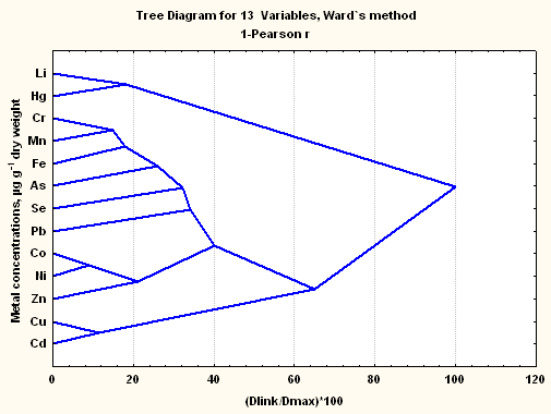
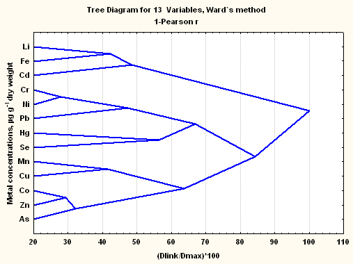
The dendrogram for the 13 residual trace elements in placentas of the NB neonate group showed four clusters (As, Se, Pb); (Cu, Ni, Hg, Co); (Cd, Fe, Mn, Cr); (Zn, Li) and for the MB neonate group three clusters (Cu, Cd, Zn, Mn); (Se, Pb, Ni, Co, Fe); (As, Cr, Hg, Li). In umbilical cord of the NB neonate group the dendrogram like tree diagram presented two clusters (Li, Fe, Ni, Co, Cr, Cd, Mn) and (Cu, Zn, As, Hg, Se, Pb) and for the MB neonate group (Figure 2) four clusters was obtained, that is to say, (Li, Hg); (Cr, Mn, Fe, As, Se, Pb); (Co, Ni, Zn); and (Cu, Cd). In the Figure 3 the tree diagram of the multi dimensional cluster of the variables is shown for the case of the amniotic membrane of the MB neonate group, which revealed three clusters (As, Zn, Co, Cu, Mn); (Se, Hg, Pb, Ni, Cr) and (Cd, Fe, Li).
The phenomenological clustering of the elements indicates that the most enriched residual trace element in placenta, umbilical cord and amniotic membrane tissues, that is to say Li, Cr, Ni, Hg and Se, presents a multidimensional similarity behaviour with essential and not essential elements that could be determined by intrinsic environmental exposure conditions to that they could have been subjected the mothers until the moment of the conception and in the period of gestation, that which could shoot inter elemental synergies. These tissues would be “good biomarker tissues” of essential and not essential trace elements in primates and human beings [20]. The binding of metals to these tissues can interfere with its physiologic functions, such as the trace elements and nutriments transport necessary for the foetal growth and its development [41], that which can lead to multimetal imbalances. During the pregnancy the quantities of metals transferred from the mother to the foetus are not constant, the biggest quantities are transferred during the last trimester [15, 37, 42].
In the case of the amniotic membranes of the MB group Figure 2 Li presents more similarity with Fe than with Cd; Cr with Ni more than with Pb; Hg with Se; Mn with Cu; and Co with Zn more than with As. In turn, in placenta tissues (Figure not shown) Li shows similarity with Hg, Cr with As, Ni with Co more than with Fe, and Se with Pb. On the other hand, in the tissues of umbilical cord Figure 1 Li presents similarity with Hg; Cr with Mn more than with Fe, As, Se, and Pb; Co with Ni more than with Zn; and Cu with Cd. In the biological systems the multiplicity of potential antagonistic or synergistic metal – metal interactions, they are important to understand the toxicity of heavy metals as Ni, Cd, Pb, Hg and As and the role of trace element such as Li, Cr, Mn, Fe, Co, Cu, Zn and Se. However, multifetal imbalances of the subtle trace element levels and the anomalous presence of heavy metals could shoot teratogenic effects and adverse effects for the neurological normal development of the human beings [43, 44].
With the purpose of exploring the two-dimensional multivariate clustering of the concentrations of the more enriched trace element in the tissues of amniotic membrane, placenta and umbilical cord of the NB and MB groups, regarding the mothers responses to the demographic variables considered in the questionnaire, the concentrations were regrouped considering this time that the most representative concentrations correspond to the mean or the median one, according to if the data distribution function follows a normal or lognormal distribution, respectively Table 6.
The Figure 3 a shows the two-way clustering of the concentrations of Li, Cr, Mn, Co, Ni, Cd, Hg, Pb, As and Se with the demographic variables in placenta of the NB newborn group, and the Figure 4b shows the two-way clustering for these same trace elements including to the copper concentration also with the demographic variables in placenta of the MB newborn group.
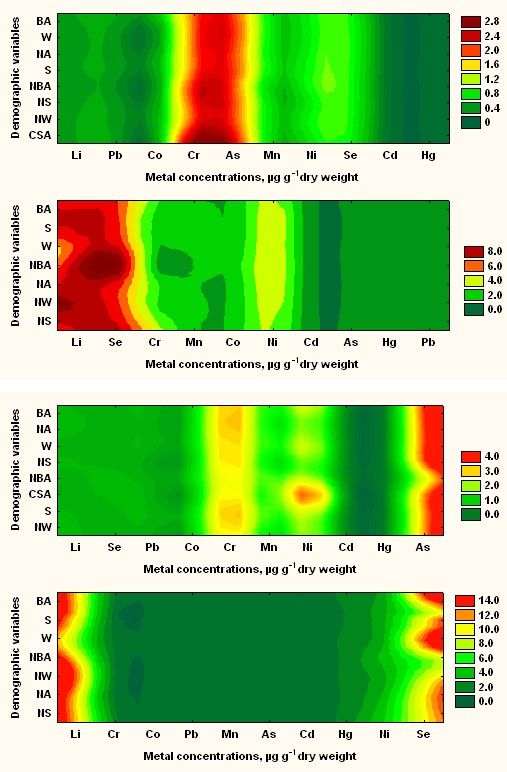
Figures 3a & 3b: Two-Way Joining Plot Residual between Placentas Traces Metal Concentrations and Mother’s Demographic Variable for Normal and Malformed Newly Born Baby’s Groups.
Figures 4a,b: Two-Way Joining Plot between Residual Umbilical Cords Traces Metal Concentrations and Mother’s Demographic Variable for Normal and Malformed Newly Born Baby’s Groups.
The monotonous distribution behaviour of some trace elements in the above two-dimensional clustering approach indicates that the enrichment tendency of this metals is to be independent of the demographic variables considered in the work, which could imply that more traverse factors that those discriminated against by these variables would be more important for the risk of the normal reproductive human development due to heavy metals exposure in the region of Antofagasta. Among these factors it is necessary up to now to highlight the exposure to the trace element matrix of the human consumption water due to the source used with this end [39], the consumption of agricultural products cultivated in the region of Antofagasta in soils enriched in heavy metals [45], the inhalation of trace element and heavy metals from atmospheres made scarce by emissions of the mining activity [1], global problems associated to the consumption of metal polluted marine foods [2, 33], and climatic factors that mobilize heavy metals from polluted soils.
Lead and arsenic did not present remarkable enrichments in placentas and umbilical cords of the MB group regarding these same tissues of the NB group, and neither inside the MB group, see Tables 7 and 8. Lead it is a heavy metal that has been present per years in punctual areas of the region of Antofagasta in Chile, included sectors of the Antofagasta city, due to the transport and later storing of sulphide metal concentrates rich in Pb in traffic through the port of Antofagasta [46, 47] which has affected the health of children of the most lead exposed sectors of the Antofagasta city. The case of Arsenic is well known and has to do predominantly with the endogenous enrichment of the element in the underground waters of the region of Antofagasta, in particular the Loa river waters that it is the main source for human consumption whose impact over the health of people is credited in the literature [48]. The concentration of arsenic in the tissues of placenta and umbilical cord of the MB newborns group collected among the years 1996 – 2000, they turned out to be significantly smaller than (p = 0.05) the arsenic concentrations measured in the same tissues of the NB newborns group collected among the years 1992 – 1993, that which would be mainly consequence of the decrease of the arsenic concentrations in the drinkable water, due to the optimization of the arsenic depression plants in operation in the region of Antofagasta. The current concentrations of total As in the drinkable water of the city of Antofagasta move among 15,0 to 30,0 ppb, that which satisfies Chilean guide of 50 ppb, but not the OMS guide of 10 ppb. Besides As, other trace element like Li, Cr, B presents enrichments in the aquatic ecosystem of the Loa river that it is the primary continental source of industrial and human consumption water [49]. In a water monitoring carried out in August of the year 2005 for the Agricultural Service and Cattleman from Chile in eight downstream water sampling stations, the range of concentrations of total As moved between 0.980 and
Another important component of the heavy metal stress of the ecosystems and the human beings of the region of Antofagasta in Chile, are the diffuse and punctual emissions coming from the copper mining industry. Otherwise, the city of Antofagasta is subjected to the climatic eolic power, typical of the dessert ecosystems that which spread through the city particulate materials enriched in heavy metals. Also, the liberated Hg from the dental amalgams and their enrichment in foods of marine origin, presently they are recognized like an important source of negative impact for the foetal normal development of pregnant women and the human being health [44, 50].
According to the results of the Tables 7&8, individually Hg appears as an environmental stressor of particular importance, since its concentration values in placental matrices of the MB group turned out to be 20 to 30 higher times than in the placental matrices of the NB group, indicating according to the concentration quotients among tissues, a clear tendency to pass through the amniotic membranes as much to the placenta as to the umbilical cord, being distributed dominantly in this last tissue, that which implies that the development new-born has been subjected to the toxic impact of Hg whose effects are broadly credited in the literature [51], mainly related with malformations and neuropathological effects.
In this work the application of the cluster techniques revealed that Li and Se show up statistical similarity with Hg Figures 1&2. So, the bioinorganic antagonism of selenium on several heavy metals, such as Hg and their antioxidant action as cofactor of the enzyme glutathione peroxidase, it is at the moment a grateful fact [52], however selenium is considered the most toxic essential trace element and in excess it is also a teratogenic [23]. Otherwise, in the last years Li has been considered essential since it protects the cells of the brain against the glutamate and Ca excess, and it is important in the transport and distribution of the vitamin B12, and therefore of Co, low levels of Li cause abnormal imbalances in the cells of the brain, neurological perturbations, difficulties of the learning in the children, bias to criminality and to heart attacks [53].
Also, it has been informed that the enrichment of Hg causes decrease of the Li concentration, that which has been considered as a factor in the neurological [54, 55, 56] illnesses. However, Li is also considered a potential theratogenic when it is administered as Lithium Carbonate in treatments of mental disorders [4]. Presently work none of the mothers was under this medication. Actually, it has been determined that Li is important for biological molecular processes in that multipurpose enzyme like the inositol monophosphatase (IMP) and the glycogen synthase kinase – 3 are involved (GSK – 3) [57].
Essentials trace element works as activators of enzyme systems or cofactors or being part of metalloenzymes, several of which are antioxidants that they protect to the cells of pathophysiological processes, and functional well – characterized metalloproteins as transferrin, hemoglobin, ceruloplasmin and ferritin [52, 58, 59]. From this point of view Mn, Fe, Co, Cu and Zn are essential elements for the metabolism of the human beings, which are often bound to protein via sulphydryl groups of amino acids as cysteine and methionine and imidazole group of histidine. The results of the concentration quotients of the Table 7 indicate with clarity the relative deficiency of these elements in umbilical cords of the congenital malformed MB group regarding those of the NB group, in this sense the cases of Mn and Zn stand out. On the other hand, the results of the concentration quotients of the Table 8, amniotic membrane / umbilical cord and placenta / umbilical cord of the congenital malformed MB newly born group, allows to infer that these essential elements have not been passed over efficiently to the umbilical cord and therefore to the foetus, presumably for inhibition of the transport of metallic essential ions that could imply to the biosynthesis of chaperone proteins [60]. Otherwise, the relative deficiency of Fe, Mn, Cu and Zn imply that the antioxidant capacity of the cells could have been depressed during the foetal development of the congenital malformed MB newly born group. In cells of mammals the enzymatic defence in front of the oxidative stress makes it mainly the enzymes catalase (CAT; Fe, Mn), superoxide dismutase (SOD; Mn, Cu-Zn) and glutathione peroxidase (GPx; Se) [52]. The deficiency in Co associates with that of vitamin B12 [53].
In this particular impairment of essential trace element and accumulation of residual heavy metals in the tissues of the newly born of the MB group, problems of inhibition of the ligand’s biosynthesis involved in the of homeostasis mechanisms of the most enriched heavy metals can be involved, in particular as the inhibition of thioligands due to problems with the metabolomics of the sulphur and with the enzymatic metallomic of the essential elements. Since Hg is not quickly eliminated from the tissues because it has been strongly bound by sulphydryl groups of enzymes, a thioproteins (metallothioneins, MT) decrease takes place in the organism, that which includes to the glutathione. Due to the decrease of the enzyme glutathione – S – transferase a decrease of the expression of MT takes place.
For the in risk newly born due to the impact of the impairment of essential trace element and the heavy metals enrichment during their intrauterine life, the situation is not restricted alone to the gestational teratogenic effects [61]
but rather also to that later already as children experience neuropsychological dysfunctions and develop pathologies like the autism [29, 43, 44, 50, 62]. In Table 9 are described the malformations that showed up in the MB newly born group. Previous results indicate us that in this group showed up a particular tendency to experience multimetal trace element imbalances for elements like Li, Cr, Mn, Co, Cu, Zn, Se and an enrichment in heavy metals as Ni and Hg [63, 64, 65] that could cause gestational teratogenic effects during the intrauterine life Lee MS, et al. [66] of the foetuses and alterations of the normal neurological development.
For some time, the narrow relationship between the environmental concentrations of essential and non-essential trace element and its concentrations in human tissues and the impact that it implies on the human being’s quality healthy life is recognized [8, 43]. Soils have a profound impact on the causation and geographical distribution of human disease and well – being Steffan JJ, et al. [67, 68], which in the last years has been rationalized by means the Medical Geology, a globally emerging discipline, which dealing with the relationship between natural geological factors Cerrillos L, et al. [69] and health in human and animals and with understanding the influence of environmental factors on the geographical distribution of such health problems, so can be considered as complementary to environmental medicine [70, 71, 72].
Of the results of this work, it could infer that the impact of the medical geology factors over the human beings begins during the development of the intrauterine life, so the status of essential and non essential trace elements on the newborns should depends on the essential and non essential trace elements status of the mothers.
Conclusions
The impairment of essential trace element and heavy metals enrichment affects the intrauterine healthy life of the Antofagasta region newly born. The situation is not restricted alone to gestational teratogenic effects, but rather also to those later already as children or young adults’ undergone neuropsychological dysfunctions like autism, diabetes, cancer and cardiovascular pathologies. The results indicate us that the MB group of babies showed up a particular tendency to experience multi metal trace element imbalances for elements like Li, Cr, Mn, Co, Cu, Zn, Se and an enrichment in heavy metals like Ni and Hg that could cause gestational teratogenic effects during the intrauterine life of the fetuses and alterations of the normal neurological development.
The impact of the medical geology factors over the human beings begins during the intrauterine life development, so the newborn essential and no essential metals status fundamentally depends of the mother trace elements status. The metal impact imprinting propagation from the mother to fetus it is a true problem. The variety of independent and dependent factors in that essential and toxic metals are involved, its relative substance amounts, intrinsic metal properties and biochemical behavior acting here and now over the fetus in the intra-uterine stage, affect their normal development and quality of its future life.
The most enriched elements were Li, Zn, Fe and Se. The concentrations of Li, Ni and Se they are distributed homogeneous and dominantly in umbilical cord. Li, Cr, Ni, As and Se and their inter-element multiple relationships, they could be associated to children’s births with malformations in the Region of Antofagasta-Chile. The tissues metal distribution was most dependent of more traverse “geo- medical imprinting factors” than demographic variables.
The most dangerous metals for the healthy life of NB children group are Chromium and Arsenic and the principal metals involved in malformed outcomes of MB children group resulting be Lithium and Selenium. Therefore, they could be “good bio-markers” for toxic metal impact over environmental health and quality of life in the Antofagasta Region-Chile.
Acknowledgements
This research was supported by resources of the University of Antofagasta through grants PI-1347-03, PI- 1331-07, and resources generated by the Bioinorganic and Environmental Analytical Chemistry Laboratory of the University of Antofagasta, through the University of Antofagasta Technical Attendance. We also thank the collaboration of the following institutions and people: Regional Hospital of Antofagasta, Obstetrics and Gynecology Service.
References
-
Duffus JH (2002) Heavy metals-a meaningless term? (IUPAC Technical Report). Pure and Applied Chemistry 74(5): 793-807.
-
Davydova SL (1999) Heavy metals as main pollutants of the Next Century. Critical Reviews in Analytical Chemistry 28(4): 377-381.
-
Gochfeld M (1997) Factors influencing susceptibility to metals. Environ Health Perspect 105(S4): 817-822.
-
Takács S, Tatár A (1987) Trace elements in the environment and in human organs: I. Methods and results. Environmental Research 42(2): 312-320.
-
Frieden E (1985) New perspectives on the essential trace elements. Journal of Chemical Education 62(11): 917-923.
-
Horovitz CT (1988) Is the major part of periodic system really inessential for life? J Trace Elem Electrolytes Health Dis 2(3): 135-144.
-
Brewer GJ, Hill GM, Dick RD, Prasad AS, Cossack ZT (1985) Interactions of trace elements: Clinical significance. Journal of the American College of Nutrition 4(1): 33-38.
-
Anke M, Groppel B (1987) Toxic actions of essential trace elements (Mo, Cu, Zn, Fe, Mn). Trace Element-Analytical Chemistry in Medicine and Biology 4: 201-236.
-
Anke M, Groppel B, Arnhold W, Langer M, Krause U (1990) The influence of the ultra-trace element deficiency (Mo, Ni, As, Cd, V) on growth, reproduction performance and life expectancy. Trace elements in Clinical Medicine pp: 361-376.
-
Tsuchiya H, Mitani K, Kodama K, Nakata T (1984) Placental transfer of heavy metals in normal pregnant Japanese women. Arch Environ Health 39(1): 11-17.
-
Sikorski R, Paszkowski T, Milart P, Radomanski T, Szkoda J (1988) Intrapartum levels of trace metals in maternal blood in relation to umbilical cord blood values: Lead, iron, copper, zinc. International Journal of Gynecology & Obstetrics 26(2): 213-221.
-
Bocca B, Ruggieri F, Pino A, Rovira J, Calamandrei G, et al. (2019) Human biomonitoring to evaluate exposure to toxic and essential trace elements during pregnancy. Part A. concentrations in maternal blood, urine and cord blood. Environmental Research 177: 108599.
-
Pi X, Qiao Y, Wei Y, Jin L, Li Z, et al. (2018) Concentrations of selected heavy metals in placental tissues and risk for neonatal orofacial clefts. Environmental Pollution 242(Pt B): 1652-1658.
-
Iyengar GV, Rapp A (2001) Human placenta as a dual biomarker for monitoring fetal and maternal environment with special reference to potentially toxic trace elements. Part 1: Physiology, function and sampling of placenta for elemental characterization. Science of the Total Environmental 280(1-3): 195-206.
-
Iyengar GV, Rapp A (2001) Human placenta as a dual biomarker for monitoring fetal and maternal environment with special reference to potentially toxic trace elements. Part 2: Essential minor, trace and other (non-essential) elements in human placenta. Science of the Total Environmental 280(1-3): 207-219.
-
Iyengar GV, Rapp A (2001) Human placenta as a dual biomarker for monitoring fetal and maternal environment with special reference to potentially toxic trace elements. Part 3: Toxic trace elements in placenta and placenta as a biomarker for these elements. Science of the Total Environmental 280(1-3): 221-238.
-
Jin L, Zhang L, Li Z, Liu JM, Ye R, et al. (2013) Placental concentrations of mercury, lead, cadmium, and arsenic and the risk of neural tube defects in a Chinese population. Reproductive Toxicology 35: 25-31.
-
Gude NM, Roberts CT, Kalionis B, King RG (2004) Growth and function of the normal human placenta. Thrombosis Research 114(5-6): 397-407.
-
Yetter JF (1998) Examination of the Placenta. American Academy Family Physician 57(5): 1045-1054.
-
Hostetler CE, Kincaid RL, Mirando MA (2003) The role of essential trace elements in embryonic and fetal development in livestock. The Veterinary Journal 166(2): 125-139.
-
Sakamoto M, Yasutake A, Domingo JL, Chan HM, Machi Kubota M, et al. (2013) Relationships between trace element concentrations in chorionic tissue of placenta and umbilical cord tissue: Potential use as indicators for prenatal exposure. Environmental Research 60: 106- 111.
-
Yiab SJ, Xiongab YW, Zhuab HL, Daiab LM, Caoab XL, et al. (2021) Environmental cadmium exposure during pregnancy causes diabetes-like phenotypes in mouse offspring: Association with oxidative stress in the fetal liver. Sci Total Environ 777: 146006.
-
Concha G, Vogler G, Lezcano D, Nermell B, Vahter M (1998) Exposure to inorganic arsenic metabolites during early human development. Toxicol Sci 44(2): 185-190.
-
Gossaia A, Lesseurb C, Farzan S, Marsitbd C, Karagasa MR, et al. (2015) Association between maternal urinary arsenic species and infant cord blood leptin levels in a New Hampshire Pregnancy Cohort. Environmental Research 136: 180-186.
-
Needleman HL (1995) Behavioural toxicology. Environ Health Perspect 103(S6): 77-79.
-
Myers GJ, Davidson PW (1998) Prenatal methylmercury exposure and children: Neurolic, developmental, and behaviour research. Environ Health Perspect 106(S3): 841-847.
-
Schuld MJ (1999) Lead toxicity: Its effects on fetal and infant development.
-
Rossipal E, Krachler M, Li F, Micetic Turk D (2000) Investigation of the transport of trace elements across barriers in human: studies of placental and mammary transfer. Acta Pediatric 89(10): 1190-1195.
-
Zheng W, Aschner M, Ghersi-Aegean JF (2003) Brain barrier systems: to new frontier in metal neurotoxicological research. Toxicology and Applied Pharmacology 192(1): 1-11.
-
Kim Y, Ha EH, Park H, Ha M, Kim Y, et al. (2013) Prenatal lead and cadmium co-exposure and infant neurodevelopment at 6 months of age: The Mothers and Children’s Environmental Health (MOCEH) study. Neuro Toxicology 35: 15-22.
-
Quevauviller Ph, Maier EA, Griepink B (1995) Quality assurence for environmental analysis. Techniques and instrumentation in Analytical Chemistry 17.
-
UNEP (PNUMA) (1984) Sampling of selected marine organisms sample preparation for trace metal analysis. Reference Methods for Marine Pollution Studies 7(2).
-
Welz B, Melcher M (1985) Decomposition of marine tissues for determination of arsenic, selenium, and mercury using hydride-generation and cold-vapor atomic absorption spectrometry. Anal Chem 57: 427- 431.
-
Román-Silva DA, Rivera L, Morales T, Avila J, Cortés P (2003) Determination of trace elements in environmental and biological samples using improved sample introduction in flame atomic absorption spectrometry (HHPN-AAS; HHPN-FF-AAS). Inter J Environ Anal Chem 83: 327-341.
-
Berndt H, Müller A (1993) Reduction of matrix interferences and improvement of detection power in flame AAS by high-performance flow/hydraulic high- pressure nebulization system for sample introduction (HPF/HHPN). Fresenius Z Anal Chem 345: 18-24.
-
Madrid Y, Cámara C (1994) Lead hydride generation atomic absorption spectrometry: An alternative to electrothermal atomic absorption spectrometry. A review Analyst 119: 1647-1657.
-
Eisenmann CJ, Miller RK (1996) Placental transport, metabolism, and toxicology of metals. Toxicology of metals. In: Louis W, Chang (Eds.), CRC Lewis Publishers, pp: 1003-1026.
-
Signes Pastora AJ, Martinez Camblor P, Bakerc E, Madana J, Guilld MF, et al. (2021) Prenatal exposure to arsenic and lung function in children from the New Hampshire Birth Cohort Study. Environment International 155: 106673.
-
Forssén UM, Herring AH, Savitz DA, Nieuwenhuijsen MJ, Murphy PA, et al. (2007) Predictors of use and consumption of public drinking water among pregnant women. J Expo Sci Environ Epidemiol 17(2): 159-169.
-
Ott WR (1995) Environmental Statistics and Data Analysis. CRC Lewis Publishers, Boca Raton New York, USA.
-
Fraga CG (2005) Relevance, essentiality and toxicity of trace elements in human health. Molecular aspects of medicine 26(4-5): 235-244.
-
Angelis P, Miller RK, Darrah TH, Katzman PJ, Pressman EK, et al. (2017) Elemental content of the placenta: A comparison between two high-risk obstetrical populations, adult women carrying multiples and adolescents carrying singletons. Environ Res 158: 553- 565.
-
Kalter H (2003) Teratology in the 20th century environmental causes of congenital malformations in human and how they were established. Neurotoxicol Teratol 25(2): 131-282.
-
Bellinger DC (2005) Teratogen update: Lead and Pregnancy. Birth Defects Res A clin Mol Teratol 73(6): 409-420.
-
Pizarro I, Gómez MM, Palacios MA, Cámara C (2003) Distribution of arsenic species in environmental samples collected of northern Chile. International Journal of Environmental Analytical Chemistry 83(10): 879-890.
-
Tuscan CD, Guilarte TR (2005) Lead neurotoxicity: from exposure to molecular effect. Brain Res Brain Res Rev 49(3): 529-554.
-
Tchernitchin AN, Lapin N, Molina L, Molina G, Tchernitchin NA, et al. (2006) Human exposure to lead in Chile. Reviews of Environmental Contamination and Toxicology 185: 93-139.
-
Hopenhayn-Rich C, Browning SR, Hertz-Picciotto I, Ferreccio C, Peralta C, et al. (2000) Chronic arsenic exposure and risk of infant mortality in two areas of Chile. Environ Health Perspec 108(7): 667-673.
-
Marshall G, Ferreccio C, Yuan Y, Bates MN, Steinmaus C, et al. (2007) Fifty -year study of lung and bladder cancer mortality in Chile related to arsenic in drinking water. J Natl Cancer Inst 99(12): 920-928.
-
Aschner JL, Aschner M (2007) Methylmercury Neurotoxicity: Exploring potential novel targets. Open Toxicol J 1: 1-9.
-
Björnberg KA, Vahter M, Petersson-Grawé K, Glynn A, Cnattingius S, et al. (2003) Methyl mercury and inorganic mercury in Swedish pregnant women and in cord blood: Influence of fish consumption. Environ Health Perspec 111(4): 637-641.
-
Gromadzinska J, Wasowicz W, Sklodowska M, Bulikowski W, Rydzynski K (1996) The influence of atmospheric chromium on selenium content and glutathione peroxidase activity in blood of Tannery workers. Environmental Health Perspectives 104(12): 1312- 1316.
-
Schrauzer GN, Shrestha KP, Flores-Arce MF (1992) Lithium in scalp hair of adults, students, and violent criminals. Effects of supplementation and evidence for interaction of lithium with vitamin B12 and with other trace elements. Biol Trace Elem Res 34(2): 161-176.
-
Schrauzer GN, De Vroey E (1994) Effects of nutritional lithium supplementation on mood. A placebo-controlled study with former drug users. Biol Trace Elem Res 40(1): 89-101.
-
Sutton DJ, Tchounwou PB, Ninashvili N, Shen E (2002) Mercury induces cytotoxicity and transcriptionally activates stress genes in human liver carcinoma (HepG2) cells. Int J Mol Sci 3(9): 965-984.
-
Aral H, Vecchio-Sadus A (2008) Toxicity of lithium to humans and the environment – A literature review. Ecotoxicology and Environmental Safety 70(3): 349-356.
-
Strunecká A, Potocka J, Sárek M (2005) How does lithium mediate you its therapeutic effect? J Appl Biomed 3: 25- 35.
-
Gambling L, Danzeisen R, Fosset C, Andersen HS, Dunford S, et al. (2003) Iron and copper interactions in development and the effects on pregnancy outcome. J Nutr 133: S1554-S1556.
-
O’Brien KO, Zavaleta N, Abrams SA, Caulfield LE (2003) Maternal iron status influences iron transfer to the fetus during the third trimester of pregnancy. Am J Clin Nutr 77(4): 924-930.
-
Lönnerdal B (2005) Trace element nutrition of infants- molecular approaches. Journal of Trace Elements in Medicine and Biology 19(1): 3-6.
-
Wang Z, Wang H, Xu ZM, Ji YL, Chen YH, et al. (2012) Cadmium-induced teratogenicity: Association with ROS- mediated endoplasmic reticulum stress in placenta. Toxicology and Applied Pharmacology 259(2): 236-247.
-
Palmer RF, Blanchard S, Stein Z, Mandell D, Miller C (2006) Environmental mercury release, special education rates, and autism disorder: an ecological study of Texas. Health & place 12(2): 203-209.
-
Xieab X, Dingb G, Cuia C, Chena L, Gaoa Y, et al. (2013) The effects of low-level prenatal lead exposure on birth outcomes. Environmental Pollution 175: 30-34.
-
Hinwooda AL, Callana AC, Ramalingama M, Boycea MJ, Heyworth J, et al. (2013) Cadmium, lead and mercury exposure in non smoking pregnant women. Environmental Research 126: 118-124.
-
Romano ME, Enquobahrie DA, Simpson C, Checkoway H, Williams MA (2016) Maternal body burden of cadmium and offspring size at birth. Environmental Research 147: 461-468.
-
Lee MS, Eum KD, Golam M, Quamruzzaman Q, Kile ML, et al. (2021) Umbilical Cord Blood Metal Mixtures and Birth Size in Bangladesh Children. Environmental Health Perspectives 129(5): 57006.
-
Steffan JJ, Brevik EC, Burgess LC, Cerdà A (2018) The effect of soil on human health: an overview. European Journal of Soil Science 69(1): 159-171.
-
Peña-Fernández A, Lobo-Bedmar MC, González-Muñoz MJ (2015) Annual and seasonal variability of metals and metalloids in urban and industrial soils in Alcalá de Henares (Spain). Environmental Research 136: 40-46.
-
Cerrillos L, Fernández R, Machado MJ, Morillas I, Dahiri B, et al. (2019) Placental levels of metals and associated factors in urban and sub-urban areas of Seville (Spain). Journal of Trace Elements in Medicine and Biology 54: 21-26.
-
Selinus O, Alloway B, Centeno JA, Finkelman RB, Fuge R, et al. (2005) Essentials of Medical Geology: Impacts of the Natural Environment on Public Health. Environ Health Perspect 113(11): A780.
-
Iyengar V, Wolttlez J (1988) Trace elements in human clinical specimens: Evaluation of literature data to identify reference values. Clin Chem 34(3): 474-481.
-
Abrahams PW (2006) Soil, geography and human disease: a critical review of the importance of medical cartography. Progress in Physical 30(4): 490-512.
- Evaluation of Skin Aging Preventive Effects of Cherry Blossom Petal Extracts Through Antioxidant and Anti-Glycation Activities
- Is Cell Death Responsible for False Positive Results of In Vivo Comet Assay?
- Pattern of Gonadal Hormones in Oral Testosterone-Supplimented Male Wistar Rats with Diabetes-Induced Hypogonadism
- Re-Evaluation of the Genotoxicity of Currently Used Food Dyes in Mouse Multiple Organs Via Continuous Administration by Drinking Using the Comet Assay
- Pharmacogenetics of Type 2 Diabetes Mellitus: Linking Genetic Variability to Drug Efficacy and its Cardiovascular Outcomes
- Exploratory Proteomic Profiling of SARS-CoV-2 Infected THP-1 Macrophages Reveals Alterations in Inflammatory Response and Cellular Metabolism