Molecular-Engineered Nanoparticle’s Intensive Use in Nanotechnology Challenges like Nanotoxicity and Breakthrough Opportunities
This critical review focuses on Engineered nanoparticles (ENPs) types, properties, toxicities, applications, and future scopes. ENPs are specially designed chemical substances or materials with a particle size between 1 and 100 nm in at least one dimension, while the term Engineered nanomaterials (ENMs) is used collectively for ENPs and nanostructured materials. They can be classified into different classes based on their properties, shapes, or sizes. The different groups include fullerenes, metal NPs, ceramic NPs, and polymeric NPs. NPs possess unique physical and chemical properties due to their high surface area and nanoscale size. NPs like carbon nanotubes (CNTs), quantum dots (QDs), fullerenes, nanocomposites, metal-oxide nanoparticles (TiO2, ZnO, and iron oxides), and fibrous nanomaterials (NMs). Exposure to such ENP increases as nanotechnology advances, though it is an emerging field with a wide range of applications as an interdisciplinary, integrated science with huge potential. Aims to present a comprehensive review of nanotoxicity to human health and impact on the environment, future aspects breakthrough opportunities. Nanowaste as a new and challenging research topic is also highlighted. NPs’ intensive use, persistence, and retention in the environment need to evaluate nanogenotoxicity to humans and other organisms. ENMs used in therapeutic targeted drug delivery, their toxicokinetic, surface chemistry, mechanism of cellular interactions, effect on chromosomes, and DNA started being studied by researchers, however, not fully understood. Nano genotoxicity assessment should be done thoroughly following regulatory guidelines in order to safeguard humans from its harmful effects like cancer and reduce fertility. ENM retention time in the environment should be traced keeping in mind Eco nanotoxicity, bioaccumulation, and biomagnification in aquatic and forest ecosystems. The unique properties of NPs can be utilized fully by knowing all useful characteristics for breakthrough inventions in health care, cancer treatment, anti-aging (longevity), aeronautics, illuminating diodes, anofertilizers and Artificial Intelligence (AI)-powered robotics for noble innovations for human use. Finally, the challenges of nanowaste management and gaps in breakthrough opportunities are highlighted to reflect the need for more serious action and advancement in nanoscience with novel inventions.
Introduction
Engineered nanoparticles (ENPs) are specially designed chemical substances or materials with a particle size between 1 and 100 nm in at least one-dimension [1, 2], while the term Engineered nanomaterials (ENMs) is used collectively for ENPs and nanostructured materials. Nanoparticle (NP), an ultrafine unit with dimensions measured in nanometres (nm; 1 nm = 10−9 meter). The prefix “nano” is derived from the Greek word “nanos” meaning “dwarf” and is used for tiny particles in science. In scientific terms, it means one billionth of something (10−9). The concept of a nanometer as one billionth of a meter was first proposed by Richard Zsigmondy, 1925 Nobel Prize Laureate [3]. He was able to measure the size of gold NPs by an imaging technique. Research can be carried out in the nanoscale by the use of a Transmission Electron Microscope (TEM) built by Max Knoll and Ernst Ruska in 1931 [4]. The width of a human hair is about 80,000 nm. The width of a single blood cell is about 7000 nm while the width of a water molecule is 0.3 nm [5].
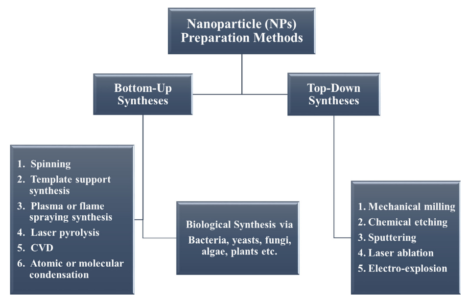
Nanoparticles can be naturally occurring, derived from a volcanic eruption, lightning causing forest fires, and ocean water evaporation (Sea salt aerosols) [6]. Nanoscale organisms, commonly known as nano-organisms are found all around us and even inside our bodies for example, nanobacteria, viruses as well as fungi, algae, and yeast that can produce NPs in their bodies. Nanobacteria, Magnetotactic bacteria, and novel nano-organisms, called nanobes, are gaining interest among nanotechnology researchers as they are found during off-shore petroleum exploration on Triassic and Jurassic sandstones in Western Australia [7]. Combustion like diesel soot, atmospheric or engineered, and created by humans using different methods of synthesis Figure 1.
![Figure 1: Typical synthetic methods of NPs for the (a) top-down and (b) bottom-up approaches [8].](/fulltextimages/11586/fig_1.png)
Simple combustion during cooking, in vehicles, fuel oil and coal for power generation Linak WP, et al. [9], airplane engines, chemical manufacturing, welding, ore refining, and smelting are some of the anthropogenic activities that lead to NP formation [10]. NMs such as carbon NPs De Volder MFL, et al. [11], TiO2 NPs Weir A, et al. [12], and hydroxyapatites Sadat-Shojai M, et al. [13] are present in commercial cosmetics, sporting goods, sunscreen, and toothpaste. Thus, these synthetic NPs are a new genre of NPs that may induce adverse environmental and human health effects.
Depending on the overall shape these materials can be 0D, 1D, 2D, or 3D [14]. The ISO standard similarly defined two-dimensional nano-objects (i.e., nanodiscs and nanoplates) and one-dimensional nano-objects (i.e., nanofibres and nanotubes) [Encyclopaedia Britannica]. NPs have unique properties because of their quantum size and large surface area-to-volume ratio compared with their larger counterparts. Moreover, ENPs unique properties may find a wide range of practical applications including improved thermal and/or electrical conductivity, substantially harder and stronger materials (yet lighter than metals such as steel), improved catalytic activity and advanced optical properties, changed surface chemistry, cellular uptake, and proliferation abilities, in medicine, engineering, and environmental remediation.
![Figure 3: Electron microscope images show how NPs can penetrate and relocate to various sites inside a phagocytic cell line. (A) Untreated phagocytic cell line (RAW 264.7). Cells were treated with (B) ultrafine particles ((<100 nm) (C) TiO2, (D) fullerol, (E) COOH–polystyrene nanospheres, and (F) NH2polystyrene nanospheres. NP exposure was conducted by treating the cells with 10 μg/mL NPs (<100 nm) for 16 h. Labels: M = mitochondria, P = particles Xia T, et al. [42], copyright 1969, American Chemical Society.](/fulltextimages/11586/fig_3.png)
NPs can be classified into any of various types, according to their size, shape, and material properties Figure 2. Some classifications distinguish between organic and inorganic NPs; the first group includes dendrimers, liposomes, and polymeric NPs, while the latter includes fullerenes, QDs, and gold NPs. Other classifications divide NPs according to whether they are carbon-based like carbon nanotubes, ceramic, semiconducting, or polymeric. In addition, NPs can be classified as hard (e.g., titania [titanium dioxide], silica [silica dioxide] particles, and fullerenes) or as soft (e.g., liposomes, vesicles, and nanodroplets) [Encyclopaedia Britannica]. It can exist as zero-valent metal nanoparticles (Ag, Fe, and Zn), metal-oxide NPs (TiO2, ZnO, and iron oxides), QDs, CNTs, nanocomposites and fibrous NMs [15]. There are reports of more than 800 consumer products already available containing NMs. The most studied NPs include titanium dioxide, alumina, zinc oxide, carbon black, CNTs, and buckminsterfullerene. Yet lighter than metals such as steel, improved catalytic activity, and advanced optical properties. Hence, human exposure is occurring and is yet to increase drastically in the coming years.
NPs are complex molecules composed of three layers i.e. (a) surface layer, which may be functionalized with a variety of small molecules, metal ions, surfactants, and polymers. (b) shell layer, which is a chemically different material from the core in all aspects, and (c) the core, which is essentially the central portion of the NP and usually refers to the NP itself [16]. In addition, NPs have three major interrelated physical properties: (i) highly mobile free state (e.g., in the absence of some other additional influence, a 10-nm-diameter nanosphere of silica has a sedimentation rate under gravity of 0.01 mm/day in water); (ii) specific enormous surface areas (e.g., a standard teaspoon, or about 6 ml, of 10-nm-diameter silica nanospheres has more surface area than a dozen doubles-sized tennis courts; 20 percent of all the atoms in each nanosphere will be located at the surface); and (iii) may exhibit as quantum effects. Thus, NPs have a vast range of compositions, depending on the product applications.
“Nanoscale technologies” also known as “nanotechnologies” include fields of nanoscience, as diverse as surface science, organic chemistry, molecular biology, semiconductor physics, energy storage Hubler A [17, 18], engineering Elishakoff I, et al. [19], microfabrication David L [20], and molecular engineering [21]. It is an emerging field of research with a wide range of applications in health care, medicine, cancer therapy, the nutraceutical industry, cosmetics (anti-aging creams, lipstick, sunscreen), paint, aerospace engineering, nano-electronics, environmental remediation, and food industry Singh N, et al. [22], computing, robotics, and whole aspects of life. they can be in any form such as tubes, rods, wires, or spheres, with more elaborate structures devised, such as nano-onions and nano peapods [23, 24]. Today, first-generation products of nanotechnology such as paints, coatings, nanostructured metals, ceramics, and so on are already on the market [25]. Second (transistors, chemical and biological sensors, amplifiers, targeted drugs, actuators, adaptive structures), third (guided assembling, 3D networking and new hierarchical architectures, robotics, and evolutionary developments), and fourth (new devices based on molecules) generation nanoproducts are in development [26]. Fifth-generation nanoproducts like light-emitting diodes, cells, and AI-powered nano transformers are in the pipeline. s. The NPs can be employed for drug delivery Lee VH [27], chemical and biological sensing Barrak H, et al. [28], gas sensing Mansha M, et al. [29, 30, 31], CO2 capturing Ganesh M, et al. [32, 33] and other related applications [34]. Nanotechnology may be able to create many new materials and devices with a vast range of applications, such as nanomedicine, nanoelectronics, biomaterials energy production, and consumer products. However, it raises many of the same issues including concerns about the toxicity and environmental impact of NMs [35].
This review article focuses on ENP types, properties, toxicities, applications, and future scopes. The ongoing debate on the future implications of nanotechnology, its toxicity, its impact on the environment, and its effects on the global economy were discussed critically. Moreover, this article highlights breakthrough opportunities and future aspects.
ENPs Challenges like Nanotoxicity
The easy availability of NPs to the human body also increases through different modes of inhalation exposure, skin, oral, or intravenous routes. With the growing nanotechnology industry, occupational exposure of the workforces, medical sources of exposure include dental prosthesis and orthopaedic implant wear debris. In this way, human exposure is occurring frequently and is yet to increase drastically in the future. Hence, it is necessary to analyze the toxicity of these NMs. It becomes necessary to know the mechanism of action of these NPs at the cellular level whether it is causing damage to DNA e.i., nanogenotoxicity, altering metabolic reactions at the physiological level, or causing any alteration at the level of gene expression. The toxicokinetics of NMs are still not well understood. Nanogenotoxicology regulatory guidelines need to be validated so that their toxic effects on human and other animal bodies can be thoroughly analyzed. The effects of such compounds on plant systems or the environment i.e., nanoecotoxicology, need to be considered because plants humans, and other animals are all interconnected through the food chain. European Union (EU) funded projects follow recently published best recommendations in nanogenotoxicology research, they created a set of criteria that address assay-specific reliability and relevance for risk assessment purposes.
Thus, irreversible oxidative stress, organelle damage, asthma, and cancer can be caused by NPs depending on their composition. The general acute toxic effects caused by exposure to NPs and nanostructured materials include reactive oxygen species generation, protein denaturation, mitochondrial disconcertion, and perturbation of phagocytic functions. Uptake by the reticuloendothelial system, nucleus, neuronal tissue, and the generation of neoantigens that cause possible organ enlargement and dysfunction are common chronic toxic effects of NPs [36].
| Nanomaterial properties | Risk description | |
|---|---|---|
| 1 | agglomeration or aggregation | Weakly bound (agglomeration) and fused particles are significant risk criteria as they lead to poor corrosion resistance, high solubility, and phase change of NMs’. This further leads to deterioration and the structure maintenance becomes challenging [37,38]. |
| 2 | reactivity or charge | NPs can be charged either by functionalization or spontaneous degradative reactions. Chemical species and their charge-related critical functional groups will be a significant factor in the specific functionality and bioavailability of NMs [39]. |
| 3 | impurity | Inherently, NPs interact with impurities due to their high reactivity. Due to this reason, encapsulation becomes a prime necessity for solution-based NP synthesis (chemical route). In the encapsulation process, the reactive nano-entities are encapsulated by nonreactive species to provide stability to the NPs. |
| 4 | contaminant dissociation | The contamination of residual impurities in the NP is considered a major risk factor. For example, sulfur impurities may present in iron oxide NPs depending on the precursor used for their production (FeCl3 or Fe2(SO4)3). Similarly, nickel, yttrium, or rubidium metal impurities may be present in the carbon nanotubes (CNTs) Bortoleto GG, et al. [40,41] that are adsorbed on the CNT surface. |
| 5 | size | Reactivity and agglomeration of NPs is mostly dependent on their particle size. It is well known that the process of agglomeration will happen at slower rates in smaller particles. After the synthesis of the NPs, it is impossible to retain their original size. Hence, encapsulation becomes highly inevitable in NP synthesis. The exceptional size-dependent chemistry of NPs is distinguished from classical colloid chemistry by categorizing NPs according to their particle size [39]. |
| 6 | recycling and disposal | NMs are not bound to any hard-and-fast safe disposal policies. The experimental results of NP exposure are not available and their potential toxicity issues are still under question. Hence, the uncertainty of a nanomaterial’s effect is yet to be developed for permanent disposal and recycling policies. |
Table 1: Summary of five basic nanomaterial properties and their potential risks and challenges Jaison Jeevanandam, 2018.
It is also noteworthy that the chemical composition and shape of the particle are the main factors contributing to NP toxicity, other than size and aging, electrostatic charges, van der Waals forces, interfacial tension effects, and steric interaction of NPs bind with cellular components and cause cell death Figure 3.
![Figure 4: Overview of the demonstrated modes of action and end-points upon induction of primary and secondary genotoxicity by NMs [72].](/fulltextimages/11586/fig_4.png)
Figure 3: Electron microscope images show how NPs can penetrate and relocate to various sites inside a phagocytic cell line. (A) Untreated phagocytic cell line (RAW 264.7). Cells were treated with (B) ultrafine particles ((<100 nm) (C) TiO2, (D) fullerol, (E) COOH–polystyrene nanospheres, and (F) NH2polystyrene nanospheres. NP exposure was conducted by treating the cells with 10 μg/mL NPs (<100 nm) for 16 h. Labels: M = mitochondria, P = particles Xia T, et al. [42], copyright 1969, American Chemical Society.
A wide variety of NPs can create reactive oxygen species and cause cellular damage via lipid peroxidation, protein alteration, DNA disruption, signaling function interference, and gene transcription modulation [43].
According to Jaison Jeevanandam and his team 2018, toxicological data, the toxicity of NMs depends on various factors:
Dose and Exposure Time Effect The number of NMs that penetrate the cells directly depends on the molar concentration of NPs in the adjacent medium multiplied by the exposure time [43].
Aggregation and Concentration Effect There are many contradictory reports on the toxicity of NPs at different concentrations. Increasing the NP concentration promotes aggregation. Most NP aggregates are micrometers in size, so a significant quantity of aggregated NPs may not penetrate cells thereby losing their toxicity.
Particle Size Effect NPs show a size-dependent toxicity. Ag NPs with ≈a 10 nm diameter show a higher capacity to penetrate and disturb cellular systems of many organisms than Ag+ ions and Ag NPs of larger diameters (20–100 nm) [44].
Particle Shape Effect NPs exhibit shape-dependent toxicity, that is, different toxicity levels at different aspect ratios. For example, asbestos fibers of 10 µm length can cause lung cancer, shorter asbestos fibers (5–10 µm) can cause mesothelioma and 2 µm length fibers can cause asbestosis [45].
Surface Area Effect Typically, the toxicological effect of NPs increases with decreasing particle size and increasing surface area. It can also be noted that nano and microparticles with the same mass dose react differently with the human cells. Crystal Structure Effect Based on the crystal structure, NPs may exhibit different cellular uptake, oxidative mechanisms, and subcellular localization [46]. For example, the two crystalline polymorphs of TiO2 (rutile and anatase) show different toxicity. In the dark, rutile NPs (200 nm) lead to DNA damage via oxidation, while anatase NPs (200 nm) do not induce DNA damage in dark conditions [47].
Surface Functionalization Effect The surface properties of NPs have shown drastic effects relating to translocation and subsequent oxidation processes [48, 49].
Pre-Exposure Effect The cellular phagocytic activity can be stimulated by shorter exposure time or the pre-exposure of lower NP concentrations [35]. This pre-exposure results in the adaptability of the human body against NPs to some degree [50].
The data gathered is collectively provided in the eNanoMapper database [51]. Mutagenicity is a key endpoint to detect hazards in all products developed including NMs due to important human health consequences [44, 45, 46, 52, 53]. The genotoxicity of ENMs, in vivo studies Pfuhler S, et al. [47, 48, 49, 50, 51, 52, 53, 54] and in vitro studies Thongkam W, et al. [55, 56, 57, 58, 59, 60, 61, 62, 63] and review highlighting future perspectives were reported Carriere M, et al. [64, 65, 66]. Bacterial gene mutation assays were omitted due to limited particle uptake and less informative for NMs [67]. Comet assay in vitro single-cell gel electrophoresis was also not included due to protocol variation and lack of standardization, high possibility of getting false positive results as NMs can interfere with DNA after lysis. For the in vivo studies, all routes of exposure were accepted [68]. Some of the nanogenotoxicity assessment inclusion criteria that need to be taken into account are given as follows:
Inclusion criteria
In vitro Studies Performed Using Validated Assays
- Mammalian cell micronucleus (MN) assay
- Chromosomal aberration (CA) assay
- Mammalian gene mutation assays: tests using hypoxanthine–guanine phosphoribosyltransferase (Hprt) or xanthine phosphoribosyltransferase (Xprt), and thymidine kinase (Tk) genes, including the mouse lymphoma assay (MLA)a
In vivo Studies Performed Using Validated Assays
- In vivo comet assay
- Mammalian erythrocyte MN assay
- Mammalian CA assay
- Gene mutation assays In vivo studies performed by inhalation, oropharyngeal aspiration, intratracheal instillation, dermal, oral exposure, or any type of injection [69].
Criteria for the Assay-Specific Evaluation of the Validated in Vitro Mammalian Cell Genotoxicity Assays
All in vitro Assays
- Adequate dose range: Doses with no or low cytotoxicity and moderate cytotoxicity should be included. For non- toxic materials, a maximum dose of ~250 µg/ml is considered adequate
- 2. If the result is positive only at highly cytotoxic doses (close to 50–60% cytotoxicity for micronucleus test or 10–20% survival for gene mutation tests) the conclusions of the study must be reevaluated
- 3. Cellular uptake should be confirmed Mammalian cell micronucleus (MN) test
- 4. Background MN frequency must be provided
- 5. The concurrent positive controls must elicit a statistically significant increase in MN frequency
- Concurrent cytotoxicity assessment must be performed as described in OECD TG487 (with Cyt-B CBPI or RI; without Cyt-B RPD or RICC)
- A minimum of 2000 cells have been scored per concentration. A minimum of two replicates or independent experiments have been performed
- If Cytochalasin B is used, it is added≥6 h after starting the exposure to allow uninhibited cellular uptake. Tests with positive results are accepted regardless of this criterion
- There are no major problems in the study design (e.g. less than 3 doses with up to 50–60% cytotoxicity, only cytotoxic doses tested, harvesting schedule does not fall within 1.5–2 cell cycles, treatment time less than 3 h, inappropriate solvent without vehicle control, too high solvent concentration)
- Treatment schedule: The cells have completed at least one cell cycle in the presence of the NM so that NMs taken up by the cells may come into direct contact with the DNA when the nuclear membrane breaks down during mitosis. Tests with positive results are accepted regardless of this criterion Mammalian chromosomal aberration (CA) test
- Background CA level must be provided
- Positive control induces a statistically significant increase in CAs
- Concurrent cytotoxicity assessment is performed as described by the OECD guideline (RPD or RICC, MI is acceptable for primary lymphocytes)
- A minimum of 300 metaphases per concentration (unless clearly positive response) are analysed. A minimum of two replicates or independent experiment are performed
- There are no major problems in the study design (e.g., less than 3 doses with up to 50–60% cytotoxicity, only cytotoxic doses tested, clearly inadequate treatment time, harvesting schedule does not fall within 1.5 cell cycles, inappropriate solvent without vehicle control, too high solvent concentration)
- Treatment schedule: The cells have completed at least one cell cycle in the presence of the NM so that NMs taken up by the cells may come into direct contact with the DNA when the nuclear membrane breaks down during mitosis. Tests with positive results are accepted regardless of this criterion [69].
For in vivo studies, Drosophila melanogaster is a noble test system for biological experiments (since 75% of the genes responsible for human diseases are known to have homologs) by feeding the 3rd instar larvae with different concentrations of NMs [70]. Co-NPs were able to induce genotoxic activity in the wing-spot assay of D. melanogaster, mainly via the induction of somatic recombination [71]. There is supporting literature showing many of the ENMs cause genotoxic effects, such as chromosomal abnormalities (like fragmentation), DNA strand breakages, point mutations, oxidative DNA stress, and gene expression profile alterations as mentioned in Figure 4 [72]. Moreover, these substances induce cytotoxicity, oxidative stress, and inflammatory responses [73, 74, 75]. NPs can easily cross the cell and nuclear membranes by diffusion or transport through Nuclear Pore complexes, thereby reaching the nucleus where they can interact directly with the DNA inducing damage, factors like retention time, cellular accumulation, and subsequent consequences need to be considered. DNA damage induction due to genetic alterations in the absence of cell death leads to carcinogenesis or if damage happens to germ cells can also affect fertility, and can transfer adverse health effects to the subsequent generations [76, 77].
Scientific Committee on Consumer Safety (SCCS) could not resolve the use of NM, SiO2 in cosmetics due to a lack of sufficient data [78]. However, the eNanoMapper database provided at least 200 entries related to the toxicity of this particular substance. Titanium Dioxide (TiO2) is the most highly used. There is available literature on several NMs having adverse effects on the environment also (e.g., marine Mussels) and human health [79].
In terms of social, economic, and scientific advances, it also has some disadvantages. The hazards of NPs originate from their size, shape, chemistry, and so on, do not have absolute control over the technology based on NPs. Since our knowledge of NPs is extremely limited, producers, consumers, and anyone in touch with nanoproducts should be very careful [25]. Nanotechnology will bring dramatic changes to every area of humanity and the environment. Moreover, it is also bringing changes in manufacturing processes and may result in unemployment. However, the effects of uncontrollably releasing NPs into the biosphere have not been made clear. Nanotechnology is currently extremely expensive. Directly handling NPs and the production of unintended NPs as by-products as nanowaste are two ways of exposure. Since NPs have a high surface-to-volume ratio, it is probable that they can take part in body reactions such as protein synthesis and, at worst, damage DNA structure. Thus, they may result in unwanted products, therefore, nanotechnology has the possible capability to degenerate the human body at an atomic and/or molecular level and to destroy it. In the study carried out on workers exposed to NPs, it was concluded that the workers had fatal diseases after a few years of exposure [80]. The British government now has commissioned a report prescribing studies on how to determine the biological hazards of NPs [81]. NPs have been utilized in the food industry in order to enhance food quality. However, the long-term side effects of the nano additives are unknown. NPs brought into some solid materials can improve their mechanical attributes such as stiffness, hardness, and flexibility. The fuel consumed by vehicles is mostly dependent on the weight of the vehicle. The weight of a vehicle can be decreased thanks to nanotechnology engineering and fuel consumption can be lowered. One of the agents that can be used in soil remediation is iron oxide. In order for the application to be effective, a large number of NPs of iron oxide should be added, but the effects of releasing large numbers of NPs are not known. Nitrogen fixation can be improved by functionalized NPs in order to enhance the quality of soil, especially in land with low fertility. Using nanotechnology, more compact and smaller devices are likely to emerge. This may lead to undetectable eavesdropping tools which are potential risks related to privacy. There is a broad consensus that nanotechnology presents a variety of ways to improve life, but what if this technology fell into the wrong hands? The gap between developed and developing countries will increase. This brings about economic and social crises. In other words, developed countries will benefit more from nanotechnology while developing countries suffer much more from the risks associated with it. The threat is the “grey-goo” scenario introduced by E. Drexler in which nanobots are revolutionized in such a way that they have self-replicating abilities. Gaining this ability, they will consume anything around to make their copies and this will be the end of humankind. In science, ethics is an area where the effects of processes and conclusions are brought under discussion in terms of the environment and living beings. As the process becomes more complicated, the ethics issue also becomes knotty [25]. This is chiefly due to a lack of knowledge. Because our knowledge is limited, it is quite difficult to have complete control over this scientific process and its consequences. In the case of nanotechnology, coined as “Nano-ethics” Robison WL [82], the ethical issue becomes even more complex. Is nano-ethics new or completely different from other scientific ethics? The questions about risk assessment and potential implications are not new [83]. What makes ethical considerations of nanotechnology complex is that almost all branches of science such as physics, chemistry, biology, and all engineering areas are converged in nanotechnology. Since nanotechnology has great promise, it naturally has a great interest in both the public and the media. As the public has discussed the benefits and risks of nanotechnology, the ethical side of nanotechnology has also drawn special attention. Armin Grunwald had a detailed discussion about the ethics of nanotechnology [84]. It is quite clear that our knowledge about nano-toxicity is not adequate.
However, although both the knowledge and awareness of potential because of the extraordinary behaviour of NPs, they can easily penetrate living organisms and change the functions of some parts either in desirable or undesirable ways. However, taking all possible implications into account, risks can be minimized or rather reduced to an acceptable level. If nanotechnology is used without care, the results may be quite destructive and irreversible. Some research has shown that some NPs can be toxic and easy inhalation of toxic materials directly or indirectly can adversely affect cells and degenerate or hinder their functions. This kind of vital information should be shared with the public immediately. To make an informed they should not aim only at their self-interest. Dangerous or harmful products should not be presented to the public. Possible contributions that nanotechnology may provide to solve some basic problems such as clean water, a clean environment, and starvation should be thoroughly investigated. Similarly, potential risks should be carefully analyzed. However, not easy to decide whether nanotechnology brings us more benefits than damages, so a detailed risk assessment should be carried out.
![Figure 5: (A) Single-walled carbon nanotubes (SWCNT); (B) Cancer cell targeting antibodies are bound to SWCNT loaded with the drug of choice; (C) Active drug delivery occurs when the antibody binds to the specific antigen on the cancer cells: (D) Broader illustration depicting active drug delivery [91].](/fulltextimages/11586/fig_5.png)
The intensive use of these nano-enabled products results in the release of NPs in the environment, resulting in a new type of waste termed nanowaste eg., particulate matter (PM), suspended PM is more dangerous because it can be easily inhaled during breathing and can accumulate inside lungs ultimately elicit different diseases. Nanaowaste management is now an active area of research.
Applications of Various Nanoparticles
NPs are broadly classified into various categories depending on their morphology, size, and chemical properties. Such NPs with specialized chemical properties can be designed to perform specific functions with a wide array of applications listed as follows:
i) Carbon-Based NPs These NMs contain carbon and are found in morphologies such as hollow tubes, ellipsoids, or spheres. Fullerenes (C60) and carbon nanotubes (CNTs) represent two major classes of carbon-based NPs. Fullerenes contain NMs that are made of globular hollow cages such as allotropic forms of carbon. They have created noteworthy commercial interest NPs 909 due to their electrical conductivity, high strength, structure, electron affinity, and versatility [85]. These materials possess arranged pentagonal and hexagonal carbon units, while each carbon is sp2 hybridized. CNTs are elongated, tubular structures, 1–2 nm in diameter Ibrahim KS [86], carbon nanofibers, carbon black, graphene (Gr), and carbon onions are included under the carbon-based NMs category [36]. L. These can be predicted as metallic or semiconducting reliant on their diameter telicity (Aqel). Due to their unique physical, chemical, and mechanical characteristics, these materials are not only used in pristine form but also in nanocomposites for many commercial applications such as fillers Saeed K, et al. [87, 88], efficient gas adsorbents for environmental remediation Ngoy JM, et al. [89], and as a support medium for different inorganic and organic catalysts [90]. Nanocarriers such as CNTs Figure 5 & QDs Figure 6 show excellent fluorescent properties and are useful candidates for imaging techniques and fluorescence-guided surgery. Another promising NP is biomimetic NMs that mimic the functionality of biological components used in targeted drug delivery using CNTs as reported by K. Tapasya, 2022.
![Figure 6: Quantum Dots in Targeted Tissue Fluorescence Imaging [91].](/fulltextimages/11586/fig_6.png)
![Figure 7: The main advantages of nanoencapsulation of nutraceuticals in the food field. “GRAS” is a Food and Drug Administration designation that means “Generally Recognized as Safe” [134].](/fulltextimages/11586/fig_7.png)
In 2001 focused on colloidal semiconducting CdSe and CdSe/ZnS QDs and their high photoluminescence properties [92]. The synthesis of crystalline Ag nanowires Sun Y, et al. [93] was the highest in 2002, followed by the demonstration of Si nanowire field-effect transistors Cui Y, et al. [94] in 2003. the application of NMs in medicine, from cytotoxicity studies of QDs Derfus AM, et al. [95] to gold nanorods that could identify cancer cells [96] to how the shape and size of gold NPs affected uptake in cells [97]. Marked a focus on solar cell designs based on inorganic−organic nanostructured materials and hybrid halide perovskites [98, 99]. In 2002 and high efficiency air filters were based on silver nanowire percolation networks Jeong S, et al. [100] in 2017.
ii) Metal NPs (Inorganic-Based NMs) Metal NPs are purely made of the metal’s precursors. These NMs include metal and metal oxide NPs and NMs. Due to well-known localized surface plasmon resonance (LSPR) characteristics, these NPs possess unique optoelectrical properties. NPs of the alkali and noble metals i.e., Cu, Ag, and Au NPs have a broad absorption band in the visible zone of the electromagnetic solar spectrum. The facet, size, and shape- controlled synthesis of metal NPs is important in present day cutting-edge materials [101]. Due to their advanced optical properties, metal NPs find applications in many research areas. Gold NPs coating is widely used for the sampling of SEM. Gold-quercetin into poly (DL-lactide-co-glycoside) NPs can induce cell autophagy and apoptosis in neuroglioma cells of humans by activating LC3/ERK/Caspase-3 and suppression of AKT/mTOR signalling [102]. Moreover, with the help of NMs hydrophobic drugs like Quercetin can be easily delivered, and also enhances stability, encapsulation efficiency, and improved accumulation at tumor sites. Hence, quercetin NPs become a promising antitumor therapeutic agent [103].
Metallic NPs can be introduced for various diagnostic and therapeutic purposes [104, 105]. Polyethylene glycol (PEG) of oxaliplatin-gold NPs reveals the high ability to penetrate the nucleus of cancer cells in the lungs [106]. AuNPs offer clear advantages as a nanocarrier for drug and gene delivery by increasing the retention time, improving the therapeutic effects, minimizing the side effects, protecting the delivery molecule, and holding high specific targeting [107].
-Silicon NPs: Silica NPs were first discovered in 1992. Silica- based NPs are generally regarded as safe by the United States FDA [108, 109, 110, 111].
iii) Ceramics NPs Ceramics NPs are inorganic non-metallic solids, synthesized via heat and successive cooling. They can be found in amorphous, polycrystalline, dense, porous, or hollow forms [112]. Therefore, these NPs are getting great attention from researchers due to their use in applications such as catalysis, photocatalysis, photodegradation of dyes, and imaging applications. [113].
iv) Semiconductor NPs Semiconductor materials possess properties between metals and nonmetals and therefore they found various applications in the literature due to this property [114, 115]. Semiconductor NPs possess wide bandgaps and therefore showed significant alteration in their properties with bandgap tuning. Therefore, they are very important materials in photocatalysis, photo optics, and electronic devices [116]. As an example, a variety of semiconductor NPs are found exceptionally efficient in water splitting applications, due to their suitable bandgap and band-edge positions [117].
v) Polymeric NPs (Organic-Based NMs) These are normally organic based NPs and, in the literature, a special term polymer nanoparticle (PNP) collective used for it. They are mostly nanospheres or nanocapsular shaped [118]. The former are matrix particles whose overall mass is generally solid and the other molecules are adsorbed at the outer boundary of the spherical surface. In the latter case, the solid mass is encapsulated within the particle completely [119]. The PNPs are readily functionalized and thus find bundles of applications in the literature [120, 121]. Polymeric NPs, solid lipid NPs, these colloidal particles can easily carry an active therapeutic agent like a drug, gene, protein, or vaccine, which may be adsorbed, dissolved, encapsulated, covalently attached, or electrostatically bound to the NMs.
vi) Lipid-Based NPs (Organic-based NMs) These NPs contain lipid moieties and are effectively used in many biomedical applications. Generally, a lipid NP is characteristically spherical in diameter ranging from 10 to 1000 nm. Like polymeric NPs, lipid NPs possess a solid core made of lipids and a matrix contains soluble lipophilic molecules. Surfactants or emulsifiers stabilized the external core of these NPs [122]. Lipid nanotechnology Mashaghi S, et al. [123] is a special field, which focuses on the designing and synthesis of lipid NPs for various applications such as drug carriers and delivery Puri A, et al. [124] and RNA release in cancer therapy [125].
Besides characterizing freely diffusing molecules and NPs of diverse types, Antonin Hlavacek, and their team speculate on homogeneous immunochemical assays and ratio metric nanosensors for high-throughput microfluidics [126]. Liposomal mRNA vaccines against the virus responsible for COVID-19 [127]. There are already cases where liposomal formulations have been used for the treatment of GBM in clinical applications [128].
vii) Composite-Based Nanomaterials The composites may be any combination of carbon- based, metal-based, or organic-based NMs with any form of metal, ceramic, or polymer bulk materials.
NMs are going to be administered as diagnostic aids, drug or gene carriers/delivery, or targeted therapeutic (drugs, nucleic acids, proteins) treatments for cancerous tumour cells, heart failure, neurological disease, and malaria for patients by various routes. The main limitations to drug delivery are the transfer across the blood-brain barrier (BBB), proper distribution, and delivery to the targeted site. Nanogels represent a biocompatible and biodegradable drug delivery system formed by a nano‐sized polymeric network, obtained by the swelling under the action of a suitable solvent [129]. Nanogels can be formulated in suitable solvents in order to be responsive to different stimuli, temperature Theune LE, et al. [130], pH Farazi S, et al. [131], and magnetic field [132].
In addition, Flavonoid-based nanomedicine was showing promising strategies to develop new and effective antitumor medication as a therapeutic agent [133]. Nanomedicine was exploited effectively to solve these problems and to enhance the effectiveness of flavonoids. Successful flavonoid-based formulations were designed and tested on multiple cancer types as a single therapy or in combination with conventional medications. Flavonoid-based nanomedicine is a promising strategy to develop new and effective anticancer medication that may support conventional treatment [133].
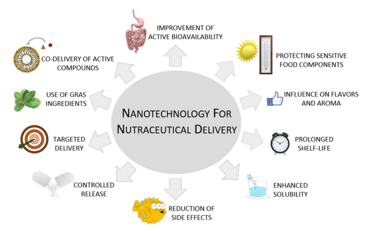
There are also nano nutraceuticals that can overcome the limitations of conventional nutraceuticals’ low water solubility, less absorptivity, low stability, and high susceptibility to light and oxygen, and possible chemical modifications after their administration to the human body as shown in Figure 7 [134]. The most important parameters to be taken into account during formulations of nanoneutraceuticals and features of nanocarriers influencing the efficiency of the delivered drugs are mentioned in Table 2.
| Nanocarrier features | Advantages | Limitations |
|---|---|---|
| Nature of nanosystems (polymeric, lipidic, metal-based, miscellaneous) | Increased efficiency | Possibility of conformational modification |
| Size distribution | Improved stability | Possibility of immunotoxicity |
| Biocompatibility and biodegradability | Enhanced bioavailability | Absence of guidelines and standardized protocols |
| Encapsulation efficiency | - | Toxic effects depending on carrier features |
| Drug release profile | - | Absence of guidelines and standardized protocols |
| Targeting the surface of the system | - | - |
Table 2: Examples of the most important parameters to be taken into account during formulations of nanonutraceuticals and feature
NMs can also be used for water and wastewater treatment [15]. There are prospects of using various NMs as eco-friendly consumer satisfaction intelligent food packaging systems, and parcels.
Nano formulations have shown promising preclinical success in delivering therapeutics for the treatment of CNS disorders which are otherwise inherently impermeable to the BBB in-vitro BBB model (DIV BBB) and the microfluidics BBB model on chip (μBBB) [135]. If the intravenous administration route is used: it is known that less than 1% of the administrated nanocarriers reach solid tumors [136].
Various advanced NMs designed to develop better nanocarrier systems are used to face different diseases such as cancer, heart failure, and malaria as shown in Figures 8 & 9 [107].
![Figure 9: Numerous incorporations of nanocarriers with drug molecules. (A) Functionalization of metallic gold nanoparticles (AuNPs) by interacting on the surface with the drug. (B) The nanohydrogels formed an aggregation of mesopore and macropore nanoparticles trapping the drug molecule in which the release occurs under absorption of water. (C) Polymeric nanocarriers exhibited nanoencapsulation of the drug molecule. (D) High dispersion of the drug molecule into the solid lipid nanocarriers [107].](/fulltextimages/11586/fig_9.png)
Figure 8: Different nanocarriers used in drug delivery systems. (A) Polymeric nanoparticles; (B) nanostructured lipid carriers; (C) solid lipid nanoparticles; (D) metallic nanoparticles; (E) liposomes; (F) nanohydrogels; (G) dendrimers; (H) cyclodextrin; and liquid crystalline system (I) lamellar; (J) hexagonal; (K) cubic. Copyright 2016, MDPI and ACS Style.
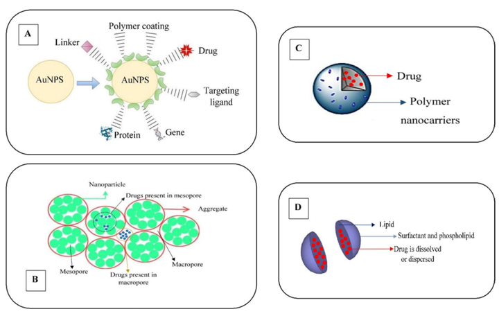
Figure 9: Numerous incorporations of nanocarriers with drug molecules. (A) Functionalization of metallic gold nanoparticles (AuNPs) by interacting on the surface with the drug. (B) The nanohydrogels formed an aggregation of mesopore and macropore nanoparticles trapping the drug molecule in which the release occurs under absorption of water. (C) Polymeric nanocarriers exhibited nanoencapsulation of the drug molecule. (D) High dispersion of the drug molecule into the solid lipid nanocarriers [107].
The understanding of the combination of nanocarriers and DNA/RNA gene delivery and RNA interference (RNAi)-based therapy Figure 10 may potentially improve therapeutic efficacy and have brought increasing attention to understanding and tackling complex genetically related diseases, such as cancer, cardiovascular and pulmonary diseases, autoimmune diseases and infections mention in Figures 11 & 12 [137].
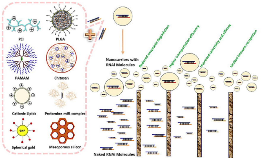
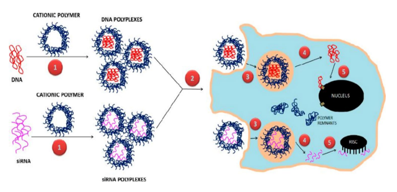
Figure 11: Design of polymeric gene delivery process. Polymeric nanocarriers for DNA and siRNA delivery: (1) polyplexes are formed by combining anionic DNA and siRNA with cationic polymers. (2) cellular uptake of polyplexes via various endocytic routes, (3) enclosure and subsequent release of polyplexes from endo-lysosomal compartments, (4) release of free DNA and siRNA from polyplexes leaving behind polymer remnants, and (5) transfer of DNA to the nucleus for expression by nuclear membrane transport proteins and binding of siRNA by the RNA-induced silencing complex (RISC). Copyright 2019, MDPI and ACS Style.
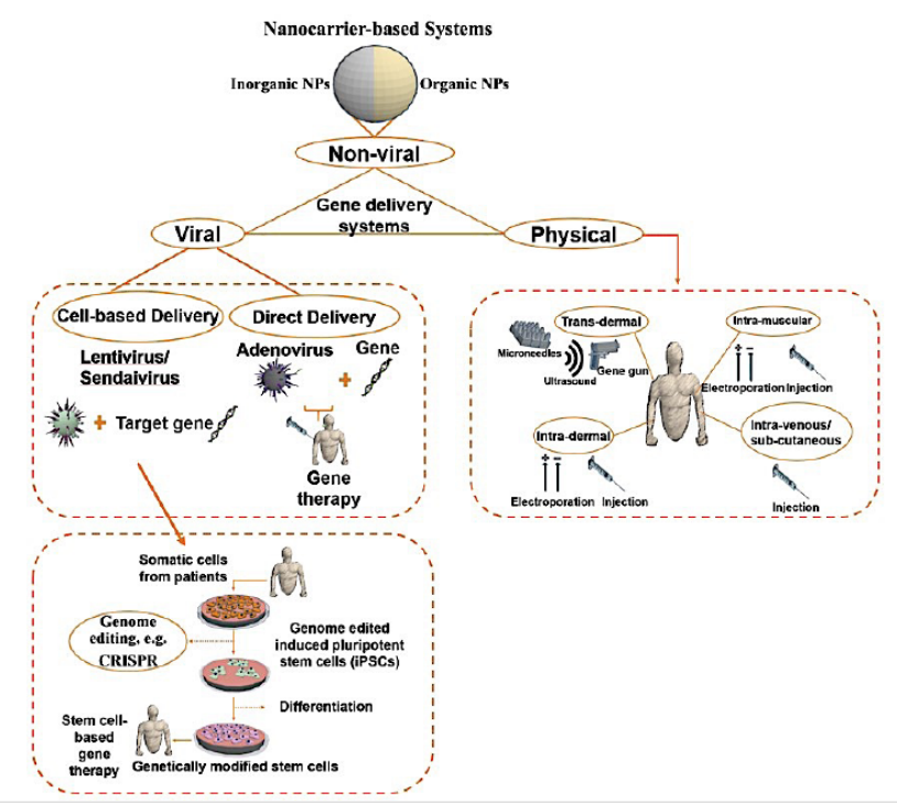
Figure 12: Gene delivery approaches; viral, non-viral, physical systems. There are mainly 3 approaches to delivering foreign genetic material into specific cells: physical methods, viral vectors, and non-viral vector methods. The physical methods usually deliver genes through gene guns, electroporation, and ultrasound methods, while viral delivery methods mainly involve cell- based delivery and direct delivery. Among the nonviral delivery systems, NP-based systems are emerging in the field of biology and nanomedicine.
Artificial responsive sub-nanofluidic ion channels have been constructed to control ion permeating intelligently further improving gating performance and having applications in real-world devices [138].
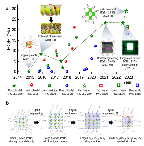
Colloidal metal halide perovskite nanocrystals (PNCs)
have high luminescence efficiency highly luminescent PNCs with high conductivity in the film can be achieved by various ligand-engineering and crystal-engineering methods. They can contribute to the commercialization of PNCs and PNC- LEDs in industrial displays and solid-state lighting as shown in Figure 13 [139].
Breakthrough ideas about nanomachines [140, 141]. Direct nanotechnology is used for applications in which nano-sized objects are directly employed, such as NPs in drug delivery systems. According to Ramsden 2009, indirect nanotechnology refers to any device containing a nano- sized device such as a cell phone, computer, and so on. But by creating a nanostructure atom by atom, it is quite possible to create strong permanent magnets. The unique properties of gold NPs (Au NPs) obtained by synthesizing NPs with different sizes and shapes make them promising candidate particles in nanomedicine, nanobiology, and photonics by Huang and El-Sayed 2010. By tailoring the sizes of NPs, it becomes possible to obtain a negative temperature coefficient for electrical conductivity as per Snow and Wohltjen 1998. Thus, the metal of interest behaves like a semiconductor. Size tailoring also has significant effects on optical properties. If the size is shorter than the electron means free path (distance taken by an electron between two subsequent collisions), intensive absorbance in near UV light occurs. Another impressive example is related to collective coherent electron oscillations, known as surface plasmon resonance. NPs, commonly referred to as 3D structures, can be helpful in the remediation of water. They are becoming promising candidates for removing a variety of pollutants from water in waste treatment plants [142]. Nanotechnology offers great potential for faster computers, effective power systems, and life-saving treatment while it also brings about an economic crisis, security, health, and environmental problems. With the ability that nanotechnology presents, self-assembling molecules will become real and this may be very helpful for sustainable development. According to Winston and Edelbach 2014, it becomes possible to take the opportunity to live longer without aging problems. Nanotechnology is a candidate to bring new perspectives to medicine. Using tiny robots, “nanobots,” artery blockages can be cleared away and surgeries can be faster and more precise. Also, reconstructing human parts cell by cell becomes possible by revolutionizing technology. Many fatal diseases can be diagnosed at an early stage so that their advance can be prevented and they can be easily cured. Recently, some treatment and diagnosis methods have come into play. These new techniques take advantage of the magnetic properties of NPs. One of the most common tools is magnetic particle imaging (MPI). Risks in this case are toxicity and the duration of NPs in one’s immune system. Thus, NPs must not be toxic and must be removed after completing their tasks. NPs of iron oxide can be used in the remediation of soil and water which is very beneficial especially for the food industry and, in turn, for human health. Although devices based on nanostructures have superior properties over previous devices. Using NPs can provide profits for tools used in metrology such as scanning probe microscopy (SPM), optical microscopes, electron microscopes, and telescopes. Another application of NMs is in the paper industry. Millions of tons of paper are globally produced annually. Besides improving paper quality, secret messages can be written using NPs so that the messages cannot be read by conventional means. Atomic weapons, bullet-proof and more comfortable military uniforms, and novel weapons can be created easily and accurately using this technology. Sensors based on nanotechnology have drawn great attention in security. Using these kinds of chemical sensors, it becomes easier to detect volatile chemicals or explosives. It is easier to remotely disable explosives thanks to nanotechnology. This technology can also be employed in textiles. Textiles with desired properties such as self-cleaning, flame resistance, wear resistance, and so forth can be obtained by using nanotechnology. Textiles can also be modified so that they can emit sweet fragrances. By combining NPs with ordinary materials, their mechanical, electrical, and optical properties can be improved. The population of the world is increasing day by day. Solar cell efficiency and energy storage capacity can be increased thanks to nanotechnology, and, of course, we will not need to pay for solar energy, thus the overall cost will be extremely decreased. This means that technology will need new generating and storage systems. These kinds of systems will become available by revolutionizing technology. Because the energy supplied by the sun is clean, we will have a clean environment and the life-span of living things will be significantly improved. Smart buildings where all of the energy used is supplied by solar panels, most modern wars have close ties with the energy available, especially petroleum, and nanotechnology will end these wars. Silicon technology has almost reached its limit. By decreasing the size of components at an atomic or molecular level by nanotechnology, the limit can be significantly enhanced. Nanotechnology is likely to have profound impacts on the economy.
A comprehensive study and evaluation will enable us to take precautions and minimize risk impacts. The Organization for Economic Co-operation and Development (OECD), International Organization for Standardization (ISO), and United Nations Educational, Scientific and Cultural Organization (UNESCO) are examples of international organizations that concentrate on human health, safety, and the environmental aspects of nanoproducts [143]. There are also some non-governmental organizations such as the International Society of Doctors for the Environment (ISDE) and Friends of the Earth (FOE) who focus on public understanding, management, and the assessment of nanotechnology. Both governmental and non-governmental organizations are quite effective in policy-making processes and public opinion toward nanotechnology some kinds of nanoproducts have already become available on the free market, accelerating scientific research on the merits and demerits of NPs becomes an immediate need. As with old scientific inventions like fire, the wheel, steam engines, silicon chips, and so on, new technology, “nano-tech,” has a great impact on society, the economy, politics, and so on.
Different countries have adopted regulations via legislation, laws and guidelines by several government organizations to mitigate the risks associated with NMs. However, there is no specific international regulation, no internationally agreed upon protocols, or legal definitions for production, handling or labeling, testing toxicity, and evaluating the environmental impact of NPs. Currently, the USA and the European Union (EU) have strong regulatory bodies and guideline legislation to control the potential risks of NMs. It can be noted that cosmetics face regulations and moderation from USFDA’s Federal Food, Drug, and Cosmetic Act (FFDCA), Personal Care Products Council (PCPC), Voluntary Cosmetic Registration Progam (VCRP), EU cosmetics product notification portal (CPNP), REACH, Scientific Committee on Consumer Safety (SCCS) and International Cooperation on Cosmetic Regulation (ICCR) (36 Jaison Jeevanandam).
Conclusion
In conclusion, ENP toxicity profiling is a highly demanded research area worldwide with a huge scope and applications. As compared to naturally occurring NMs, ENMs have a higher risk of human exposure and more chances of causing acute toxic effects to humans, other living organisms, and the environment as a whole. Therefore, there is an urgent need for regulatory nano-specific guidelines for nanogenotoxicity assessments as well as for understanding the toxicokinetics of different NMs. In order to avoid the potentially toxic effects of ENM’s implementation of regulations and laws by government agencies in many countries becomes mandatory. Extensive research in the field of nanotoxicology is highly encouraged to identify and avoid toxic NPs, nanowastes. From this review article, it can be noted that ENPs after undergoing toxicity guidelines for nanosafety can change the whole outlook and perspective of nanoscience. A noble NM should have the criteria like diameter >100 nm, possess high scalability, and easy to degrade, low cost and high productivity, biocompatibility, nontoxic, and without initiating pathological processes like inflammation and thrombosis, highly sufficient targeting and ability to penetrate multiple biological barriers, like BBB stable in the blood and resistant to be cleared by RES, good carrier of loaded molecules, loaded drugs/gene could be released smoothly to the targeted site and achieve significant therapeutic effect for the diseases. ENPs’ unique advanced properties can be utilized for designing noble nanoproducts for use in nanomedicine, nanobots, engineering, agriculture, and other diverse fields.
Acknowledgements
My sincere gratitude to Prof. Raghumani Singh Ningthoujam, Senior Scientist, Chemistry Division, BARC, Trombay for allowing me to access his lab facilities and to synthesize upconversion NaGdF4.10Yb2Er magnetic fluorescent inorganic NP by thermolysis method using Green Chemistry route, Silver (Ag) and Gold (Au) NPs within a short period of time. My fascination with nanoparticles is because of him. Also, I would like to thank Diana Jill, editor of the “Advances in Clinical Toxicology” journal for trusting and giving me this opportunity to write this review article.
References
-
Laurent S, Forge D, Port M, Roch A, Robic C, et al. (2010) Magnetic iron oxide nanoparticles: synthesis, stabilization, vectorization, physicochemical characterizations, and biological applications. Chem Rev 110: 2574-2574.
-
ISO/TS 80004-1 (2010) Nanotechnology–Vocabulary– Part 1: Core Terms. International Organization for Standardization: Geneva, Switzerland.
-
Hulla J, Sahu S, Hayes A (2015) Nanotechnology: History and future. Human & ExperimentalToxicology 34(12): 1318-1321.
-
Krumeich F (2017) Transmission Electron Microscopy, Material Science.
-
Sahoo SK, Parveen S, Panda JJ (2007) The present and future of nanotechnology in human health care. Nanomedicine 3(1): 20-31.
-
Buseck PR, Posfai M (1999) Airborne minerals and related aerosol particles: Effects on climate and the environment. PNAS 96: 3372-3379.
-
Uwins PJR, Webb RI, Taylor AP (1998) Novel nano- organisms from Australian sandstones. Am Mineral 83: 1541-1550.
-
Khan I, Saeed K, Khan I (2019) Nanoparticles: Properties, applications and toxicities. Arabian Journal of Chemistry 12(7): 908-931.
-
Linak WP, Miller CA, Wendt JOLJ (2000) Air Waste Manage Assoc 50: 1532-1544.
-
Rogers F, Arnott P, Zielinska B, Sagebiel J, Kelly KE, et al. (2005) Real-Time Measurements of Jet Aircraft Engine Exhaust. Air Waste Manage Assoc 55(5): 583-593.
-
De Volder MFL, Tawfick SH, Baughman RH, Hart AJ (2013) Carbon nanotubes: present and future commercial applications. Science 339(6119): 535-539.
-
Weir A, Westerhoff P, Fabricius L, Hristovski K, Von Goetz N (2012) Titanium dioxide nanoparticles in food and personal care products. Environ Sci Technol 46(4): 2242-2250.
-
Sadat-Shojai M, Atai M, Nodehi A, Khanlar LN (2010) Hydroxyapatite nanorods as novel fillers for improving the properties of dental adhesives: Synthesis and application. Dent Mater 26(5): 471-482.
-
Tiwari JN, Tiwari RN, Kim KS (2012) Zero-dimensional, one-dimensional, two-dimensional and three- dimensional nanostructured materials for advanced electrochemical energy devices. Prog Mater Sci 57: 724- 803.
-
Monika S, Tiwari M, Tiwari T, Ahamed MIN (2021) Review Report of Wastewater Treatment Using Nanomaterials. Wesleyan Journal of Research 14(4).
-
Shin WK, Cho J, Kannan AG, Lee YS, Kim DW (2016) Cross-linked composite gel polymer electrolyte using mesoporous methacrylate-functionalized SiO2 nanoparticles for lithium-ion polymer batteries. Sci Rep 6: 26332.
-
Hubler A (2010) Digital quantum batteries: Energy and information storage in nanovacuum tube arrays. Complexity 15(5): 48-55.
-
Shinn E (2012) Nuclear energy conversion with stacks of graphene nanocapacitors. Complexity 18(3): 24-27.
-
Elishakoff I, Pentaras D, Dujat K, Versaci C, Muscolino G, et al. (2012) Carbon Nanotubes and Nano Sensors: Vibrations, Buckling, and Ballistic Impact, ISTE-Wiley, London, UK, pp: 421.
-
David L (2013) Gap size dependence of the dielectric strength in nano vacuum gaps. IEEE Transactions on Dielectrics and Electrical Insulation 20(4): 1467-1471.
-
Rajiv S, Santosh S, Sugandha S (2010) Nanotechnology: The Future Medicine. Journal of Cutaneous and Aesthetic Surgery 3(1): 32-33.
-
Singh N, Manshian B, Jenkins GJS, Griffiths SM, Williams PM, et al. (2009) The DNA damaging potential of engineered nanomaterials. Biomaterials 30(23-24): 3891-3914.
-
Ballestri M (2001) Liver and kidney foreign bodies granulomatosis in a patient with malocclusion, bruxism, and worn dental prostheses Gastroenterology 121(5): 1234-1238.
-
Sahoo SK, Parveen S, Panda JJ (2007) The present and future of nanotechnology in human health care. Nanomedicine 3(1): 20-31.
-
Kumar V, Dasgupta N, Ranjan S (2018) Nanotoxicology Toxicity Evaluation, Risk Assessment and Management, © 2018 by Taylor & Francis Group, LLC CRC Press, International Standard Book Number-13: 978-1-4987- 9941-6.
-
Roco MC (2005) International perspective on government nanotechnology funding in 2005. J Nanopart Res 7: 707- 712.
-
Lee VH (2011) Advanced Drug Delivery Reviews: advancing science, improving therapy. Adv Drug Deliv Rev 63(1-2): 1-2.
-
Barrak H, Saied T, Chevallier P, Laroche G, M’nif A, (2016) Synthesis, characterization, and functionalization of ZnO nanoparticles by N-(trimethoxysilylpropyl) Nanoparticles 927 ethylenediamine triacetic acid (TMSEDTA): investigation of the interactions between phloroglucinol and ZnO@TMSEDTA. Arab J Chem.
-
Mansha M, Qurashi A, Ullah N, Bakare FO, Khan I, et al. (2016) Synthesis of In2O3/graphene heterostructure and their hydrogen gas sensing properties. Ceram Int 42(9): 11490-11495.
-
Rawal I, Kaur A (2013) Synthesis of mesoporous polypyrrole nanowires/nanoparticles for ammonia gas sensing application. Sens Actuators A Phys 203: 92-102.
-
Ullah H, Khan I, Yamani ZH, Qurashi A (2017) Sonochemical driven ultrafast facile synthesis of SnO2 nanoparticles: growth mechanism structural electrical and hydrogen gas sensing properties. Ultrason Sonochem 34: 484-490.
-
Ganesh M, Hemalatha P, Peng MM, Jang HT (2017) One pot synthesized Li, Zr doped porous silica nanoparticle for low temperature CO2 adsorption. Arab J Chem 10: S1501-S1505.
-
Ramacharyulu PVRK, Muhammad R, Praveen Kumar J, Prasad GK, Mohanty P (2015) Iron phthalocyanine modified mesoporous titania nanoparticles for photocatalytic activity and CO2 capture applications. Phys Chem Chem Phys 17: 26456-26462.
-
Shaalan M, Saleh M, El-Mahdy M, El-Matbouli M (2016) Recent progress in applications of nanoparticles in fish medicine: a review. Nanomed. Nanotechnol. Biol Med 12: 701-710.
-
Buzeafirst C, Pachecofirst II, Robbiefirst K (2007) Nanomaterials and nanoparticles: Sources and toxicity. Biointerphases 2(4): MR17-MR71.
-
Jeevanandam J, Barhoum A, Chan YS, Dufresne A, Michael K, et al. (2018) Review on nanoparticles and nanostructured materials: history, sources, toxicity and regulations. Beilstein J Nanotechnol 9: 1050-1074.
-
Kennedy AJ, Hull MS, Steevens JA, Dontsova KM, Chappell MA (2008) Factors influencing the partitioning and toxicity of nanotubes in the aquatic environment. Environ Toxicol Chem 27(9): 1932–1941.
-
Wang Y, Li Y, Pennell KD (2008) Influence of electrolyte species and concentration on the aggregation and transport of fullerene nanoparticles in quartz sands. Environ Toxicol Chem 27(9): 1860-1867.
-
Tervonen T, Linkov I, Figueira JR, Steevens J, Chappell M, et al. (2009) Risk-based classification system of nanomaterials. Journal of Nanoparticle Research 11: 757-766.
-
Bortoleto GG, de Oliveira Borges SS, Bueno MIM (2007) X-ray scattering and multivariate analysis for classification of organic samples: A comparative study using Rh tube and synchrotron radiation. Anal Chim Acta 595(1-2): 38-42.
-
Chen Q, Saltiel C, Manickavasagam S, Schadler LS, Siegel RW, et al. (2004 ) Aggregation behavior of single-walled carbon nanotubes in dilute aqueous suspension. Colloid Interface Sci 280(1): 91-97.
-
Xia T, Kovochich M, Brant J, Hotze M, Sempf J, et al. (2006) Comparison of the abilities of ambient and manufactured nanoparticles to induce cellular toxicity according to an oxidative stress paradigm. Nano Lett 6(8): 1794-1807.
-
Buzea C, Pacheco II, Robbie K (2007) Nanomaterials and nanoparticles: sources and toxicity. Biointerphases 2(4): MR17-MR71.
-
Dusinska M, Boland S, Saunders M, Juillerat-Jeanneret L, Tran L, et al. (2015) Towards an alternative testing strategy for nanomateri als used in nanomedicine: lessons from NanoTEST. Nanotoxicology 9(1): 118-132.
-
Dusinska M, Tulinska J, El Yamani N, Kuricova M, Liskova A, et al. (2017) Immunotoxicity, genotoxicity and epigenetic toxicity of nanomate rials: New strategies for toxicity testing. Food Chem Toxicol 109(1): 797-811.
-
Stone V, Pozzi-Mucelli S, Tran L, Aschberger K, Sabella S, et al. (2014) ITS-NANO-prioritising nanosafety research to develop a stakeholder driven intelligent testing strategy. Part Fibre Toxicol 11(1): 9.
-
Pfuhler S, Downs TR, Allemang AJ, Shan Y, Crosby ME (2017) Weak silica-nanomaterial-induced genotoxicity can be explained by indirect DNA damage as shown by the OGG1-modified comet assay and genomic analysis. Mutagenesis 32(1): 5-12.
-
Cordelli E, Keller J, Eleuteri P, Villani P, Ma-Hock L, et al. (2017) No genotoxicity in red blood cells upon 3- or 6-month inhalation exposure to CeO2 or BaSO4 nanomaterials. Mutagenesis 32(1): 13-22.
-
Catalan J, Rydman E, Aimonen K, Hannukainen K-S, Suhonen S, et al. (2017)Genotoxic and inflammatory effects of nanofibrillated cellulose in murine lungs. Mutagenesis 32(1): 23-31.
-
Li Y, Yan J, Ding W, Chen Y, Pack LM, et al. (2017) Genotoxicity and gene expression analyses of liver and lungs of mice treated with titanium dioxide nanoparticles. Mutagenesis 32(1): 33-46.
-
Wallin H, Kyjovska ZO, Poulsen SS, Jacobsen NR, Saber AT, et al. (2017) Surface modification does not influence the genotoxic and inflammatory effects of TiO2 nanoparticles after pulmonary exposure by instillation in mice. Mutagenesis 32(1): 47-57.
-
Rahman L, Wu D, Johnson M, William A, Halappanavar S (2017) Toxicogenomics analysis of mouse lung responses following exposure to titanium dioxide nanomaterials reveal their disease potential at high doses. Mutagenesis 32(1): 59-76.
-
Di Y, Aminot Y, Schroeder DC, Readman JW, Jha AN (2017) Integrated biological responses and tissue- specific expression of p53 and res genes in marine mussels following exposure to benzo(α)pyrene and C60 fullerenes, either alone or in combination. Mutagenesis 32(1): 77-90.
-
Gorrochategui E, Li J, Fullwood NJ, Ying GG, Tian M, et al. (2017) Diet-sourced carbon-based nanoparticles include lipid alterations in tissue of zebrafish (Danio rerio) with genomic hypermethylation changes in brain. Mutagenesis 33(1): 91-103.
-
Thongkam W, Gerloff K, Berlo DV, Albrecht C, Schins RPF (2017) Oxidant generatin, DN damage and cytotoxicity by a panel of engineered nanomaterials in three different human epithelial cell lines. Mutagenesis 32(1): 105-115.
-
El Yamani N, Collins AR, Runden-Pran E, Fjellsbo LM, Shaposhnikov S, et al. (2017) In vitro Genotoxicity testing of four reference metal nanomaterials, titanium dioxide, zinc oxide, cerium oxide and silver: towards a robust and reliable hazard assessment. Mutagenesis 32(1): 117-126.
-
Di Bucchianico S, Cappellini F, Bihanic FL, Zhang Y, Dreij K, et al. (2017) Genotoxicity of TiO2 nanoparticles assessed by mini-gel comet assay and micronucleus scoring with flow cytometry. Mutagenesis 32(1): 127-137.
-
Proquin H, Rodrlguez-Ibarra C, Moonen C, Urrutia Ortega IM, Briede JJ, et al. (2017) Titanium dioxide food additive (E171) induces ROS formation and genotoxicity: contribution of micro and nano-sized fractions. Mutagenesis 32(1): 139-149.
-
Li Y, Doak SH, Yan J, Chen DH, Zhou M, et al. (2017) Factors affecting the in vitro micronucleus assay for evaluation of nanomaterials. Mutagenesis 32(1): 151-159.
-
Biola-Clier M, Beal D, Caillat S, Libert S, Armand L, et al. (2017) Comparison of the DNA damage response in BEAS-2B and A549 cells exposed to titanium dioxide nanoparticles. Mutagenesis 32(1): 161-172.
-
Borghini A, Roursgaard M, Andreassi MG, Kermanizadch A, Moller P (2017) Repair activity of oxidatively damaged DNA and telomere length in human 1 lung epithelial 2 cells after exposure to multiwalled carbon nanotubes. Mutagenesis 32(1): 173-180.
-
Oner D, Moisse M, Ghosh M, Duca RC, Poels K, et al. (2017) Epigenetic effects of carbon nanotubes in human monocytic cells. Mutagenesis 32(1): 181-191.
-
Gabelova A, El Yamani N, Alonso TI, Buliaková B, Srančíková A, et al. (2017) Fibrous shape underlies the mutagenic and carcinogenic potential of nanosilver while surface chemistry affects the biosafety of iron oxide nanoparticles. Mutagenesis 32(1): 193-202.
-
Carriere M, Sauvaigo S, Douki T, Ravanat JL (2017) Impact of nanoparticles on DNA repair processes: current knowledge and working hypotheses. Mutagenesis 32(1): 203-213.
-
Nelson BC, Wright CW, Ibuki Y, Moreno-Villanueva M, Karlson HL, et al. (2017) Emerging Metrology for High- Throughput Nanomaterial Genotoxicology. Mutagenesis 32(1): 215-232.
-
Evans SJ, Clift MJ, Singh N, de Oliveira Mallia J, Burgum M, et al. (2017) Critical review of the current and future challenges associated with advanced in vitro systems towards the study of nanoparticle (secondary) genotoxicity. Mutagenesis 32(1): 233-241.
-
Clift MJD, Raemy DO, Endes C, Ali Z, Lehmann AD, et al. (2013) Can the Ames test provide an insight into nano-object mutagenicity? Investigating the interaction between nano-objects and bacteria. Nano toxicology 7(8): 1373-85.
-
George JM, Magogotya M, Vetten MA, Buys AV, Gulumian M (2017) From the cover: An Investigation of the Genotoxicity and Interference of Gold Nanoparticles in Commonly Used In Vitro Mutagenicity and Genotoxicity Assays. Toxicol Sci 156(1): 149-166.
-
Siivola KM, Burgum JM, Merino BS, Clift MJD, Doak SH, et al. (2022) A systematic quality evaluation and review of nanomaterial genotoxicity studies: a regulatory perspective. Particle and Fibre Toxicology 19: 59.
-
Demir E, Demir FT, Marcos R (2022) Drosophila as a Suitable In Vivo Model in the Safety Assessment of Nanomaterials. Adv Exp Med Biol 1357: 275-301.
-
Vales G, Demir E, Kaya B, Creus A, Marcos R (2012) Genotoxicity of cobalt nanoparticles and ions in Drosophila. Nanotoxicology 7(4): 462-468.
-
Singh N, Manshian B, Jenkins GJ, Griffiths SM, Williams PM, et al. (2009) NanoGenotoxicology: the DNA damaging potential of engineered nanomaterials. Biomaterials 30(23-24): 3891-3914.
-
Papageorgiou I, Brown C, Schins R, Singh S, Newson R, et al. (2007) The effect of nano- and micron-sized particles of cobalt–chromium alloy on human fibroblasts in vitro. Biomaterials 28(19): 2946-2958.
-
Gurr JR, Wang SSR, Chen CH, Jan KY (2005) Ultrafine titanium dioxide particles in the absence of photoactivation can induce oxidative damage to human bronchial epithelial cells. Toxicology 213(1-2): 66-73.
-
Park EJ, Yi J, Chung KH, Ryu DY, Choi J, et al. (2008) Oxidative stress and apoptosis induced by titanium dioxide nanoparticles in cultured BEAS-2B cells. Toxicol Lett 180(3): 222-229.
-
(2020) Nanomaterials and genotoxicity—a literature review. Swedish Chemicals Agency, Report No: 361218.
-
Kohl Y, Rundén-Pran E, Mariussen E, Hesler M, El Yamani N, et al. (2020) Genotoxicity of nanomaterials: advanced in vitro models and high throughput methods for human hazard assessment—a review. Nanomaterials 10(10): 1911.
-
Bernauer U, Bodin L, Chaudhry Q, Coenraads PJ, Dusinska M, et al. (2021) The SCCS scientifc advice on the safety of nanomaterials in cosmetics. Regul Toxicol Pharmacol 126: 105046.
-
Al-Subiai SN, Arlt VM, Frickers PE, Readman JW, Stolpe B, et al. (2012) Merging nano-genotoxicology with eco- genotoxicology: An integrated approach to determine interactive genotoxic and sub-lethal toxic effects of C-60 fullerenes and fluoranthene in marine mussels, Mytilus sp. Mutat Res 745(1-2): 92-103.
-
Revell PA (2006) The biological effects of nanoparticles. Nanotechnol Percept 2: 283-298.
-
Tran CL, Donaldson K, Stones V (2005) A Scoping Study to Identify Hazard Data Needs for Addressing the Risks Presented by Nanoparticles and Nanotubes. London: Institute of Occupational Medicine.
-
Robison WL (2004) Nano-Ethics. Amsterdam: IOS Press.
-
MacDonald C (2004) Nanotech is Novel; the Ethical Issues are not.
-
Grunwald A (2005) Nanotechnology—A new field of ethical inquiry?. Sci Eng Ethics 11(2): 187-201.
-
Astefanei A, Nu´n˜ez O, Galceran MT (2015) Characterisation and determination of fullerenes: a critical review. Anal Chim Acta 882: 1-21.
-
Ibrahim KS (2013) Carbon nanotubes-properties and applications: a review. Carbon Lett 14: 131-144.
-
Saeed K, Khan I (2016) Preparation and characterization of singlewalled carbon nanotube/nylon 6,6 nanocomposites. Instrum Sci Technol. 44: 435-444.
-
Saeed K, Khan I (2014) Preparation and properties of single-walled carbon nanotubes/poly (butylene terephthalate) nanocomposites. Iranian Polymer Journal 23: 53-58.
-
Ngoy JM, Wagner N, Riboldi L, Bolland O (2014) A CO2 capture technology using multi-walled carbon nanotubes with polyaspartamide surfactant. Energy Procedia 63: 2230-2248.
-
Mabena LF, Ray SS, Mhlanga SD, Coville NJ (2011) Nitrogen-doped carbon nanotubes as a metal catalyst support. Appl Nanosci 1: 67-77.
-
Tapasya K, Kumar AS, Dharmarajan A, Parvathi VD (2022) *Nanocarriers: The Promising Future to Cancer Diagnostics and Treatment. Biomedical & Pharmacology Journal 15(2): 785-802.
-
Talapin DV, Rogach AL, Kornowski A, Haase M, Weller H (2001) Highly Luminescent Monodisperse CdSe and CdSe/ZnS Nanocrystals Synthesized in a Hexadecylamine−Trioctylphosphine Oxide− Trioctylphospine Mixture. Nano Lett 1(4): 207-211.
-
Sun Y, Gates B, Mayers B, Xia Y (2002) Crystalline Silver Nanowires by Soft Solution Processing. Nano Lett 2(2): 165-168.
-
Cui Y, Zhong Z, Wang D, Wang WU, Lieber CM (2003) High Performance Silicon Nanowire Field Effect Transistors. Nano Lett 3(2): 149-152.
-
Derfus AM, Chan WCW, Bhatia SN (2004) Probing the Cytotoxicity of Semiconductor Quantum Dots. Nano Lett 4(1): 11-18.
-
El-Sayed IH, Huang X, El-Sayed MA (2005) Surface Plasmon Resonance Scattering and Absorption of anti- EGFR Antibody Conjugated Gold Nanoparticles in Cancer Diagnostics: Applications in Oral Cancer. Nano Lett 5(5): 829-834.
-
Chithrani BD, Ghazani AA, Chan WCW (2006) Determining the Size and Shape Dependence of Gold Nanoparticle Uptake into Mammalian Cells. Nano Lett 6 (4): 662-668.
-
Noh JH, Im SH, Heo JH, Mandal TN, Seok SI (2013) Chemical Management for Colorful, Efficient, and Stable Inorganic− Organic Hybrid Nanostructured Solar Cells. Nano Lett 13(4): 1764-1769.
-
Frost JM, Butler KT, Brivio F, Hendon CH, Schilfgaarde MV, et al. (2014) Atomistic Origins of High-Performance in Hybrid Halide Perovskite Solar Cells. Nano Lett 14(5): 2584-2590.
-
Jeong S, Cho H, Han S, Won P, Lee H, et al. (2017) High Efficiency, Transparent, Reusable, and Active PM2.5 Filters by Hierarchical Ag Nanowire Percolation Network. Nano Lett 17(7): 4339-4346.
-
Dreaden EC, Alkilany AM, Huang X, Murphy CJ, El- Sayed MA (2012) The golden age: gold nanoparticles for biomedicine. Chem Soc Rev 41: 2740-2779.
-
M Lou, Zhang LN, Ji PG, Feng FQ, Liu JH, et al. (2016) Quercetin nanoparticles induced autophagy and apoptosis through AKT/ERK/Caspase-3 signaling pathway in human neuroglioma cells: in vitro and in vivo. Biomed Pharmacother 841-849.
-
Zang X, Cheng M, Zhang X, Chen X (2021) Quercetin nanoformulations:a promosing strategy for tumor therapy. Food Funct 15: 6664-6681.
-
Klębowski B, Depciuch J, Parlińska-Wojtan M, Baran J (2018) Applications of noble metal-based nanoparticles in medicine. Int J Mol Sci 19(12): 4031.
-
Mody VV, Nounou MI, Bikram M (2009) Novel nanomedicine-based MRI contrast agents for gynecological malignancies. Adv Drug Deliv Rev 61(10): 795-807.
-
Brown SD, Nativo P, Smith J, Stirling D, Edwards PR, et al. (2010) Gold nanoparticles for the improved anticancer drug delivery of the active component of oxaliplatin. J Am Chem Soc 132(13): 4678-4684.
-
Hamimed S, Jabberi M, Chatti A (2022) Nanotechnology in drug and gene delivery. Naunyn- Schmiedeberg’s Archives of Pharmacology pp: 769-787.
-
Caltagirone C, Bettoschi A, Garaua A, Montis R (2015) Silica-based nanoparticles: a versatile tool for the development of efficient imaging agents. Chem Soc Rev 14: 4645-4671.
-
Kesse S, Boakye-Yiadom KO, Ochete BO, Opoku- Damoah Y, Akhtar F, et al. (2019) Mesoporous Silica Nanomaterials: versatile Nanocarriers for Cancer Theranostics and Drug and Gene Delivery. Pharmaceutics 11(2): 77.
-
Lai N, Lin C, Ku P, Chang L, Liao K, et al. (2014) Hollow mesoporous Ia3d silica nanospheres with singleunit-cell-thick shell: spontaneous formation and drug delivery application. Nano Res 7(10): 1439-1448.
-
Kresge CT, Leonowicz ME, Roth WJ, Vartuli JC, Beck JS (1992) Ordered mesoporous molecular sieves synthesized by a liquid-crystal template mechanism. Nature 359: 710-712.
-
Sigmund W, Yuh J, Park H, Maneeratana V, Pyrgiotakis G (2006) Processing and structure relationships in electrospinning of ceramic fiber systems. J Am Ceram Soc 89(2): 395-407.
-
Thomas S, Harshita BSP, Mishra P, Talegaonkar S (2015) Ceramic nanoparticles: fabrication methods and applications in drug delivery. Curr Pharm Des 21(42): 6165-6188.
-
Ali S, Khan I, Khan SA, Sohail M, Ahmed R, et al. (2017) Electrocatalytic performance of Ni@Pt core– shell nanoparticles supported on carbon nanotubes for methanol oxidation reaction. J Electroanal Chem 795: 17-25.
-
Khan I, Abdalla A, Qurashi A (2017) Synthesis of hierarchical WO3 and Bi2O3/WO3 nanocomposite for solar-driven water splitting applications. Int J Hydrogen Energy 42(5): 3431-3439.
-
Sun S, Murray CB, Weller D, Folks L, Moser A (2000) Monodisperse FePt nanoparticles and ferromagnetic FePt nanocrystal superlattices. Science 287(5460): 1989-1992.
-
Hisatomi T, Kubota J, Domen K (2014) Recent advances in semiconductors for photocatalytic and photoelectrochemical water splitting. Chem Soc Rev 43(22): 7520-7535.
-
Mansha M, Khan I, Ullah N, Qurashi A (2017) Synthesis, characterization and visible-light-driven photoelectrochemical hydrogen evolution reaction of carbazole-containing conjugated polymers. Int J Hydrogen Energy 42(16): 10952-10961.
-
Rao JP, Geckeler KE (2011) Polymer nanoparticles: preparation techniques and size-control parameters. Prog Polym Sci 36(7): 887-913.
-
Abd Ellah NH, Abouelmagd SA (2016) Surface functionalization of polymeric nanoparticles for tumor drug delivery: approaches and challenges. Expert Opin Drug Deliv 14(2): 201-214.
-
Abouelmagd SA, Meng F, Kim BK, Hyun H, Yeo Y (2016) Tannic acid-mediated surface functionalization of polymeric nanoparticles. ACS Biomater Sci Eng 2(12): 2294-2303.
-
Rawat MK, Jain A, Singh S, Mehnert W, Thunemann AF, et al. (2011) Studies on binary lipid matrix based solid lipid nanoparticles of repaglinide: in vitro and in vivo evaluation. J Pharm Sci 100(6): 2366-2378.
-
Mashaghi S, Jadidi T, Koenderink G, Mashaghi A (2013) Lipid nanotechnology. Int J Mol Sci 14(2): 4242- 4282.
-
Puri A, Loomis K, Smith B, Lee JH, Yavlovich A, et al. (2009) Lipid-based nanoparticles as pharma ceutical drug carriers: from concepts to clinic. Crit Rev Ther Drug Carrier Syst 26(6): 523-580.
-
Gujrati M, Malamas A, Shin T, Jin E, Sun Y, et al. (2014). Multifunctional cationic lipid-based nanoparticles facilitate endo somal escape and reduction-triggered cytosolic siRNA release. Mol Pharm 11(8): 2734-2744.
-
Hlavacek A, Krivankova J, Prikryl J, Foret F (2019) Photon-Upconversion Barcoding with Multiple Barcode Channels: Application for Droplet Microfluidics. Analytical Chemistry 91(20): 12630-12635.
-
Odom TW (2021) Celebrating 20 years of Nano Letters. Nano Letters 21(1): 1-2.
-
Iturrioz-Rodríguez N, Bertorelli R Ciofani G (2021) Lipid-Based Nanocarriers for The Treatment of Glioblastoma. Advanced NanoBioMed Research 1(2).
-
Paolino D, Mancuso A, Cristiano MC, Froiio F, Lammari N, et al. (2021) Nanonutraceuticals: The New Frontier of Supplementary Food. Nanomaterials 11(3): 792.
-
Theune LE, Charbaji R, Kar M, Wedepohl S, Hedtrich S, et al. (2019) Critical parameters for the controlled synthesis of nanogels suitable for temperature‐triggered protein delivery. Mater Sci Eng: C (100): 141-151.
-
Farazi S, Chen F, Foster H, Boquiren R, McAlpine SR, et al. (2020) Real time monitoring of peptide delivery: In vitro using high payload pH-responsive nanogels. Polymer Chemistry 11(2): 425-432.
-
Novikau IS, Minina ES, Sánchez PA, Kantorovich SS (2020) Suspensions of magnetic nanogels at zero field: Equilibrium structural properties J Magn Magn Mater 498: 166152.
-
Wamidh HT, Abuawad A, Thiab S, Alshweiat A, Mahmod AS (2022) Flavonoid-based nanomedicines to target tumor microenvironment. OpenNano 8.
-
Paolino D, Mancuso A, Cristiano MC, Froiio F, Lammari N, et al. (2021) Nanonutraceuticals: The New Frontier of Supplementary Food. Nanomaterials (Basel) 11(3): 792.
-
Mc Carthy DJ, Malhotra M, O’Mahony MA, Cryan JF, O’Driscol CM (2015) Nanoparticles, and the Blood- Brain Barrier: Advancing from In-Vitro Models Towards Therapeutic Significance. Pharm Res 32(4): 1161-1185.
-
Wilhelm S, Tavares AJ, Dai Q, Ohta S, Audet J, et al. (2016) Analysis of nanoparticle delivery to tumours. 1(5).
-
Shen H, Huang X, Min J, Le S, Wang Q, et al. (2019) Nanoparticle Delivery Systems for DNA/RNA and their Potential Applications in Nanomedicin. Current Topics in Medicinal Chemistry 19(27): 2507-2523.
-
Zhao C, Hou J, Hill M, Freeman B, Wang H, et al. (2023) Enhanced Gating Effects in Responsive Sub-nanofluidic Ion Channels. Accounts of Materials Research 4(9): 786- 797.
-
Young-Hoon K, Tae-Woo L (2023) Engineering Colloidal Perovskite Nanocrystals and Devices for Efficient and Large-Area Light-Emitting Diodes. Accounts of Materials Research 4(8): 655-667.
-
Ozin GA, Manners I, Fournier-Bidoz S, Arsenault A (2005) Dream nanomachines. Adv Materials 17(24): 3011-3018.
-
Balzani V, Credi A, Silvi S, Venturi M (2016) Artificial nanomachines based on interlocked molecular species: Recent advances. Chem Soc Rev 35: 1135-1149.
-
Jangid P, Inbaraj MP (2008) Application of nanoparticles in waste water treatment. ScienceDirect 43: 2877-2881.
-
ISO Technical Committee (TC) 229 (2006) Nanotechnologies—The Ethics and Politics of Nanotechnology. UNESCO.
- Evaluation of Skin Aging Preventive Effects of Cherry Blossom Petal Extracts Through Antioxidant and Anti-Glycation Activities
- Is Cell Death Responsible for False Positive Results of In Vivo Comet Assay?
- Pattern of Gonadal Hormones in Oral Testosterone-Supplimented Male Wistar Rats with Diabetes-Induced Hypogonadism
- Re-Evaluation of the Genotoxicity of Currently Used Food Dyes in Mouse Multiple Organs Via Continuous Administration by Drinking Using the Comet Assay
- Pharmacogenetics of Type 2 Diabetes Mellitus: Linking Genetic Variability to Drug Efficacy and its Cardiovascular Outcomes
- Exploratory Proteomic Profiling of SARS-CoV-2 Infected THP-1 Macrophages Reveals Alterations in Inflammatory Response and Cellular Metabolism