N-Myristoyltransferase: A Novel Therapeutic Target for Cancer, Inflammation and Radiation Response Modifications
N-Myristoyltransferase (NMT) is a well conserved monomeric protein that catalyzes myristoyl group transfer to various target proteins carrying consensus amino acid sequence motif GXXXS/T (where ‘X’ is any amino acid). NMT mediated myristoylation promotes protein-protein and protein-membrane interactions, leading to diverse signaling including cell proliferation, carcinogenesis, inflammation and immune responses. However, inter-networking linkage between NMT over-expression and protein myristoylation under oxidative stress coupled with inflammation and carcinogenesis is completely missing and needs further investigation. Various types of tumor and cancer tissue showed significant increase in NMT expression. Therefore, inhibition of NMT expression either by using chemical NMT inhibitors or gene silencing could be a promising approach in controlling carcinogenesis. Various scientific evidence suggests that NMT mediated myristoylation promotes membrane association of some cell cycle and cell proliferation regulatory proteins and thus may contribute to cell cycle regulation and cell survival homeostasis. Several NMT inhibitors have been evaluated and tested for their probable anticancer potential. Molecular linkage between oxidative stress and carcinogenesis has been established in several studies. In this review, the authors cover the role and therapeutic potential of NMT in cancer, inflammatory disease and radiation response modulation.
Introduction
N-Myristoyltransferase (NMT; EC 2.3.1.97) is a functional protein that catalyzes myristoylation at N- terminal glycine of consensus motif GXXXS/T (where X is any amino acid) residue of various proteins [1, 2, 3]. As the result of myristoylation, NMT regulates molecular signaling associated with cell cycle regulation, cell survival and apoptosis in non-cancerous cells. Interestingly, over-expression of NMT leads to higher degree of myristoylation, which further promotes uncontrolled cell proliferation and thus, contributes to carcinogenesis Aronheim A, et al. [4, 5]. NMT appears to be ubiquitous throughout from eukaryotes, i.e., Candida albicans Devadas B, et al. [6], Saccharomyces cerevisiae Duronio RJ, et al. [7] and parasitic protozoa (plasmodium species, Leishmania major and Trypanosoma brucei) Olaleye TO, et al. [8], insects (Drosophila melanogaster) Ntwasa M, et al. [9], including plants (Arabidopsis thaliana) and human [10, 11].
NMT mediated transfer of myristate from myristoyl- CoA to the N-terminal glycine of consensus Thinon E,, et al. [5, 11] motif of target proteins occurs during both co-translational and post-translational processes. NMT ensures covalent amidic bond linkage between the myristate group and glycine residue of the target protein [1]. Though myristoylation is an irreversible process James G, et al. [12], but exceptions to this modification have also been reported [13]. Exposure of consensus motif followed by removal of adjacent methionine residue due to proteolytic cleavage is an essential prerequisite requirement for co-translational myristoylation [2, 5, 11, 14]. During post-translational myristoylation, proteolytic cleavage is necessary to expose subsequent glycine residue for myristoylation [11]. Methionine excision is not mandatory to expose glycine for myristoylation. Rather, random cleavage in the peptide linkage to expose glycine is a prerequisite requirement for post–translational myristoylation [11]. By transferring the lipidic myristate group to the target proteins, myristoylation enhances lipophilicity of modified proteins, leading to their membrane localization, protein-protein interaction and ligand-induced conformational [5, 11]. Different endogenous enzymes and regulatory proteins including protein kinase C Verge`res G, et al. [15], protein kinase A, nitric oxide synthase Cardena GG, et al. [16], NADH-cytochrome b5 reductase Borgese N, et al. [17], recoverin Ames JB, et al. [18], most of the G protein a subunit Wedegaertner PB, et al. [19, 20, 21, 22], Fac, Jun, Fus and oncoprotein Src, Myc, etc., are potential substrates for myristoylation [10, 11].
Although myristoylation is a regulated reaction, it also requires involvement of various other enzymes or proteins, including N-methionylaminopeptidase, acyl-CoA-binding proteins, fatty acid synthetase and long chain acyl-CoA synthetase etc., during co-translational myristoylation Felsted RL, et al. [23, 24, 25, 26]. However, no report is available indicating direct involvement of NMT with the protein synthesis machinery. Two NMT isoforms, i.e., NMT-1 and NMT- 2 have been reported in human, which represent significant divergence (76-77%) from each other in terms of amino acid sequences Giang DK, et al. [10, 11], suggesting that they evolve from two distinct families of enzymes. Inter-enzyme comparison indicated a greater level of homology (~95%) between mouse and human NMTs amino acid sequences and thus, justified their interspecies conservation throughout the evolution process [10]. NMT mediated myristoylation plays a key role in several kinds of processes in the cells, including carcinogenesis and inflammation [27]. Myristoylation of the oncoproteins i.e. src, myc, MARCKS McLaughlin S, et al. [27, 28] Gip2 etc., promotes their membrane association. Membrane anchoring further induced molecular signaling, resulting in uncontrolled cell proliferation and carcinogenesis.
Recently, the role of NMT expression in inflammation has also been reported in cattle lungs. Experimental evidence demonstrated that NMT-/- gene knock-out netrophiles showed short life span compared to the control neutrophils carrying normal NMT+/+ gene alleles [29]. Further study revealed that NMT expression is associated with the neutrophils’ longer survival in the inflamed tissues to combat infections for a longer time. However, the expression pattern and role of NMT in infected tissue is not reported. A correlation between NMT expression and increased life span, cell survival and myelopoisis has been reported [29].
Although myristoylation is ubiquitous among the eukaryotes, it ranges from yeast to humans. However, its significance in diseases and non-diseases scenarios is still not completely understood. The role of myristoylation in cancer progression and the search for NMT inhibitors to combat aggressive cancer promotion as well as to inhibit parasitic infections is the prime focus of research [30]. However, very limited information is available about the role and necessity of myristoylation in inflammation and normal life processes [29].
In view of the foregoing, this review is focused on the molecular mechanism of myristoylation of target proteins that lead to carcinogenic, apoptotic and inflammatory responses. This review also highlighted some prominent technical disconnects and undiscovered scientific facts and gaps arise in the area of myristoylation leading to carcinogenesis, apoptosis and inflammation. Further, novel futuristic applications of myristoyl transferase inhibitors and their negative regulators in normal and cancerous cells are also critically discussed.
Myristoylation
The reaction of transfer of 14-carbon saturated fatty acid, i.e., myristate from myristyl-CoA to N-terminal glycine residue of target peptides/proteins catalyzed by myristoyltransferase is referred as myristoylation reaction [2, 10, 31, 32]. Myristoylation occurs during both Co- translational and post-translational periods in eukaryotic organisms [11]. Co-translational transfer of the myristoyl group is known to occur followed by cleavage of N-terminal methionine residue of growing peptides/proteins [33]. Removal of N-terminal methionine provides an opportunity for the next adjacent N-terminal glycine residue to interact with myristyl-CoA and get myristoylated with the help of NMT
(Figure 1). While post-translational myristoylation usually occurs after caspase-3 and caspase- 8 mediated cleavage of preceding amino acid moiety, not necessarily methionine, resulting in glycine residue exposed with myristyl-CoA- NMT complex [13, 34]. Viruses and bacteria are deficient in the NMT gene and thus, unable to perform myristoylations [30]. Several proteins of different protein families including cAMP-dependent serine/threonine kinases Carr SA, et al. [35], retroviral poly-protein precursors HIV-1pr55, v-Src, viral capsid Bryant M, et al. [36, 37] components and α-subunit of signal transducing G proteins, calcinurin Aitken, et al. [38] etc. undergo myristoylation Aitken A, et al. [38, 39], as shown in Table 1. The addition of the myristoyl group to the target site of the proteins is usually undergone through a nucleophilic addition-elimination reaction. Briefly, in the first step, myristoyl CoA is positioned itself within the active binding site of NMT in such a way that phenylalanine170 and leucine171 residues of NMT are set on opposite sites of the carbonyl group of mristoyl-CoA [31]. This type of molecular arrangement polarizes the carbonyl group of mristoyl-CoA (the carbon atom gets charged positively) and becomes labile to nucleophilic attack by the glycine residue of the proteins going to be myristoylated. Following myristoyl CoA binding, NMT re-orients to allow peptide binding at its catalytic site. Thereafter, NMT deprotinate its NH3+ group and attacks the carbonyl group of Myristoyl CoA. As the result of various chemical re-arrangements, a tetrahedral intermediate is formed and stabilized by the interaction between a positively charged oxy-anion and the negatively charged alkoxide anion. Finally, free CoA is released and subsequent conformational changes in the NMT provide a favorable chemical environment to release modified myristoylated peptide and thus, the myristoylation process is completed Rajala RV, et al. [40, 41, 42] as shown in Figure 1 [43, 44, 45, 46, 47, 48, 49, 50, 51, 52, 53, 54, 55, 56, 57, 58, 59, 60, 61, 62, 63, 64, 65, 66, 67, 68, 69, 70, 71, 72, 73, 74, 75].
![Figure 1: Co (A) and post (B) translational myristoylation process of targeted proteins: During co-translational myristoylation (A), MGXXX(S/C/T) motif undergoes methionyl amino-peptidase mediated cleavage specifically between the methionine and glyciine site. The result of cleaved methionine is removed and N terminal Glycine residue remains exposed freely. In the subsequent step, N-myristoyltransferase1/2 transfers the myristoyl group at glycine residue of a consensus motif [GXXX(S/ C/T)]. During post-translational myristoylation (B), the target protein is specifically cleaved by capases in a manner where glycine residue of consensus motif [GXXX(S/C/T)]is exposed. In the second step, N-myristoyltransferase1/2 transfers the myristoyl group to the glycine and thus, myristoylation is completed. MGXXX(S/C/T); methionine-glycine-X (any amino acid)- serine/cysteine/threonine.](/fulltextimages/12106/fig_1.png)
Figure 1: Co (A) and post (B) translational myristoylation process of targeted proteins: During co-translational myristoylation (A), MGXXX(S/C/T) motif undergoes methionyl amino-peptidase mediated cleavage specifically between the methionine and glyciine site. The result of cleaved methionine is removed and N terminal Glycine residue remains exposed freely. In the subsequent step, N-myristoyltransferase1/2 transfers the myristoyl group at glycine residue of a consensus motif [GXXX(S/ C/T)]. During post-translational myristoylation (B), the target protein is specifically cleaved by capases in a manner where glycine residue of consensus motif [GXXX(S/C/T)]is exposed. In the second step, N-myristoyltransferase1/2 transfers the myristoyl group to the glycine and thus, myristoylation is completed. MGXXX(S/C/T); methionine-glycine-X (any amino acid)- serine/cysteine/threonine.
| S. No. | Protein | Physiological Role | Myristoylation Function | Biological outcome | Reference |
|---|---|---|---|---|---|
| 1. | Actin (15kDa fragment ) | Cytoskeleton structural protein, mediator of apoptosis | Post-translational myristoylation during apoptosis. | Suppressed apoptosis, reduced caspase activity, higher NMT levels. | [76] |
| 2. | Bid | Apoptosis promoting protein. | Post-translational myristoylation of Bid protein to mitochondrial membrane, it, induced release of cytochrome C and cell death. | Myristoylation help to anchored p7 and p15 fragments of BID to mitochondrial membrane. | [14] |
| 3. | MARCKS | Actin cross-linking when phosphorylated by protein kinase C | Co-translational myristoylation aids in plasma membrane association. | Help to recruit cells at inflammatory sites and thus increase inflammation. | [27] |
| 4. | G-Protein (αil, αa2, αi3, α0 and α) | Signaling GTPase. N-terminal myristoylation simply provides a hydrophobic anchoring to membrane attachment to the α,αao, and αz polypeptides | Co-translational myristoylation aids in plasma membrane association. | Myristoylation of G protein alpha subunits facilitate formation of heterotrimer and the localization of alpha to the plasma membrane and induced molecular signaling for cell survival lead to carcinogensis. | [43] |
| 5. | Gelsolin | Actin filament-severing protein | Post-translational myristoylation up- regulates anti-apoptotic signaling. | N-myristoylation of Gelsolin is required for the apoptosis inhibition. | [76] |
| 6. | p21-activated protein kinase (PAK2,) | Serine/threonine kinase participates in cell growth, mobility, Cell survival stimulator, apoptosis inhibitor in non myristoylated state. | Post-translational myristoylation facilitate plasma membrane localization resulted apoptosis up-regulation. signaling | De-myristoylation of CtPAK2 inhibits apoptosis. | [75] |
| 7. | ADP-ribosylation factor (ARF) | ARF regulates traffic across the membrane and organelle structure by recruiting cargo- sorting coat proteins. ARF also modulate membrane lipid composition and interaction with regulators of G proteins. | N-terminus myristoylation aids membrane association | Myristoylation of ARF induces conformational changes that support its association with the membrane. | [39, 58] |
| 8. | Hippocalcin | Neuronal calcium sensing protein | Contains a Ca2+/myristoyl switch that participate in Ca2+ sensing in the neuronal cells | Myristoylation of hippocalcin help in its targeting to the membrane. | [44] |
Table 1: Myristoylation of Target Proteins and Modulation of their Biological Function.
Inhibition of pp60src myristoylation decreased colony formation, cell proliferation and targeting to the membrane and thus limits the tumorigenicity.
- NMT is phosphorylated by tyrosine kinases that in turn, myristoylate Src protein tyrosine kinases
- 9. v-Src
- Signal transduction associated with cellular transformation.
- Both hNMT-1 and hNMT-
- 2 myristoylate cAMPdependent serine/ threonine kinases
- 10. cAMP-dependent serine/threonine kinases
- Phosphorylation of specific protein
- Retroviral poly protein precursors such as HIV-
- 1pr55, viral capsid components
- Myristoylated oncoproteins and viral proteins essential for assembly/ replication
- Viral capsid components.
- 11.
- Heterotrimeric guanine nucleotidebinding regulatory G proteins transduces signals across cell membranes by coupling receptors for chemical and sensory stimuli to effectors such as enzymes and ion channels
- Myristoylation ensure covalent attachment of a lipid to a protein that can stabilize the Target protein’s interaction with a the membrane.
- Subunit of many signal transducing, heterotrimeric G proteins
- 12.
- These polypeptides are translated on free polysomes and myristoylated before being transported to the plasma membrane
- 13.
- Rous sarcoma virus, p60v-src protein
- Transforming protein
Table 2: Myristoylation of Target Proteins and Modulation of their Biological Function.
Structure of N-Myristoyltransferase
Available reports demonstrated that 3-D structure of NMTs has two separate domains: i) the N-terminal domain that binds with the lipid chain and peptide recognition and ii) the second C-terminal domain that participates in myristoylation catalytic reaction. Structural comparative analysis of Saccharomyces cerevisiae NMT with other NMTs, suggested that the N-terminal domain of NMT binds with both myristoyl-CoA (MYA) and peptide substrate. However, it does not involve the subsequent steps, i.e., myristoylation catalytic reaction Duronio RJ, et al. [42, 47]. Probably, MYA demonstrate a “question mark” conformation with the NMT binding site. The 3-D crystal structure has four bends in the pyrophosphate group, i.e., the C6–C7 positions of pantatheine, the C1 position of myristate, and the C5–C6 positions of myristate, respectively Wu J, et al. [47, 48]. Along with various residues of the B’A’ loop, the N-terminal region of NMT forms a part of the MYA-binding sites. Particularly, a) Gln29 forms a hydrogen bond with the 2’ hydroxyl group of the ADP-ribose moiety via its N-e side chain atom at 2.8A0 Haun RS, et al. [49] b); His38 forms a hydrogen bond with one 3’-phosphate oxygen of ADP via its N-d side chain atom (2.6 A0), and c); The side chain O-e atom of Glu11 forms a hydrogen bond with the side chain of N-e atom of His38 (2.6 A0) d). The aromatic side chain of Phe27 makes pi-pibond interaction with the adenine moiety of ADP. All these kinds of interactions established the binding of MYA with the N-terminal domain of NMT. Moreover, the B’A’ loop of NMT has several hydrophilic interactions with the Ab loop, that may play a critical role in the binding of MYA [31, 48].
The reported structural information about the interactions between a NMT and its peptide substrates is unrevealed from the ternary structural complex with bound Arf2p-derived GLYASKLA [44]. In this complex, Gly1 was positioned 6.3 Å from the thioester carbonyl of myristoyl-CoA in the 2.5-Å resolution structure. Structural studies unveiled several elements in the active site of the NMT enzyme that facilitate the myristoyl transfer reaction. During NMT substrate binding, an oxyanion hole, formed by the backbone amides of Phe170 and Leu171, polarizes the reactive carbonyl group of bound myristoyl-CoA just before the nucleophilic attack of N-terminal Gly-1 of a peptide substrate. Further, C-terminal carboxylate of Leu455, situated within 2.9 Å from the N-terminal Gly nitrogen of the acceptor peptide, acts as the catalytic base. Leu455 deprotonates the Gly ammonium to a nucleophilic amine.
Excision of the C-terminal Met454 and Leu455 from NMT1p lead to a 300–400-fold reduction in the chemical transformation rate NMTs are reported to be highly selective towards their substrate i.e., myristoyl-CoA. However, several in vitro studies revealed that NMT can catalyze myristoylation reaction with synthetic peptides that mimics the N-terminal region of target protein with different fatty acids including tridecanoic, penta decanoic, lauric and palmitic acid. Interestingly, myristoyl-CoA concentration is very low (~5 nM) in animal cells. Palmitic acid concentrations in the cells are significantly higher than myristic acid, suggesting that interaction of cytosolic NMTs to a particular pool of myristoyl-CoA is of critical importance. The consensus amino acid sequence recognized by NMTs is Gly-X3-X4-X5-(Ser/Thr/Cys), where X represents any amino acids, except proline, aromatic or charged residues at position X6. Amino-terminal glycine residue is an absolute prerequisite requirement for myristoylation.
Any substitutions at N terminal glycine residue abrogate myristoylation. Though Ser, Thr, Cys are preferred at position X, other amino acids can also be accommodated [1, 50].
The role of myristoylation is still not entirely understood and not all myristoylated proteins have been experimentally determined. However, in humans, protein myristoylation is connected with several diseases, including cancer, genetic disorders and infection [39, 51, 52]. In humans, NMT1 represents its four distinct splicing variant isoforms ranging from 49 to 68 kDa in size [10]. The longer isoform of 496 amino acids represents the full-length protein, whereas the shorter isoform represents a translation product of 416 amino acids that initiates with a methionine at amino acid position 81 in the full-length cDNA [10, 31]. Some of these isoforms are cytosolic, whilst most NMT variants are associated with cellular membranes [42, 53]. Followed by myristoylation, the overall hydrophobicity of the proteins is enhanced and contributes to establish better association with the target locations.
N-Myristoyltransferase Expression and its Correlation with Carcinogenesis
Although, transcriptional regulation of NMT expression in different pathological conditions However, has not been investigated. However, it’s over expression has been correlated with cancer progression.
Myristoylated proteins are known to associate with molecular signaling processes like cell proliferation, differentiation, myelpoiesis and cell growth [54]. Over expression of NMT has been reported in human colorectal tumors and adenocarcinomas as well as in early carcinogenesis like non-malignant colonic polyps [55]. A co-linear relationship has been established between NMT expression and gallbladder, oral squamous cell carcinomas, breast carcinoma and brain tumours. Myristoylation of diverse proteins have been reported in different kind of cancers [40, 56]. However, no direct relationship between specific protein’s myristoylation with types of cancer has been established.
N-Myristoyltransferase and Colorectal Adenocarcinomas
Colorectal cancer is the second leading cause of malignant death world-wide. In the European countries, colorectal cancer accounts for the second most common cause of mortality. While, in the USA alone, it is the fourth most common cause of malignancy. Several studies have shown that NMT expression patterns qualify NMT as a clinical diagnosis biomarker as well as a therapeutic target for colon cancer [54, 55, 56, 57, 58, 59].
Myristoylated Alanine Rich protein Kinase C substrate (MARCKS) has been implicated in the pathogenesis of various highly metastatic epithelial malignancies [27]. In murine colon carcinoma CT26 cells, MARCKS-depletion decreases motility, penetration and induces an aberrant prolonged mitotic process. Significant metastases were developed in a syngeneic model of colon metastasis by MARCKS-depleted CT26 in comparison to CT26-tumor challenged mice [28].
Therefore, MARCKS plays an important role in the progression of colorectal cancer. Significantly higher NMT activity in rat colonic tumors, (polyps and stage B1 tumors) was reported compared to normal colonic mucosa, suggesting that NMT could be used as a diagnostic/prognostic biomarker candidate for colorectal cancer early detection [57, 59]. Further, altered expression of NMT in the peripheral blood of colon cancer patients has offered an advantage to using it as a blood based colorectal detection biomarker [59]. In addition, surprisingly, in colorectal patients, NMT was found predominantly in the nuclei of the bone marrow (BM) mononuclear cells. While in the control bone marrow specimens, it remained cytoplasmic [59]. Therefore, a striking difference in the NMT expression and its altered localization in bone marrow cells of colon cancer patients offer to establish NMT as a prominent screening or diagnosis biomarker of colon cancer [60]. Increased expression of NMT in colon cancer cell lines has been implicated with elevated c-Src levels. Similarly, a significant increase in the levels of myristoylated tyrosine kinases, pp60c-src and pp60c-yes, was found in colonic pre-neoplastic lesions and neoplasms as compared to normal non-cancerous colon cells. A previous study has revealed that pp60c-src over expression enhanced kinase activity in progressive and metastasis stages of human colorectal cancer. The blockages of pp60c- src N-myristoylation in colonic cell lines depression colony formation leads to reduced cell proliferation and thus limit cancer progression. Generally, tyrosine kinase p60src is predominantly localized in the cytoplasm. Followed by myristoylation, it moves towards the inner surface of the membrane, localize there and transmits proliferative signals to the nucleus via transducer proteins including Ras [39, 58, 60, 61].
NMT Over Expression and Oral Squamous Cell Carcinoma (OSCC)
OSCC is the most frequent and prevalent type of malignancy of the head and neck region. In OSCC, N-myristoyltransferase catalyzes the myristoylation of proteins associated with proliferative signal transduction, transformation, differentiation which lead to oncogenesis [23, 59]. The increased NMT activity in OSCC has been reported in previous studies [59]. A significant increase in the immune staining for the N-myristoyltransferase inhibitor protein 71 (NIP71) was reported in the OSCC Shrivastav, et al. [59, 62], suggesting a regulatory relationship between NMT and NIP71 during tumorigenesis or progression of OSCC. It may be possible that the elevated expression activity of NIP71 is a cellular self-regulatory effort of the cells to combat NMT mediated transformations. An earlier study indicated that heat shock cognate protein 70 (hsc70), a constitutively active form of hsp70, has significant homology with N-myristoyltransferase inhibitor protein 71 [59, 63]. Further, Hsp70 is one of the prime molecular markers of OSCC, suggesting a role of NMT in oral cancer. Reports also suggested that NIP71/hsc70 and NMT interact, resulting in hsc 70 chaperon activities including protein-protein interaction initiation. The role of hsc-70 has also been explored in the nuclear localization of NMT. Therefore, it could be concluded that NIP71/hsc70 over-expression is the prime factor which control elevated NMT activity in tumor cells [59, 64]. Subsequent reports provided additional strength to the conclusion that NMT knock-down cells demonstrated a high rate of apoptosis [7, 9]. Thus, the novel relationship between NIP71/hsc70 and NMT decides the fate of the cells by taking decisions to promote or inhibit carcinogensis or cellular transformation [63].
Role of N-Myristoylation in Gall Bladder Cancer
A correlation between N-myristoylation, p60src activity and cellular transformation, has been established [40, 59, 64]. V-src has been implicated in increasing constitutive activation of Jak-2 and Tyk-2 and resistance against cisplatin in human gall bladder carcinomas [65]. It may be speculated that an increase in p60c-src expression in gall bladder cancer patients obviously requires increased N-myristoylation level that further facilitates myristoylated pp60c-src targeting to the cytoskeletion. Cellular transformation generally occurs either through Src or Ras mediated pathway. However, in gall bladder carcinoma, a moderate to high level increase in NMT expression was reported, suggesting that these cells selectively adopted Src mediated pathways for cellular transformation [40]. A possible correlation between NMT and p53 expression in gall bladder carcinoma cases has been studied using immune histochemistry in both in situ and in invasive tumor tissue [66]. These studies concluded that increased NMT expression in gallbladder tumors is associated with poor clinical outcomes as evidenced by their mean survival times. Enhanced expression of NMT was reported in the cells carrying the mutated p53 gene, suggesting that wild-type p53 protein may act as a negative regulator of NMT expression [40, 65, 66].
Role of N-Myristoylation in Breast Cancer and Hepatocarcinoma
Breast cancer is the fifth most common cause of cancer death and contributes approximately 10.4% of all cancer incidences among women globally. Elevated NMT activity has been reported in various human breast cancer cell lines [52, 67, 68]. The breast cancer tissue array revealed higher expression levels of NMT in malignant breast tissues compared with normal breast cells. A clear gradation of infiltrating ductal carcinoma breast tissues was performed for grade I, II, and III, on the basis of NMT immune staining. These studies demonstrated over-expression of Akt/PKB resulted in NMT1 phosphorylation in breast cancer cells. Elevated NMT activity was also observed in various cancers showing reduced Akt/PKB expression [23, 56]. Previous studies demonstrated an approximately<50% reduction in NMT expression in the HepG2 cells carrying active Akt/ PKB gene transcript as compared to parental normal HepG2 cells. Later on, reduced NMT activity in HepG2-CA-Akt/PKB recombinant cells was found to be associated with NMT1
phosphorylation. Likewise, carcinogenic or metastatic breast cancer cell lines such as MDA-MB-231, MDA-MB-435, and Hs 578T indicated decreased myristoylation level probably due to phosporylation of NMT [56, 59].
Differential Molecular Signaling Induced by Myristoylation of Cancer Promoter and Suppressor Proteins
Myristoylation of proteins may contribute to differential signaling output. Interestingly, myristoylation of some oncoproteins like Src, Myc, p53, GPCR, Fac etc., modulates their membrane localization, leading to carcinogensis [5, 11]. However, as the result of myristoylation of tumor suppressor protein, i.e., Fus-1, lost its tumor suppression activity [69, 70, 71]. Therefore, in view of recent evidence, it could be concluded that myristoylation is an event which can regulate cancer fate in both ways: i.e., it may enhance or suppress carcinogenesis, depending upon the myristoylation of specific target proteins. Cellular transformation of the cells requires Src protein myristoylation. Myristoylation of Src protein enhanced phosphorylation and increased kinase activity, leading to cellular proliferation and subsequent transformation [71]. Non-myristoylated Src is known to decrease its phosphatase and kinases activities and thus limit its membrane localization and cell proliferative activity [45, 71]. Therefore, increased myristoylation of c-Src enhances cell proliferation and thus, forces normal cells to undergo transformation [61]. This alteration directs c-Src localization on cellular membranes and thus accelerates its transforming functional activity. Interestingly, various colorectal cancer and gallbladder tumor exhibit induced N-myristoyltransferase expression levels [45]. These findings suggest that targeting/ blocking N-myristoyltransferase expression, which mediates myristoylation of several proteins leading to carcinogenesis, may be a potential strategy to inhibit carcinogenesis. Whereas, on the other hand, myristoylation of Fus1 is a prerequisite requirement for tumor suppression. Non myristoylated Fus-1 protein was found to lose its tumor suppressing activity [69]. Therefore, myristoylation acts as a facilitating factor for tumor growth suppression. In normal cells, only myristoylated Fus1 protein is detected, while, in cancer cells, non-myristoylated Fus 1 protein was also present in significant amounts. However, non-myristoylated Fus-1 protein is known to have reduced half-life as compared to wild-type Fus1 and lost its characteristic localization at cellular membranes. The non-myristoylated Fus-1 protein is also reported to be degraded by protease more efficiently compared to myristoylated protein [69]. In view of the above, due to the diverse nature of both myristoylation and cancer progression, inhibition of NMT in cancer cells has a potential negative or positive impact on proliferation and cell death. Conclusively, anticancer therapies targeting NMT expression inhibition being developed may be useful in a specific subset of cancers, but the possibility of provocation of other kind of cancer cannot be ruled out.
Role of Myristoylation in Apoptosis and Correlation with Carcinogenesis
Myristoylation is an established integral part of programmed cell death regulation. Apoptosis can be induced by either mitochondrial or death receptor mediated activation [72, 73, 74]. Receptor mediated apoptosis is initiated by the formation of the death-inducing protein signaling complex, composed by numerous protein partners including caspases-3. Caspase 3 cleaves a number of proteins that subsequently undergo NMT mediated myristoylation. Similarly, the pro-apoptotic BH3-interacting death domain (Bid) agonist is one of the various proteins myristoylated by NMT and translocates to the mitochondria where it promopts the release of cytochrome C leading to apoptosis [14]. In continuation, gelsolin Utsumi SN, et al. [21] and p21- activated kinase 2 (PAK2) are three other proteins that are myristoylated followed by caspase-3 mediated cleavage. Myristoylation of these proteins modulates apoptosis. Both isoforms of NMT (i.e. NMT 1 and NMT2) are reported to be cleaved during apoptosis [57]. PAK-2 (p21-activated protein kinase 2) is the most prominent substrate of caspase-3 found to be myristoylated in apoptotic cells at post translational event. PAK-2 is a serine/threonine kinase, activated by small GTPases and cleaved by caspases-3 during apoptosis [75]. Myristoylated PAK-2 passes molecular signals and promotes apoptotic events, but it does not promote mitochondrial dysfunction directly. Experimental evidence suggests that isozymes [72] of NMT have different functions in apoptosis induction and progression [14, 21]. An increase in apoptosis has been reported upon either NMT1 or NMT 2 inhibition. However, the effect is more prominent with NMT2 inhibition compared to NMT1 [21]. Further, expression of anti-apoptotic proteins such as Bcl-XL and Mcl-1[50] was reported to be decreased in response to NMT2 knock down, but the effects of losing NMT1 were not as significant [7, 9]. Pro-apoptotic protein Bid has been reported to be cleaved by caspase-8 [74]. The cleaved p15 fragment of Bid was reported to be myristoylated post-translationally by NMT during apoptosis. The p15 fragment is known to be re- localized to the mitochondrial membrane as a non-covalent complex, where it facilitates oligomerisation of another pro- apoptotic protein, Bak. Thus, the myristoylated p15-Bid-Bak complex initiates mitochondrial dysfunction and release of cytochrome C into cytoplasm [5, 14]. Similarly, post- translational myristoylation is a mandatory requirement for the translocation of Bid, caspase-cleaved actin and gelsolin towards mitochondrial membrane [21]. However, biological functions of these myristoylation modifications (Figure 2) processes are still not completely understood. Caspase cleavage site is located on the N terminal side of both NMTs.
NMT1 act as a substrate for caspase-3 and caspase-8, while NMT2 acts as a substrate for caspase-3 only. Therefore, caspases play an important role in the post-translational myristoylation of target proteins but also regulate the rate of NMT synthesis and its localization. Caspase 3/8-cleaved NMT1/ NMT2 and thus altered their subcellular localization during apoptosis. Most importantly, significant suppression in capases expression levels was reported. This may support higher level NMT accumulation in cancer cells [1, 21, 57]. Thus, increased level of NMT accelerates myristoylation of other proteins and thus, contributes to cancer progression (Figure 2).
![Figure 2: Concomitant relationship between myristoylation induced carcinogenic signaling and apoptosis: Oncogenic transformation down regulates caspase 3 expression in the cells that contributes to higher NMT expression and thus, increased myristoylation of target proteins such as Myc/Src. Myristoylated proteins that interact with membrane lipids and activate G protein mediated signaling, leading to cell proliferation and carcinogenesis progression. Concomitantly, in normal cells, during apoptosis, caspase 8 and subsequently caspase 3 are up-regulated. As caspase-3 utilizes NMT as its substrate. Therefore, caspase-3 mediated NMT cleavage inhibits myristoylation of target proteins and thus, functional impairment of NMT contributes to enhanced apoptosis in normal cells. PAK2; p21-activated protein kinase, ctBid; C-terminal fragment of BH3 interacting domain death agonist, MAP Kinase; Mitogen-activated protein kinase 1, GTP; Guanosine triphosphate, GDP; Guanosine di Phosphate, Pi; Inorganic Phosphate, BID; BH3 interacting domain death agonist, Apaf-1; Apoptosis protease-activating factor-1, Cyt C; Cytochrome C, TRADD; Tumor necrosis factor receptor type 1-associated DEATH domain, FADD; Fas-Associated protein with Death Domain, Raf: Rapidly Accelerated Fibrosarcoma, ERK1/2; Extracellular Signal regulated kinases [76].](/fulltextimages/12106/fig_2.jpeg)
$$ \mathrm {M} _ {\mathrm {m}} ^ {\prime} $$
| Cas | pase-3 |
$$
\dots \dots \dots \dots \dots \dots \dots \dots \dots \dots \dots \dots \dots \dots \dots \dots \dots \dots \dots \dots \dots \dots \dots \dots \dots \dots \dots \dots \dots \dots \dots \dots \dots \dots \dots \dots \dots \dots \dots \dots \dots \dots \dots \dots \dots \dots \dots \dots \dots \dots \dots \dots \dots \dots \dots \dots \dots \dots \dots \dots \dots \dots \dots \dots \dots \dots \dots \dots \dots \dots \dots \dots \dots \dots \dots \dots \dots \dots \dots \dots \dots \dots \dots \dots \dots \dots \dots \dots \dots \dots \dots \dots \dots \dots \dots \dots \dots \dots \dots \dots \dots \dots \dots \dots \dots \dots \dots \dots \dots \dots \dots \dots \dots \dots \dots \dots \dots \dots \dots \dots \dots \dots \dots \dots \dots \dots \dots \dots \dots \dots \dots \dots \dots \dots \dots \dots \dots \dots \dots \dots \dots \dots \dots \dots \dots \dots \dots \dots \dots \dots \dots \dots \dots \dots \dots \dots \dots \dots \dots \dots \dots \dots \dots \dots \dots \dots \dots \dots \dots \d Figure 2: Concomitant relationship between myristoylation induced carcinogenic signaling and apoptosis: Oncogenic transformation down regulates caspase 3 expression in the cells that contributes to higher NMT expression and thus, increased myristoylation of target proteins such as Myc/Src. Myristoylated proteins that interact with membrane lipids and activate G protein mediated signaling, leading to cell proliferation and carcinogenesis progression. Concomitantly, in normal cells, during apoptosis, caspase 8 and subsequently caspase 3 are up-regulated. As caspase-3 utilizes NMT as its substrate. Therefore, caspase-3 mediated NMT cleavage inhibits myristoylation of target proteins and thus, functional impairment of NMT contributes to enhanced apoptosis in normal cells. PAK2; p21-activated protein kinase, ctBid; C-terminal fragment of BH3 interacting domain death agonist, MAP Kinase; Mitogen-activated protein kinase 1, GTP; Guanosine triphosphate, GDP; Guanosine di Phosphate, Pi; Inorganic Phosphate, BID; BH3 interacting domain death agonist, Apaf-1; Apoptosis protease-activating factor-1, Cyt C; Cytochrome C, TRADD; Tumor necrosis factor receptor type 1-associated DEATH domain, FADD; Fas-Associated protein with Death Domain, Raf: Rapidly Accelerated Fibrosarcoma, ERK1/2; Extracellular Signal regulated kinases [76].
N-Myristoyltransferase Inhibitors of Indigenous and Synthetic Origin
Myristoylation of proteins is an epigenetically regulated process. Several functional outcomes like carcinogenesis, apoptosis, cell proliferation and survival etc., of myristoylated proteins are dependent on NMT expression and activity [77, 78]. Apart from that, several human pathogens such as pathogenic fungi and parasites require NMT expression for their own survival. While some viruses and bacteria use host $$ \mathrm {C O} _ {2} + \mathrm {H} _ {2} \mathrm {O} \rightarrow \mathrm {H} _ {2} \mathrm {O} _ {2} + \mathrm {C O} _ {2} $$ NMT to myristoylate their own proteins that help them not only to survive but to proliferate in the host environment. However, NMTs exhibited a preference for myristoyl-CoA compared to other acyl-CoA derivatives. However, it also represents diverse specificity towards divergent peptide substrates.
Interestigly, siRNA mediated interference in NMT 1 gene expression leads to tumor growth inhibition [79]. Therefore, NMT is a potential target for the development of novel anti-cancer therapeutics.
Quantitative RT-PCR analysis of human NMT 1 gene expression showed its up-regulation in breast, colon and lung cancer by 1.8 to 3.7-fold respectively [80]. A variety of compounds have been reported to inhibit NMT activity. Enolase is an indigenous potent inhibitor of the N-myristoylation reaction in vitro [81]. It is a glycolytic pathway enzyme that catalyzes conversion of 2-phosphoglycerate to phosphoenol pyruvate. Myristoylation reaction intermediate i.e., Myristoyl- CoA:Protein-N-myristoyltransferase is a novel target for anticancer, antiparasitic and antifungal agents [53, 77, 81]. N-myristoyltransferase inhibitors like benzofurans and benzothiazole derivatives have been investigated for their in vivo antifungal activity [62, 77, 81, 82, 83]. Though, various leads have been implicated to develop NMT inhibitors in vitro. However, no rational drug based on NMT inhibition has been developed for clinical use. However, bio-informatic based homology modeling and/or molecular dynamic computer simulation based studies can help in virtual screening of some new lead compounds [84, 85, 86]. In a study on bovine brain NMT, several synthetic substrates analogs i.e., acyl-CoA and non-CoA myristoyl analogs were tested in vitro for possible NMT inhibition [62]. The first class of NMT inhibitors includes S-(2-ketopentadecyl)-CoA, S-(2- bromotetradecanoyl)- CoA and S-(3-(epoxymethylene) dodecanoyl)-CoA and the multi substrate derivative N-(2-S- CoA-tetradecanoyl) glycinamide exhibited higher inhibitory activity [86, 87, 88]. A variety of compounds carrying NMT inhibitory activity have been evaluated in Leishmania major and Trypanosoma brucei using bio-informatics tools [77]. Several NMT inhibitors are listed in Table 2.
| S. No. | NMT Inhibitors | Targeted protein/Cells | Functional outcome | References |
|---|---|---|---|---|
| 1. | NMT inhibitor Protein 71 (NIP71), homologous to heat-shock cognate protein (HSC70) | Oral Squamous cell carcinoma (OSCC) | Inhibition of tumourogenesis | [52,56] |
| 2. | γ- γ form of Enolase | Saccharomyces cerevisiae | A putative marker of small cell lung carcinoma | [52] |
| 3. | Putative adenylate kinase 2 (PAK-2) | P. falciparum | Inhibit growth of P. falciparum upon NMT inhibition | [75] |
| 4. | Small interfering RNAs (siRNA) | Colon cancer HT-29 cells and ovarian carcinoma SKOV-3 cells | Inhibit colon cancer cells growth | [79] |
| 5. | HSC70 and/or enolase | Neutrophils and macrophages in inflamed lung | Decrease NMT activity in inflammation responsive cells | [52,56] |
| 6. | An imidazole-substituted tripeptide | Candida albicans | Antifungal activity | [23,62] |
| 7. | CP-030890-27 | Trypanosoma brucei and Leishmania major | Anti-protozoan activity | [77,107] |
| 8. | CP-014553 | Plasmodium falciparum | Anti-malarial activity | [107] |
| 9. | S-(2-ketopentadecyl)-CoA | Plasmodium falciparum/ Trypanosoma brucei/ Leishmania major/ Saccharomyces cerevisiae | Anti-parasitic activity | [47,82] |
| 10. | S-(2-bromotetradecanoyl)-CoA | |||
| 11. | S-(3-(epoxymethylene)dodecanoyl)-CoA | |||
| 12. | The multisubstrate derivative N-(2-S- CoA-tetradecanoyl) glycinamide | |||
| 13. | 1-bromo-2-pentadecanone | |||
| 14. | Compound 1 | Hela Cells | Inhibition of human NMT in cell culture condition. | [11] |
| 15. | Pyrazole sulfonamide inhibitors of TbNMT | T. brucei, and T. cruzi | Inhibition of T. brucei, and T. cruzi cell growth. | [77] |
| 16. | Pyrazole sulfonamide DDD85646 | Trypanosoma sp. | Trypanocidal: Directly inhibit cell proliferation | [84] |
| 17. | Pentamidine and melarsoprol in the T. b. | T. brucei | T. brucei growth inhibition | [77,82] |
| 18. | LELP-optimised NMT inhibitor | P. falciparum, and L. donovani | Parasitic NMT isozyme inhibition leads to growth inhibition | |
| 19. | Benzofuron, Benzothiazoles, Benzothiophenes, Indoles, Benzothiophenes | P. falciparum and P. vivax | Anti-malarial activity | [111] |
| 20. | Quinolines | C. albicans, and P. falciparum | Anti-fungal and anti- malarial activities |
Table 4: N-Myristoyltransferase (NMT) Inhibitors and Their Protein Targets.
N-Myristoylatransferase (NMT) and Inflammation
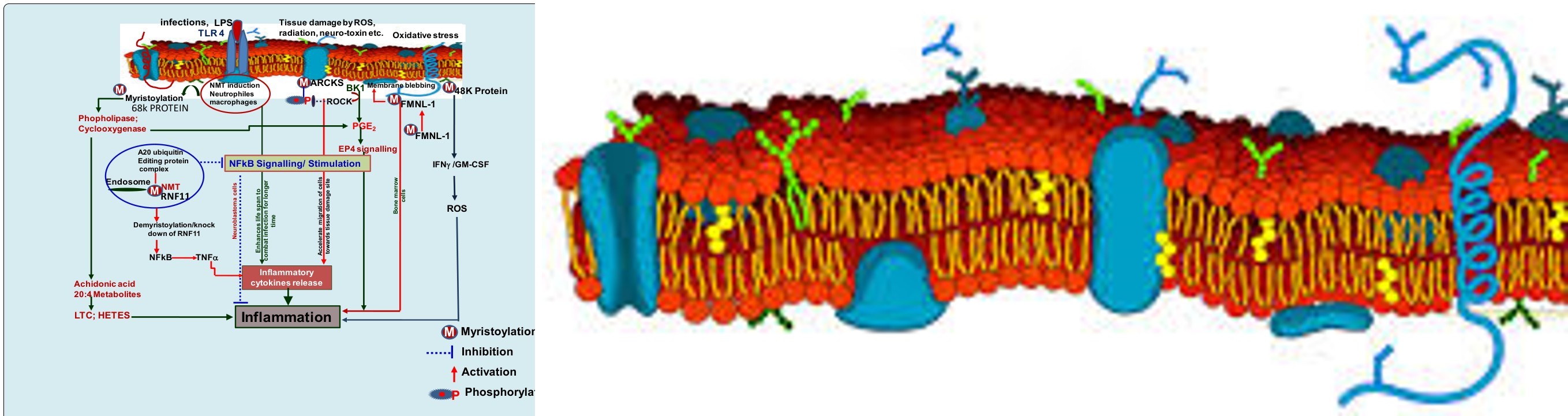
Inflammation occurs because of tissue injuries, immune insults and infections. Cytotoxic neutrophiles and macrophages respond to infection and moved towards the infection site of the tissue to combat the infectious agents. Expression and activity of NMT in phagocytic cells was reported to be increased in lung inflammation caused by bacterial infection [29]. NMT1 activities are also known to be induced in neutrophils and macrophages upon lipo- polysaccharide (LPS) treatment [29]. NMT1 induction reported in inflamed tissue was not due to bacterial presence in the inflamed tissue because bacteria do not have an NMT1 gene. NMT1 expression was reported to be increased in inflamed lung along with decreased functional activity due to induction of its indigenous inhibitor, i.e., a 48 kD protein enolase [52, 89].
Immuno-electron microscopy revealed that NMT1 is predominantly localized in the nucleus of leukocytes within inflamed lungs [29]. Nuclear localization of inflammation regulatory proteins like TLR-4 and integrin avb3 in leukocytes and endothelial cells of inflammed lungs are found consistent with NMT1.
Although its exact functional outcome is not clearly understood. However, its shuttling between cytoplasm and nucleus perhaps depends upon the requirement of myristoylation of cytoplasmic or nuclear proteins involved in cell proliferation, inflammation and carcinogenesis [29]. Experiments demonstrated a ~30% reduction in the lifespan of normal and activated neutrophils in which the NMT1 gene was knocked down [90]. Therefore, increased gene expression and functional activity of NMT1 in activated neutrophils and their decreased lifespan following NMT1 inhibition provide novel insights in the role of NMT1 in the regulation of neutrophil life span.
NFkB transcription factor plays a crucial role in cell proliferation, differentiation, adaptive and innate immune response, and inflammation [91]. Defects in NF-κB cellular and molecular signaling, such as continuous stimulation or activation, may be associated with induced inflammation and chronic neuro-inflammation that lead to neurodegenerative diseases. Proteins like A20 (TNF-α-induced protein3) act as NFkB inhibitors [91]. However, the regulatory function of A20 depends on its interaction with RING finger protein11 (RNF11). RNF11 is known as an integral component of the A20 ubiquitin-editing protein complex, that negatively regulates NF-κB signaling in human monocytic cell lines [92]. Interestingly, it was reported that myristoylation of RNF11 functional motifs is an essential event for its interaction with A20 ubiquitin-editing protein complex and NF-κB regulation [91]. Therefore, myristoylation of RNF11 proteins is essentially required for negative control of NFkB signaling in neuro-inflammation (Figure 3).
Forminsone proteins involved in polarized processes contribute to inflammation. Formin like 1 (FMNL1) protein is specifically expressed in hematopoietic lineage cells and over-expressed in malignant cells of different origin [93]. This restricted expression suggested that FMNL1 to be an attractive target for novel immune-therapies in malignant and inflammatory diseases. A splice variant of FMNL1 induces polarized non-apoptotic membrane blebbing that lead to inflammation. Membrane localization of FMNL1 essentially depends on its N-terminal myristoylation [94]. Thus, N-myristoylation is a regulatory mechanism for FMNL1 membrane trafficking and potentially responsible for diverse polarized processes including inflammation in hematopoietic lineage-derived cells (Figure 3).
| NFkB S | ignalling/ Sti | mulation | |
| combat infection for longer time Accelerate migration of cells |
Figure 3: Myristoylation Induced Inflammatory Signaling Pathways: Myristoylation of FMNL-1 protein induces its anchoring with membrane and activation of BK1 mediated EP4 signaling lead to inflammation. Further, myristoylation of 68k protein in neutrophiles and macrophages induces phospholipase and cyclooxygenase signaling. The metabolic product of cyclooxygenase reaction i.e. archidonic acid, LTC and HETES induced inflammation. However, myristoylated RNF11 protein form a complex with A20 ubiquitinin editing protein in the endosomes of neuronal cells which block NFkB mediated inflammatory responses. Therefore, myristoylation diversely modulates inflammatory response in specific situations. MARCKS; Myristoylated alanine-rich C-kinase substrate, BK1; Bradykinin-1, ROCK; Rho-associated protein kinase, FMNL- 1; Formin like protein, ROS; Reactive oxygen species, IFNg; interferon gamma, GM-CSF; granulocyte macrophages colony stimulating factor, PGE2; Prostaglandin E2, RNF 11; RING finger protein 11, NFkB; Nuclear factor kuppa beta, TNFa; tumor necrosis factor, LTC; Leukotriene, HETES; hydroxyeicosatetraenoic acids, EP 4; Prostaglandin E receptor 4.
Protein myristoylation is a main regulatory step during signal transduction in macrophages [95]. The role of myristoylation in arachidonic acid metabolism and in interferon-γ signaling leading to inflammation has been deduced [96]. Macrophages predominantly produce arachidonic acid (20:4) metabolites, which are prime mediators of inflammation. At the time of macrophage interactions with pathogens, phospholipases are activated that result in the release of metabolites producing 20:4 metabolite from membrane phospholipids and induced inflammation (Davies et al., 1984). The free 20:4 is further oxygenated by cyclooxygenase and produces prostaglandin E2 (PGE2) and prostacyclin (PGI2). It can also be converted into leukotriene C (LTC) and hydroxyeicosatetranoic acids (HETEs) with the help of a lipoxygenase pathway (Davies et al., 1984). The reaction end products like prostaglandins and prostacyclin are known as pro-inflammatory agents (Figure 3).
Bacterial lipopolysaccharide (LPS) has been reported to enhance 20:4 metabolism in macrophages. LPS has also been reported to promote covalent attachment of myristic acid to a set of macrophage proteins. The time of treatment and concentration of LPS regulates myristoylation and subsequently 20:4 cascading in macrophages. LPS-primed macrophages show greatly increased phosphorylation of the
68K protein upon protein kinase C activating phorbol esters treatment [97]. It is proposed that the myristoylation of the 68K protein promotes its attachment to the membrane. Where, myristoylated 68K protein binds with activated protein kinase C (PKC), and thus contributes to mobilization and oxygenation of 20:4 metabolites from macrophages [98]. Similarly, inflammatory response in the activated macrophages induces lymphokine interferon (IFN-γ) expression and activity which promotes the myristoylation of a 48 K protein. Membrane attachment of myristoylated 48K protein accelerates microbicidal activity of macrophages and inflammation in infected tissues. The Molecular signaling pathway depicting association between myristoylation and inflammation regulation is presented below Figure 3.
A Novel Strategy to Combat Carcinogenesis and Radiation Toxicity in Normal Tissue Using Differential Myristoylation Regulation
• Though complete molecular signaling is not very clear, but myristoylation undisputedly is an integral part of cancer progression. Several proteins Table-1 undergo myristoylation resulting in cell membrane attachment leading to down-stream signaling and cancer progression.
Similarly, myristoylation of various target proteins (like MARCKS, FMNL1, RNF11, 68K, 48K etc.) in neutrophils, macrophages and neuronal cells contributes to inflammation. More interestingly, inhibition of NMT either by using its inhibitor or gene knock out strategy, cell proliferation in cancerous cells and cell survival in normal cells were compromised. This phenomenon suggests that, on one hand, inhibition of NMT gene expression demonstrates a beneficial effect by blocking cell proliferating molecular signaling induced by myristoylated proteins (targeted protein myristoylation inhibition contributes to cancer progression inhibition), while, at the same time, it may be detrimental to the fate of normal cells. Expression of NMT in normal cells seems to be under the tight control of its negative regulators such as NIP71, hsc-70/HSP-70, Fus1, A20 ubiquitious protein editing complex and caspases etc. Therefore, strategically, inhibition of NMT expression in cancer cells provides an opportunity to limit their cell proliferation and cancer progression. But at the same time, NMT stimulation in normal cells can enhance their survival under environments like gamma radiation stress during radiotherapy of cancer patients. In view of the above, the strategy appears to be promising to protect tissues against cancer progression, inflammation and gamma radiation induced cytotoxicity: Though NMT antagonists/ inhibitors are established therapeutic targets to combat cancer progression. However, by increasing NMT expression using NMT agonists, pro-survival signaling can be induced in normal cells. Available literature suggests that NMT only contributes to cancer progression and not its initiation. Therefore, it should not initiate cancinogenesis in normal cells Figure 4. Myristoylation of 68K, 48K proteins, MARCKs and FMNL-1 proteins in neutrophiles and macrophages induces inflammatory response. As the result of higher inflammation, pro-inflammatory cytokines are released that may cause tissue damage at the infection site. However, inhibition of NMT in neutrophiles and macrophages compromises their life span that may cause more severe infection. In view of the above, optimized myristoylation of targeted proteins in inflammatory responsive cells needs to be maintained.
- Furthermore, myristoylation of RNF11 protein is a prerequisite requirement to make a complex with A20 ubiquitin editing protein in the endosome which blocks NFkB mediated inflammation in neuronal tissues. Therefore, upregulation of RNF11 myristoylation specifically in endosome would be a prominent therapeutic target to combat neuronal inflammation.
- NMT cellular level can be enhanced by down-regulation of NMTs negative regulators like NIP71, hsc-70/HSP- 70, Fus1, A20 ubiquitious protein editing complex and caspases etc., would result in improved cell survival which may contribute to protection of cells against ionizing radiation (Figure 4).
- NMT over-expression in the cells of hematopoietic lineage like neutrophils and macrophages is known to increase their life span and activate pro-inflammatory response by increasing inflammatory cytokines. These findings suggest that NMT up-regulation may induce a TH2 type cellular immune response necessary for immune system stimulation under gamma radiation stress and thus, may provide radioprotection.
- NMT acts as a selective substrate for caspase-3 in normal cells. Interestingly, caspase-3 expression in cancer cells is suppressed, resulting in increased NMT levels and thus over myristoylation and carcinogenesis. Thus, by decreasing capase-3 expression in normal cells, NMT expression can be enhanced, resulting in better survival against radiation-induced cellular damage (Figure 4).
- Various proteins, including cytoskeletal actin and gesolin, apoptosis regulating protein Bid, radiosenstizing protein MARCKS and RGS5 and radioresponsive protein Pak-2 are known as prominent targets of NMT [14, 21, 27, 76]. Expression level of these proteins was reported to be altered under radiation stress [98, 99, 100, 101, 102, 103, 104, 105, 106, 107]. However, information about whether myristoylation of these proteins influence radioresponse influences to radiosensitization or radioprotection is completely missing and needs focused attention.
![Figure 4: Myristoylation of 68K, 48K proteins, MARCKs and FMNL-1 proteins in neutrophiles and macrophages induces inflammatory response. As the result of higher inflammation, pro-inflammatory cytokines are released that may cause tissue damage at the infection site. However, inhibition of NMT in neutrophiles and macrophages compromises their life span that may cause more severe infection. In view of the above, optimized myristoylation of targeted proteins in inflammatory responsive cells needs to be maintained. • Furthermore, myristoylation of RNF11 protein is a prerequisite requirement to make a complex with A20 ubiquitin editing protein in the endosome which blocks NFkB mediated inflammation in neuronal tissues. Therefore, upregulation of RNF11 myristoylation specifically in endosome would be a prominent therapeutic target to combat neuronal inflammation. • NMT cellular level can be enhanced by down-regulation of NMTs negative regulators like NIP71, hsc-70/HSP- 70, Fus1, A20 ubiquitious protein editing complex and caspases etc., would result in improved cell survival which may contribute to protection of cells against ionizing radiation (Figure 4). • NMT over-expression in the cells of hematopoietic lineage like neutrophils and macrophages is known to increase their life span and activate pro-inflammatory response by increasing inflammatory cytokines. These findings suggest that NMT up-regulation may induce a TH2 type cellular immune response necessary for immune system stimulation under gamma radiation stress and thus, may provide radioprotection. • NMT acts as a selective substrate for caspase-3 in normal cells. Interestingly, caspase-3 expression in cancer cells is suppressed, resulting in increased NMT levels and thus over myristoylation and carcinogenesis. Thus, by decreasing capase-3 expression in normal cells, NMT expression can be enhanced, resulting in better survival against radiation-induced cellular damage (Figure 4). • Various proteins, including cytoskeletal actin and gesolin, apoptosis regulating protein Bid, radiosenstizing protein MARCKS and RGS5 and radioresponsive protein Pak-2 are known as prominent targets of NMT [14,21,27,76]. Expression level of these proteins was reported to be altered under radiation stress [98-107]. However, information about whether myristoylation of these proteins influence radioresponse influences to radiosensitization or radioprotection is completely missing and needs focused attention.](/fulltextimages/12106/fig_4.jpeg)
Figure 4: Schematic Representation of Myristoylation Mediated Radio-Response Modification in Normal and Cancerous Cells by NMT Negative Regulators: The present hypothesis comfortably explains the significant role of myristoylation in radio- response modulation in normal and cancerous cells. Up-regulation of NMT negative regulators in cancerous cells lead to NMT expression inhibition resulting in cell proliferation arrest. Thus, in this scenario, by applying radiotherapy in cancer patients, more cell death can be achieved. However, at the same time, by down regulation of NMT negative regulators, NMT expression can be enhanced, which may lead to higher cell growth and thus better protection against gamma radiation during radiotherapy of cancer patient. Therefore, by differential modulation of NMT negative regulators, radio-senstization in cancerous cells and radioprotection in normal calls can be achieved. P53, Tumor protein 53; NIP71, NMT inhibitor protein-71; hsc-70, heat shock cognate protein-70; HSP 70, heat shock protein-70; Akt/PKB, protein kinase B; Src, oncogene of Rous sarcoma virus; Myc, myelocytomatosis viral oncogene; GPCR, G protein coupled receptor; mP53, Mutant P53 protein; Fac, Fanconi’s anemia complementation group C protein; MARCKS, Myristoylated alanine-rich C-kinase substrate; ct-PAK-2, C terminal p21-activated protein kinase; PAK2, p21-activated protein kinase.
Myristoylation and Prominent Technical Gaps
Myristoylation of target proteins facilitates membrane anchoring, protein-protein interaction, protein phosphorylation leading to cell proliferation, embryonic differentiation, apoptosis, inflammation and immune regulation in cells of different lineages. N-myristoyltransferases (NMT1, NMT2) were found to be conserved throughout the evolutionary path and have been studied by several workers worldwide. However, the following questions still have not been answered and should be investigated.
- How is NMT’s transcriptional regulation maintained under different kind of stimuli and stresses?
- What are the factors that control NMT gene splicing and isoforms modification leading to myristoylation of specific protein targets?
- How is NMT specificity with its natural substrate, i.e. myristic acid (present >5nM/cell) is maintained while it represent cross-reactivity with abundantly available palmitic acid and other cellular protein substrates?
- How myristoylation and membrane attachment of the target proteins transmit downstream molecular signaling to progress or initiate cellular transformation, carcinogenesis, differentiation or inflammation?
- Whether, myristoylation of specific protein contributes to the specific type of cancer, or a diverse protein myristoylation can be associated with a specific type of cancer?
- Localization studies revealed NMT presence in the nucleus, cytoplasm, cell membrane, endosomes and ribosomes. However, how NMT translocation among different cellular compartments is regulated?
- Myristoylation is known to participate in tethering the G protein to the inner surface of the plasma membrane, and thus, facilitate G protein interaction with its receptor. Various proteins include catalytic subunit of cAMP4- dependent protein kinase, tyrosine kinases (pp60src, pp60yes, pp56lck, pp59fyn/syn, and c-Abl), calcineurin, myristoylated alanine-rich C kinase substrate (MARCK), some guanine nucleotide binding proteins and ADP ribosylation factors etc., undergo myristoylation. What are the exact function and purposes of myristoylation of all proteins in normal cells?
- As the NMT gene is conserved among all eukaryotic organisms, it indicates that NMT expression is essential for performing several important functions in normal cells. Therefore, the effect of NMT inhibitors on normal cell functioning needs to be further investigated.
- Whether NMT inhibition is sufficient to block cancer cells/tissue growth or not?
- Several negative regulators (like NIP-71, hsc-70, Akt/ PkB and wtp53 protein) of myristoylation have been reported. Whether these proteins differentially regulate NMT expression in normal and cancerous cells needs to be investigated.
Conclusion
Myristoyltransferase (NMT1/2) enzymes are thoroughly conserved throughout evolution in all eukaryotic organisms ranging from yeast to humans [108, 109, 110]. The exact function of NMTs in various stages of development is under investigation at various laboratories. NMT catalyzes myristoyl (C14 lipid chain compound) group transfer to the glycine residue of consensus amino acid sequences of target proteins. Myristoylated proteins then transduce down-stream signals to induce either continuous cell proliferation with carcinogenic outcome or inflammation with a desired immune response/ apoptosis.
Though myristoylation of different proteins activates diverse response in normal and cancerous cells, integral intermediate molecular factors responsible for the molecular cascade lead to the desired outcome are still unknown. Further, whether myristoylation participates in cancer progression only or it also involved in carcinogensis induction is not really understood. Transcriptional regulation of NMT gene expression against different stimuli and its translocation mechanism in different cellular compartments are a matter of concern for investigators. Caspase 3 was reported as the main regulator of NMT activity in cancer cells as NMT act as a substrate for caspase 3 in normal cells. Down regulation of caspases contribute to NMT accumulation in cancer cells leading to increased myristoylation. Therefore, either induction of caspase 3 or inhibition of NMT in cancer cells could be the prominent therapeutic target to control aggressive tumor growth. Several indigenous and chemical NMT inhibitors have been evaluated for their possible application as anticancer therapeutic agents. However, most of them are still in preclinical stage. Myristoylation of several proteins such as FMNL1, RFN11, 68K protein and 48K protein and MARCKS etc., activate various inflammatory reactions including NFkB activation, induced TNF-α, INF-γ, GM-CSF pro inflammatory cytokine expression in neutrophiles, machrophages, and neuroblastoma cells. Myristoylation of FMNL1 proteins enhances membrane blebbing and inflammation in neuroblastoma cells. Therefore, NMT mediated myristoylation modulates inflammatory response in the infected cells or tissues. Despite significant work done, several technical gaps still remained unfilled with appropriate scientific evidence to resolve prominent questions associated with myristoylation in normal and cancer cells. If gaps are filled with relevant scientific information, differential responses in normal and cancer cells can be unraveled. By understanding the mechanism of negative and positive regulators of NMT transcription and myristoylation, it is possible to achieve radioprotection in normal cells along with radio-sensitization in cancer cells. Continuous, concentrated and focused attempts to understand the molecular mechanism of myristoylation and its contribution to differential cellular response in normal and cancer cells will certainly enhance better understanding of myristoylation and its application in anticancer, ant- apoptosis and anti-inflammatory response targets discovery.
Conflict of Interest declaration: Authors declared no conflict of interest Acknowledgements: This study was supported by grants from the defence research development organization (DRDO), India.
References
-
Maneka A, Perinpanayagam, Beauchamp E, Sim JY, Yap MC, et al. (2013) Regulation of co- and post-translational myristoylation of proteins during apoptosis: interplay of N-myristoyl transferases and caspases. The FASEB Journal 27(2): 811-821.
-
Boutin JA (1997) Myristoylation. Cell Signal 9(1): 15-35.
-
Resh MD (2006) Trafficking and signaling by fatty- acylated and prenylated proteins. Nat Chem Biol 2(11): 584-590.
-
Aronheim A, Engelberg D, Li N, al-Alawi N, Schlessinger J, et al. (1994) Membrane targeting of the nucleotide exchange factor Sos is sufficient for activating the Ras signaling pathway. Cell 78(6): 949-961.
-
Megan H, Wright, William P, Tate EW (2010) Protein myristoylation in health and disease. J Chem Biol 3(1): 19-35.
-
Devadas B, Freeman SK, Zupec ME, Kuneman DW, Sikorski JA, et al. (1997) Design and Syntheses of Potent and Selective Dipeptide Inhibitors of Candida albicans Myristoyl-CoA:Protein N-Myristoyltransferase. J Med Chem 40(16): 2609-2615.
-
Duronio RJ, Towler DA, Heuckeroth RO, Gordon JI (1989) Disruption of the yeast N-myristoyl transferase gene causes recessive lethality. Science 243(4892): 796-800.
-
Olaleye TO, Brannigan JA, Roberts SM, Leatherbarrow RJ, Wilkinson AJ, et al. (2014) Peptidomimetic inhibitors of N-myristoyltransferase from human malaria and leishmaniasis parasites. Org Biomol Chem 12(41): 8132- 8137.
-
Ntwasa M, Aapies S, Schiffmann DA, Gay NJ (2001) Drosophila embryos lacking N-myristoyltransferase have multiple developmental defects. Exp Cell Res 262(2): 134-144.
-
Giang DK, Cravatt BF (1998) A second mammalian N-myristoyltransferase. J Biol Chem 273(12): 6595- 6598.
-
Thinon E, Serwa RA, Broncel M, Brannigan JA, Mann DJ, et al. (2014) Global profiling of co- and post-translationally N-myristoylated proteomes in human cells. Nature Communications 26(5): 4919.
-
James G, Olson EN (1990) Fatty acylated proteins as components of intracellular signaling pathways. Biochemistry 29(11): 2623-2634.
-
Silva AMD, Klein C (1990) A rapid posttranslational myristylation of a 68-kD protein in D. discoideum. J Cell Biol 111(2): 401-407.
-
Zha Z, Weiler S, Oh KJ, Wei MC, Korsmeyer SJ (2000) Posttranslational N-myristoylation of BID as a molecular switch for targeting mitochondria and apoptosis. Science 290(5497): 1761-1765.
-
Verge`res, G, Manenti S, Weber T, Sturzinger C (1995) The myristoyl moiety of myristoylated alanine-rich C kinase substrate (MARCKS) and MARCKS-related protein is embedded in the membrane. J Biol Chem 270(34): 19879-19887.
-
Cardena GG, Oh P, Liu J, Schnitzer JE, Sessa WC (1996) Targeting of nitric oxide synthase to endothelial cell caveolae via palmitoylation: implications for nitric oxide signaling. Proc Natl Acad Sci USA 93(13): 6448-6453.
-
Borgese N, Aggujaro D, Carrera P, Pietrini G, Bassetti MA (1996) Role for N-myristoylation in protein targeting: NADH-cytochrome b5 reductase requires myristic acid for association with outer mitochondrial but not ER membranes. J Cell Biol 135(6 pt1): 1501-1513.
-
Ames JB, Porumb T, Tanaka T, Ikura M, Stryer L (1995) Amino-terminal myristoylation induces cooperative calcium binding to recoverin. J Biol Chem 270(9): 4526- 4533.
-
Wedegaertner PB, Wilson PT, Bourne HR (1995) Lipid modifications of trimeric G proteins. J Biol Chem 270(2): 503-506.
-
Neer EJ, Smith TF (1996) G protein heterodimers: new structures propel new questions. Cell 84(2): 175-178.
-
Utsumi SN, Utsumi T (2006) Posttranslational N-myristoylation is required for the anti-apoptotic activity of human Gelsolin, the C-terminal caspase cleavage product of human gelsolin. J Biol Chem 281(20): 14288-14295.
-
Yonemoto W, McGlone M, Taylor SS (1993) N-myristylation of the catalytic subunit of cAMP- dependent protein kinase conveys structural stability. J Biol Chem 268(4): 2348-2352.
-
Felsted RL, Glover CJ, Hartman K (1995) Protein N-myristoylation as a chemotherapeutic target for cancer. J Natl Cancer Inst 87(21): 1571-1573.
-
Deichaite I, Casson LP, Ling HP, Resh MD (1988) In vitro synthesis of pp60v-src: myristylation in a cell-free system. Mol Cell Biol 8(10): 4295-4301.
-
Rudnick DA, McWherter CA, Gokel GW, Gordon JI (1993) MyristoylCoA:protein N-myristoyltransferase. Adv Enzymol 67: 375-430.
-
Johnson DR, Bhatnagar RS, Knoll LJ, Gordon JI (1994) Genetic and biochemical studies of protein N-myristoylation. Annu Rev Biochem 63: 869-914.
-
McLaughlin S, Aderem A (1995) The myristoyl- electrostatic switch: a modulator of reversible protein- membrane interactions. Trends Biochem Sci 20(7): 272- 276.
-
Matsubara M, Titani K, Taniguchi H, Hayashi N (2003) Direct involvement of protein myristoylation in myristoylated alanine-rich C kinase substrate (MARCKS)-calmodulin interaction. J Biol Chem 278(49): 48898-48902.
-
Shrivastav A, Suri S, Mohr R, Janardhan KS, Sharma RK, et al. (2010) Expression and activity of N-myristoyl transferase in lung inflammation of cattle and its role in neutrophil apoptosis. Vet Res 41(1): 9.
-
Maurer-Stroh S, Eisenhaber F (2004) Myristoylation of viral and bacterial proteins. Trends Microbiol 12(4): 178-185.
-
Farazi TA, Waksman G, Gordon JI (2001) The biology and enzymology of protein N-myristoylation. J Biol Chem 276(43): 39501-39504.
-
Resh MD (1999) Fatty acylation of proteins: new insights into membrane targeting of myristoylated and palmitoylated proteins. Biochim Biophys Acta 145(1): 1-16.
-
Olso, EN, Spizz G (1986) Fatty acylation of cellular proteins. Temporal and subcellular differences between palmitate and myristate acylation. J Biol Chem 261(5): 2458-2466.
-
Pillia S, Baltimore D (1987) Myristoylation and the post-translational acquisition of hydrophobicity by the membrane immunoglobulin heavy-chain polypeptide in B lymphocytes. Proc Natl Acad Sci USA 84(21): 7654- 7658.
-
Carr SA, Biemann K, Shozo S, Parmelee DC, Titani K (1982) n-Tetradecanoyl is the NH2-terminal blocking group of the catalytic subunit of cyclic AMP-dependent protein kinase from bovine cardiac muscle. Proc Natl Acad Sci USA 79(20): 6128-6131.
-
Bryant M, Ratner L (1990) Myristoylation-dependent replication and assembly of human immunodeficiency virus 1. Proc Natl Acad Sci USA 87(2): 523-527.
-
Weaver TA, Panganiban AT (1990) N myristoylation of the spleen necrosis virus matrix protein is required for correct association of the Gag polyprotein with intracellular membranes and for particle formation. J Virol 64(8): 3995-4001.
-
Aitken A, Cohen P, Santikarn S, Williams DH, Calder AG, et al. (1982) Identification of the NH2-terminal blocking group of calcineurin B as myristic acid. FEBS Lett 150(2): 314-318.
-
Cross FR, Garber EA, Pellman D, Hanafusa H (1984) A short sequence in the p60src N terminus is required for p60src myristylation and membrane association and for cell transformation. Mol Cell Biol 4(9): 1834-1842.
-
Rajala RV, Radhi JM, Kakkar R, Datla RS, Sharma RK (2000) Increased expression of N-myristoyltransferase in gallbladder carcinomas. Cancer 88(9): 1992-1999.
-
Gordon JI, Duronio RJ, Rudnick DA, Adams SP, Gokel GW (1991) Protein N-Myristoylation. J Biol Chem 266(14): 8647-8650.
-
Duronio RJ , Rudnick DA, Adams SP, Towler DA, Gordon JI (1991) Analyzing the substrate specificity of _Saccharomyces_ _cerevisiae_ myristoyl-CoA:protein N-myristoyltransferase by co-expressing it with mammalian G protein alpha subunits in Escherichia coli. J Biolog Chem 16: 10498-10504.
-
Chen CA, Manning DR (2001) Regulation of G proteis by covalent modification. Oncogene 20(13): 1643-1652.
-
Kobayashi M, Takamatsu K, Saitoh S, Noguchi T (1993) Myristoylation of hippocalcin is linked to its calcium- dependent membrane association properties. J Biol Chem. Sep 268(25): 18898-18904.
-
Rajala RV, Datla RS, Carlsen SA, Anderson DH, Qi Z, et al. (2001) Phosphorylation of human N-myristoyltransferase by N-myristoylated SRC family tyrosine kinase members. Biochem Biophys Res Commun 288(1): 233-239.
-
Patwardhan P, Resh MD (2010) Myristoylation and membrane binding regulate c-Src stability and kinase activity. Mol Cell Biol 30(17): 4094-4107.
-
Wu J, Tao Y, Zhang M, Howard MH, Gutteridge S, et al. (2007) Crystal structures of _Saccharomyces cerevisiae_ Nmyristoyltransferase with bound myristoyl-CoA and inhibitors reveal the functional roles of the N-terminal region. J Biol Chem 282(30): 22185-22194.
-
Bhatnagar RS, Fütterer K, Farazi TA, Korolev S, Murray CL, et al. (1998) Structure of N-myristoyltransferase with bound myristoylCoA and peptide substrate analogs. Nat Struct Biol 5(12): 1091-1097.
-
Haun RS, Tsai SC, Adamik R, Moss J, Vaughan M (1993) Effect of myristoylation on GTP-dependent binding of ADP-ribosylation factor to Golgi. J Biol Chem 268(10): 7064-7068.
-
Martin DD, Beauchamp E, Berthiaume LG (2011) Post- translational myristoylation: Fat matters in cellular life and death. Biochem 93(1): 18-31.
-
Shoji S, Kurosawa T, Inoue H, Funakoshi T, Kubota Y (1990) Human cellular src gene product: identification of the myristoylated pp60c-src and blockage of its myristoyl acylation with N-fatty acyl compounds resulted in the suppression of colony formation. Biochem Biophys Res Commun 173(3): 894-901.
-
Shrivastav A , Pasha MK, Selvakumar P, Gowda S, Olson DJH, et al. (2003) Potent inhibitor of N-myristoylation: a novel molecular target for cancer. Cancer Res 63(22): 7975-7978.
-
Duronio RJ, Reed SI, Gordon JI (1992) Mutations of human myristoyl-CoA:protein N-myristoyltransferase cause temperature-sensitive myristic acid auxotrophy in _Saccharomyces cerevisiae_. Proc. Natl. Acad. Sci. U. S. A 89(9): 4129-4133.
-
Raju RV, Moyana TN, Sharma RK (1997) N-Myristoyltransferase overexpression in human colorectal adenocarcinomas. Exp Cell Res 235(1): 145- 154.
-
Magnuson BA, Raju RV, Moyana TN, Sharma RK (1995) Increased N-myristoyltransferase activity observed in rat and human colonic tumors. J Natl Cancer Inst 87(21): 1630-1635.
-
Shrivastav S, Varma S, Senger A, Khandelwal RL, Carlsen S, et al. (2009) Over expression of Akt/PKB modulates N-myristoyltransferase activity in cancer cells. J Pathol 218(3): 391-398.
-
Selvakumar P, Smith-Windsor E, Bonham K, Sharma RK (2006) N-myristoyltransferase 2 expression in human colon cancer: cross-talk between the calpain and caspase system. FEBS Lett 580(8): 2021-2026.
-
Dehm S, Senger MA, Bonham K (2001) SRC transcriptional activation in a subset of human colon cancer cell lines. FEBS Lett 487: 367-371.
-
Shrivastav A, Sharma AR, Bajaj G, Charavaryamath C, Ezzat W, et al. (2007) Elevated N-myristoyltransferase activity and expression in oral squamous cell carcinoma. Oncology Reports 18(1): 93-97.
-
Bagrodia S, Taylor SJ, Shalloway D (1993) Myristylation is required for Tyr-527 dephosphorylation and activation of pp60c-src in mitosis. Mol Cell Biol 13(3): 1464-1470.
-
Kamps MP, Buss JE, Sefton BM (1985) Mutation of NH2- terminal glycine of p60src prevents both myristoylation and morphological transformation. Proc Natl Acad Sci USA 82(14): 4625-4628.
-
King MJ, Sharma RK (1993) Identification, purification and characterization of a membrane-associated N-myristoyltransferase inhibitor protein from bovine brain. Biochem J 291: 635-639.
-
Selvakumar P, Lakshmikuttyamma A, Shrivastav A, Das SB, Dimmock JR, et al. (2007) Potential role of N-myristoyltransferase in cancer. Prog Lipid Res 46(1): 1-36.
-
Raju RVS, Sharma RK (1999) Preparation and assay of myristoyl-CoA: protein N-myristoyltransferase. Methods Mol Biol 116: 193-211.
-
Murakami Y, Nakano S, Niho Y, Hamasaki N, Izuhara K (1998) Constitutive activation of Jak-2 Tyk-2 in a v-src transformed human gallbladder adenocarcinoma cell line. J Cell Physiol 175(2): 220-228.
-
Masumoto N, Nakano S, Fujishima H, Kohno K, Niho Y (1999) V-src induces cisplatin resistance by increasing the repair of cisplatin-DNA interstrand cross-links in human gallbladder adenocarcinoma cells. Int J Cancer 80(5): 731-737.
-
Ottefhoff-Kalff AE, Ruksen G, Van Beurden EACM, Hennipman A, Michels AA, et al. (1992) Characterization of protein tyrosine kinase from human breast cancer: involvement of c-src oncogene product. Cancer Res 52(17): 4773-4778.
-
Medina-Ramirez CM, Goswami S, Smirnova T, Bamira D, Benson B, et al. (2011) Apoptosis inhibitor ARC promotes breast tumorigenesis, metastasis, and chemoresistance. Cancer Res 71(24): 7705-7715.
-
Uno F, Sasaki J, Nishizaki M, Carboni G, Xu K, et al. (2004) Myristoylation of the Fus1 Protein Is Required for Tumor Suppression in Human Lung Cancer Cells. Cancer Research 64(9): 2969-2976.
-
Kondo M, Ji L, Kamibayashi C, Randle D, Sekido Y, et al. (2001) Overexpression of candidate tumor suppressor gene FUS1 isolated from the 3p21.3 homozygous deletion region leads to G1 arrest and growth inhibition of lung cancer cells. Oncogene 20(43): 6258-6262.
-
Benetka W, Mehlmer N, Maurer-Stroh S, Sammer M, Koranda M, et al. (2008) Experimental testing of predicted myristoylation targets involved in asymmetric cell division and calcium-dependent signaling. Cell Cycle 7(23): 3709-3719.
-
Kerr JF, Wyllie AH, Currie AR (1972) Apoptosis: a basic biological phenomenon with wide-ranging implications in tissue kinetics. Br J Cancer 26(4): 239-157.
-
Wyllie AH, Bellamy CO, Bubb VJ (1999) Apoptosis and carcinogenesis. Br J Cancer 80(1): 34-37.
-
Jiang X, Wang X (2004) Cytochrome C mediated apoptosis. Annu Rev Biochem 73: 87-106.
-
Vilas GL, Corvi MM, Plummer GM, Seime AM, Lambkin GR, et al. (2006) Posttranslational myristoylation of caspase-activated p21- activated protein kinase 2 (PAK2) potentiates late apoptotic events. Proc Natl Acad Sci USA 103(17): 6542-6547.
-
Utsumi T, Sakurai N, Nakano K, Ishisaka R (2003) C-terminal 15 k Da fragment of cytoskeletal actin is posttranslationally N-myristoylated upon caspasemediated cleavage and targeted to mitochondria. FEBS Lett 539(1-3): 37-44.
-
Frearson JA, Brand S, McElroy SP, Smid O, Stojanovski L, et al. (2010) N-Myristoyltransferase inhibitors as new leads to treat sleeping sickness. Nature 464(7289): 728- 732.
-
Bernal F, Valdés I, Coy-Barrera ED (2014) N-myristoyl transferase inhibitors from natural sources: A molecular docking exploration. Planta Med.
-
Ducker CE, Upson JJ, French KJ, Smith CD (2005) Two N-myristoyltransferase isozymes play unique roles in protein myristoylation, proliferation, and apoptosis. Mol Cancer Res 3(8): 463-476.
-
Chen L, Ling B, Alcorn J, Yang J (2009) Quantitative Analysis of the Expression of Human N-myristoyltransferase 1 (hNMT-1) in Cancers. The open Biomarker Journal 2: 6-10.
-
Eisenhaber F, Eisenhaber B, Kubina W, Maurer- Stroh S, Neuberger G, et al. (2003) Prediction of lipid posttranslational modifications and localization signals from protein sequences: big-Pi, NMT and PTS1. Nucleic Acids Res 31(13): 3631-3634.
-
Sheng CH, Ji H, Miao Z, Che X, Yao J, et al. (2009) Homology modeling and molecular dynamics simulation of N-myristoyltransferase from protozoan parasites: active site characterization and insights into rational inhibitor design. J Comput Aided Mol Des 23(6): 375-389.
-
Glover CJ, Hartman KD, Felsted RL (1997) Human N-myristoyltransferase amino-terminal domain involved in targeting the enzyme to the ribosomal subcellular fraction. J Biol Chem 272(45): 28680-28689.
-
Paul KS, Jiang D, Morita YS, Englund PT (2001) Fatty acid synthesis in African trypanosomes: a solution to the myristate mystery. Trends Parasitol 17(8): 381-387.
-
Janardhan KS, Appleyard GD, Singh B (2004) Expression of integrin subunits alphav and beta3 in acute lung inflammation. Histochem Cell Biol 121(5): 383-390.
-
Rouzer CA, Scott WA, Cohn ZA, Blackburn P, Manning JM (1980) Mouse peritoneal macrophages release leukotriene C in response to a phagocytic stimulus. Proc Natn Acad Sci USA 77(8): 4928-4932.
-
Perkins ND (2007) Integrating cell-signaling pathways with NF-κB and IKK function. Nat Rev Mol Cell Biol 8(1): 49-62.
-
Pranski EL, Dalal NV, Herskowitz JH, Orr AL, Roesch LA, et al. (2012) Neuronal RING finger protein 11 (RNF11) regulates canonical NF-κB signaling. Journal of Neuro inflammation 9: 67-82.
-
Yanyan Han, Elfriede Eppinger, Ingrid G, Weigand LU, Liang X, et al. (2009) Formin-like 1 (FMNL1) Is Regulated by N-terminal Myristoylation and Induces Polarized Membrane Blebbing. J Biol Chem 284(48): 33409-33417.
-
Moriya K, Yamamoto T, Takamitsu E, Matsunaga Y, Kimoto M, et al. (2012) Protein N-myristoylation is required for cellular morphological changes induced by two formin family proteins, FMNL2 and FMNL3. Biosci Biotechnol Biochem 76(6):1201-1209.
-
Rosen A, Keenan KF, Thelen M, Nairn AC, Aderem A (1990) Activation of protein kinase C results in the displacement of its myristoylated, alanine rich substrate from punctate structures in macrophage filopodia. J Exp Med 172(4): 1211-1215.
-
Davies P, Bailey PJ, Goldenberg MM, Ford Hutchinson AW (1984) The role of arachidonic acid oxygenation products in pain and inflammation. A Rev Immun 2: 335- 357.
-
Aderem AA (1988) Protein myristoylation as an intermediate step during signal transduction in macrophages: its role in arachidonic acid metabolism and in responses to interferon gamma, J Cell Sci Suppl 9: 151-167.
-
Mishra S, Bansal DD, Malhotra P, Reddy DSK, Jamwal VS, et al. (2014) Semiquinone glucoside derivative (SQGD) isolated from Bacillus sp. INM-1 protects against gammaradiation-induced oxidative stress. Environ Toxicol Pharmacol 29(12): 553-562.
-
Mishra S, Patel DD, Bansal DD, et al (2014) Semiquinone glucoside derivative provides protection against Y- radiation by modulation of immune response in murine model. Environmental Tixicology 31(4): 478-488.
-
Mishra S, Bansal DD, Malhotra P, Reddy DSK, Jamwal VS, et al (2013) Semiquinone fraction isolated from Bacillus sp. INM-1 protects hepatic tissues against radiation induced toxicity 29(12):1471-1478.
-
Kempf SJ, Buratovic S, von Toerne C, Moertl S, Stenerlöw B, et al. (2014) Ionising radiation immediately impairs synaptic plasticity-associated cytoskeletal signaling pathways in HT22 cells and in mouse brain: an in vitro/ in vivo comparison study. PLoS One 9(10): e110464.
-
Maas C, de Vries E, Tait SW, Borst J (2011) Bid can mediate a pro-apoptotic response to etoposide and ionizing radiation without cleavage in its unstructured loop and in the absence of p53. Oncogene 30(33): 3636- 3647.
-
Slee EA, Keogh SA, Martin SJ (2000) Cleavage of BID during cytotoxic drug and UV radiation-induced apoptosis occurs downstream of the point of Bcl-2 action and is catalysed by caspase-3: a potential feedback loop for amplification of apoptosis-associated mitochondrial cytochrome c release. Cell Death Differ 7(6): 556-565.
-
Rohrbach TD, Jarboe JS, Anderson JC, Trummell HQ, Hicks PH, et al. (2015) Targeting the effector domain of the myristoylated alanine rich C-kinase substrate enhances lung cancer radiation sensitivity. Int J Oncol 46(3): 1079-1088.
-
Li M, Cui F, Cheng Y, Han L, Wang J, et al. (2015) Gelsolin: role of a functional protein in mitigating radiation injury. Cell Biochem Biophys 71(1): 389-396.
-
Sakai N, Ohtsu M, Fujita H, Koike T, Kuzumaki N (1999) Enhancement of G2 checkpoint function by gelsolin transfection in human cancer cells. Exp Cell Res 251(1): 224-233.
-
Roig J, Traugh JA (1999) p21-activated protein kinase gamma-PAK is activated by ionizing radiation and other DNA-damaging agents. Similarities and differences to alpha-PAK. J Biol Chem 274(44): 31119-31122.
-
Lodge JK, Jackson-Machelski E, Toffaletti DL, Perfect JR, Gordon JI (1994) Targeted gene replacements demonstrates that myristotl-CoA: protein N-myristoyltransferase is essential for viability of Cryptococcus neoformans. Natl Acad Sci USA 91 (25): 12008-12012.
-
Chen CA, Manning DR (2001) Regulation of G proteis by covalent modification. Oncogene 20(13): 1643-1652.
-
Kobayashi M, Takamatsu K, Saitoh S, Noguchi T (1993) Myristoylation of hippocalcin is linked to its calcium-dependent membrane association properties. J Biol Chem 268(25): 18898-18904.
-
Panethymitaki CPW, Bowyer PW, Price HP, Leatherbarrow RJ, Brown KA, et al. (2006) Characterization and selective inhibition of myristoyl- CoA: protein N-myristoyltransferase from _Trypanosoma_ _brucei_ and _Leishmania major_. Biochem J 396(2): 277- 285.
-
Patwardhan P, Resh MD (2010) Myristoylation and membrane binding regulate c-Src stability and kinase activity. Mol Cell Biol 30(17): 4094-4107.
-
Wyllie AH (1997) Apoptosis: an overview. Br Med Bull 53(3): 451-465.
-
Rajala RV, Datla RS, Carlsen SA, Anderson DH, Qi Z, et al. (2010) Phosphorylation of human N-myristoyltransferase by N-myristoylated SRC family tyrosine kinase members. Biochem Biophys Res Commun 288(1): 233-239.
-
Bower PW, Gunaratne RS, Grainger M, Martinez CW, Wickramsinghe SR, et al. (2007) Molecules incorporating a benzothiazole core Scaffold inhibit the N-myristoyl transfarase of Plasmodium faliciparum. Biochem J 408(2): 173-180.
- Evaluation of Skin Aging Preventive Effects of Cherry Blossom Petal Extracts Through Antioxidant and Anti-Glycation Activities
- Is Cell Death Responsible for False Positive Results of In Vivo Comet Assay?
- Pattern of Gonadal Hormones in Oral Testosterone-Supplimented Male Wistar Rats with Diabetes-Induced Hypogonadism
- Re-Evaluation of the Genotoxicity of Currently Used Food Dyes in Mouse Multiple Organs Via Continuous Administration by Drinking Using the Comet Assay
- Pharmacogenetics of Type 2 Diabetes Mellitus: Linking Genetic Variability to Drug Efficacy and its Cardiovascular Outcomes
- Exploratory Proteomic Profiling of SARS-CoV-2 Infected THP-1 Macrophages Reveals Alterations in Inflammatory Response and Cellular Metabolism