Suppression of Hyperglycemia-Induced Insulin Resistance by Rapanone Isolated from Connarus Ruber
Although it would be ideal if insulin resistance due to a persistent hyperglycemic state could be prevented by consuming particular foods, to the best of our knowledge, no foods with such effects have as yet been reported. Connarus ruber (CR) extract, previously indicated to be effective against diabetes, was investigated in this study to ascertain whether it can inhibit the induction of insulin resistance in human hepatoma-derived HepG2 cells. Insulin resistance can be induced in cells by cultivating under hyperglycemic conditions. HepG2 cells were cultured for 24 and 48 h with 1.25 µg/mL of CR extract and its components, rapanone and embelin, at 1.25 and 5 µg/mL, followed by the induction of insulin resistance by hyperglycemic conditions. The results revealed a decrease over time in the glucose concentration of HepG2 cells, indicating that insulin resistance was inhibited in these cells. However, when HepG2 cells, already insulin resistant owing to previous exposure to hyperglycemic conditions, were cultured for 24 and 48 h with CR extract containing rapanone and embelin at <10 µg/mL, the glucose concentration creased over time, indicating that insulin resistance was not affected under these conditions. The results suggest that insulin resistance is inhibited only when CR extract exposure occurs before exposure to hyperglycemia conditions and CR cannot induce recovery from insulin resistance. Thus, this study indicates that CR extract, with rapanone and embelin as active ingredients, could be used as a functional food to reduce the risk of developing type 2 diabetes mellitus as a result of chronic hyperglycemia.
Introduction
Diabetes, which is characterized by chronic hyperglycemia, is known to be caused by impaired insulin secretion and insulin resistance [1]. Sustained hyperglycemia can result in cellular insulin resistance, leading to type 2 diabetes (T2DM), which is a chronic metabolic disease that is predicted to affect more than 366 million people worldwide by 2025 [2], with the associated medical costs expected to become a considerable problem. Currently, T2DM is mainly treated using medicines that can improve insulin resistance, promote insulin secretion, and regulate sugar absorption and excretion, with the treatment selected according to the condition, exercise, and lifestyle of the patient. However, the existing treatments are often associated with side effects; hence, there is an increased interest in the use of natural products rather than pharmaceuticals. To date, researchers have reported various natural products derived from medicinal plants, particularly triterpenes, flavonoids, and polyphenols, that exhibit hypoglycemic effects [3]. Thus, the discovery of foods that could prevent insulin resistance resulting from sustained hyperglycemic conditions is of considerable interest.
Connarus ruber (CR), a member of the Quercus family, grows near Manaus in the middle of the Amazon. Its bark extract, taken as herbal tea, has not only demonstrated effectiveness against diabetes [4, 5], but has also been clinically reported to lower the DNA damage level in the white blood cells of smokers [6]. Additionally, our previous study indicated that CR extract can inhibit the induction of insulin resistance [7]. The active substances in CR extract mainly comprise quinones (1.45 %), of which rapanone accounts for 34.8 % and embelin 36.6 %. Rapanone, with various antioxidant [8], radical scavenging [8], and melanoma cell proliferation inhibition functions [9], is cytotoxic to human cancer cells [10]. This study investigates the inhibitory
effects of both rapanone and embelin on insulin resistance.
Material and Methods
Sample Preparation
CR cortex (240 g) was placed in 18 L of distilled water and incubated at 97 ± 2°C for 15 min. CR extract (91 g) was obtained from the cortex through filtration and evaporation with a rotary evaporator.
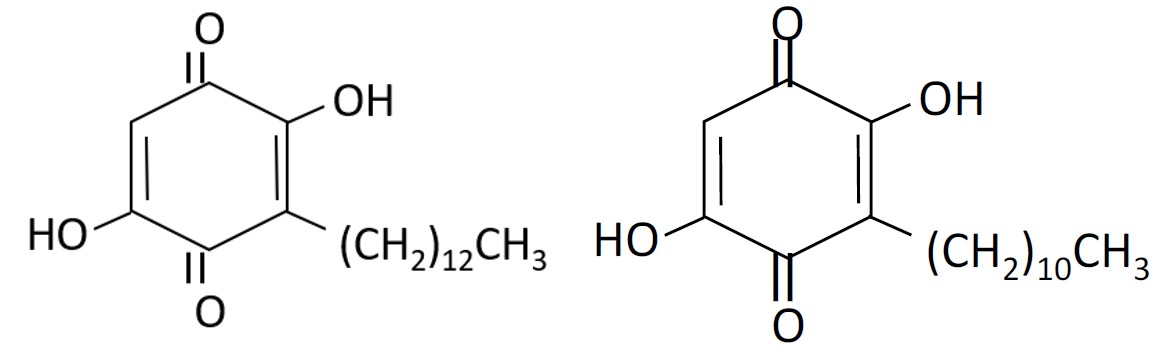
A precipitate was obtained by extracting 50 g of Connarus cortex with MeOH (3 × 1.0 L) over 3 d before evaporating until dry under a vacuum. The precipitate was dissolved in EtOAc (600 mL) and extracted with H2O (3 × 300 mL). The organic layer was evaporated until dry under a vacuum, and the residue dissolved in MeOH-H2O (1:9) (500 mL) before washing in n -Hexane (300 mL). Of the 725.5 mg precipitate obtained, 180 mg was filtrated and subjected to chromatography on an ODS packed silica gel column before elution with 0.1 % H3PO4 aq.-CH3CN (1:9) [11] to obtain 62.6 mg of rapanone (2,5-dihydroxy-3-tridecylcyclohexa- 2,5-diene-1,4-dione) (Figure 1a) and 65.2 mg of embelin (2,5-dihydroxy-3-undecylcyclohexa-2,5-diene-1,4-dione) (Figure 1b).
Cell Line
Human hepatoma HepG2 cells, obtained from the JCRB cell bank (Osaka, Japan) were, maintained in Dulbecco’s modified Eagle’s medium (DMEM: Nissui Pharmaceutical Co., Ltd.) with 10% fetal bovine serum (FBS: COSMO BIO Co., Ltd.) in 200 µg/mL streptomycin at 37 °C in a 5 % CO2 atmosphere. The cells were maintained in a logarithmic growth medium for use.
Insulin Resistance Test
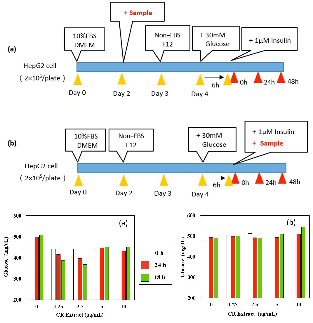
HepG2 cells were seeded in 6 cm dishes at a concentration of 2 × 10⁵ cells/mL in 5 mL of medium. Insulin resistance in HepG2 cells was induced as described by Najima et al. and Mohamadpour, et al. [12]. As depicted in Figure 2, cells were cultured for 48 h in FBS-free Ham’s F12 medium (Nissui Pharmaceutical Co., Ltd.), then glucose (30 mM) and insulin (1 µM) were added and cells were cultivated in serum-free conditions for 24 and 30h, respectively. Glucose
concentrations were measured at 0, 24, and 48 h after insulin administration using the Glucose Assay Kit-WST (DOJINDO LAB., Japan).
The CR extract concentration was determined via a cytotoxicity test, for which 100 µL of HepG2 cells were seeded in a 96-well microplate at a concentration of 5 × 10³ cells/mL, and cell viability was measured at 4, 24, and 48 h after exposure to the CR extract using a Cell Counting Kit-8 (DOJINDO LAB., Japan). Insulin resistance tests were performed when the cell viability was 80 % or higher.
Results and Discussion
Cell death was not observed at CR extract concentrations below 10 µg /mL (data not shown). As demonstrated in Figure 3a, the glucose concentration decreased in a time- dependent manner when HepG2 cells were exposed to CR extract for 24 h, followed by cultivation under hyperglycemic conditions for 24 h (Figure 2a). This indicated that exposure to CR extract inhibits the induction of insulin resistance.
However, as seen in Figure 3b, these changes were not observed when insulin resistance was induced before culture in CR extract (Figure 2b). Increased glucose concentrations in the medium resulted in insulin resistance because the intracellular glucose consumption was suppressed in the presence of insulin [13]. The glucose concentration was also observed to decrease over time when HepG2 cells were exposed to rapanone and embelin followed by cultivation under hyperglycemic conditions, as seen in Figures 4a & 4b, indicating that rapanone and embelin play important roles in inhibiting insulin resistance caused by hyperglycemic conditions. Rapanone did not show any cytotoxic effects at 5 µg/mL or less (cell viability was 80 % or more). Therefore, the possibility is excluded that decreased cell numbers due to dying cells lead to the decrease glucose in such during the 48 h treatment period for rapanone at shown dose range. Nakajima et al. reported that insulin resistance is induced when HepG2 cells reach a hyperglycemic state [13]. We previously reported that CR extract does not lead to glucose uptake by HepG2 cells in this state, even if insulin is administered [7].
Figure 3a: Effect of CR Exposure before the Induction of Insulin Resistance on Glucose Concentration in Insulin Resistant HepG2 Exposed to CR Extract at 0 and 1.25 µg/mL, the Pearson Correlation Coefficients between Culture Period and Glucose Concentration were 0.9102 and 0.9152, Respectively. Reproducibility was Confirmed in three Repeated Experiments. Figure 3b: Effect of CR Exposure after the Induction of Insulin Resistance on Glucose Concentration in HepG2 Cells Exposed to CR Extract 1.25 µg/mL followed by the Induction of Insulin Resistance, the Pearson Correlation Coefficient between Culture Period and Glucose Concentration was -0.7559. Reproducibility was Confirmed in three Repeated Experiments.
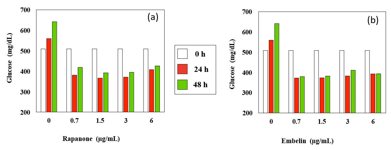
Figure 4: Effect of Rapanone (a) and Embelin (b) Exposure before the Induction of Insulin Resistance on Glucose Concentration. Induction of Insulin Resistance after 24 h of Exposure to Rapanone or Embelin. Reproducibility was Confirmed in three Repeated Experiments. (a) In HepG2 cells exposed to rapanone at 0.7, 1.5, and 3 µg/mL followed by the induction of insulin resistance, the Pearson correlation coefficients between culture period and glucose concentration were -0.5732, -0.6097, and -0.5295, respectively. (b) In HepG2 cells exposed to embelin at 0.7, 1.5, and 3 µg/mL followed by the induction of insulin resistance, the Pearson correlation coefficients between culture period and glucose concentration were -0.5407, -0.6019, and -0.5713, respectively.
This study revealed that both rapanone and embelin can suppress insulin resistance. Although embelin has two fewer carbons than rapanone, no difference was observed in the magnitude of the inhibitory effect, suggesting that both benzoquinones inhibit insulin resistance. However, further studies are required to elucidate the mechanisms by which these quinones inhibit insulin resistance. The results suggest that CR extract, with rapanone as its active ingredient, could be used as a functional food to reduce the risk of developing T2DM from chronic hyperglycemia. The purification of the functional ingredient of CR extract required for further studies.
Previous studies have indicated that the glucose transporters GLUT4 and GLUT2 are expressed in adipocytes and hepatocytes, respectively [14]. GLUT4 transfers glucose into cells, whereas GLUT2 transports glucose both inside and outside the cell depending on the intracellular glucose concentration. When insulin binds to its receptors, the intracellular glucose concentration decreases as glucose is consumed, and glucose is transported passively into the cell. Therefore, we assumed that the glucose concentration in the medium would decrease as cells took up the glucose. Nonetheless, exposure to high glucose concentrations (15– 33 mM) leads to phosphorylation of the serine residues in insulin receptor 1, reducing the electrophoretic mobility of insulin at the receptor and suppressing the signal [12]. When the insulin signal originating from GLUT2 is suppressed in the HepG2 cells, intracellular glucose consumption is no longer promoted, even in the presence of insulin [13], resulting in an increase in the glucose concentration in the medium. Moreover, insulin resistance was inhibited only when exposure to the CR extract was initiated before the onset of hyperglycemia. These results suggest that CR can affect the induction of insulin resistance but cannot initiate recovery from insulin resistance.
Acknowledgements
This research was conducted in the Hachinohe National College of Technology Materials and Biotechnology Course based on the allocation of school education expenses. The authors acknowledge the Material and Biological Engineering Course, National Institute of Technology, Hachinohe College.
Ethical Approval
It is not applicable.
References
-
Matsuoka T, Kajimoto Y, Watada H, Kaneto H, Kishimoto M, et al. (1997) Glycation dependent reactive oxygen species-mediated suppression of the insulin gene promoter activity in HIT cells. J Clin Invest 99(1): 144- 150.
-
Winder WW, Hardie DG (1999) AMP activated protein kinase, a metabolic master switch possible roles in Type 2 diabetes. Am J Physiol 277(1): 1-10.
-
Hung HY, Qian K, Morris-Natschke SL, Hsu CS, Lee KH (2012) Recent discovery of plant derived anti-diabetic natural products. Nat Prod Rep 29(5): 580-606.
-
Nwodo OF, Alumanah EO (1991) Stqudies on Abrus precatorius seed II, Antidiarrhoeal activity. J Ethnophamacology 31(3): 394-398.
-
Monago CC, Alumanah EO (2005) Antidiabetic effect of chloroform methanol extract of Abrus Precatorius Linn Seed in alloxan diabetic rabbit. J Appl Sci Environ Mgt 9: 85-88.
-
Nakamura T, Kurimoto S, Saigo K (2016) Genotoxicity suppressing effect of a connarus ruber cortex aqueous extract on DNA damage in smoker’s white blood cells a preliminary study. Journal of Clinical and Laboratory Investigation 4: 11-19.
-
Kawaguchi S, Kawasaki R, Murashige R, Yamasaki K, Kurimoto S, et al. (2020) Can connarus rubber extract inhibit the induction of insulin resistance by hyperglycemia?. MOJ Toxicology 6(1): 6-10.
-
Vega-Hernández KDL, Antuch M, Cuesta-Rubio O, Núñez- Figueredo Y, Pardo-Andreu GL, et al. (2017) Discerning the antioxidant mechanism of rapanone: A naturally occurring benzoquinone with iron complexing and radical scavenging activities. J Inorg Biochem 170: 134- 147.
-
Wróbel-Biedrawa D, Grabowska K, Galanty A, Sobolewska D, Żmudzki P, et al. (2020) Anti-melanoma potential of two benzoquinone homologues embelin and rapanone – a comparative in vitro study. Toxicol In Vitro 65: 104826.
-
Kuete V, Omosa LK, Tala VRS, Karaosmanoğlu O, Sivas H, et al. (2016) Cytotoxicity of Plumbagin, Rapanone and 12 other naturally occurring Quinones from Kenyan Flora towards human carcinoma cells. BMC Pharmacol Toxicol 17(1): 60.
-
Podolak I, Strzałka M (2008) Qualitative and quantitative LC profile of embelin and rapanone in selected Lysimachia species. Chromatographia 67: 471-475.
-
Nakajima K,Yamauchi K, Shigematsu S, Ikeo S, Komatsu M, et al. (2000) Selective attenuation of metabolic branch of insulin receptor down-signaling by high glucose in a hepatoma cell line, HepG2 Cells. The Journal of Biological Chemistry 275(27): 20880-20886.
-
Hresko RC, Heimberg H, Chi MM, Mueckler M (1998) Glucosamine-induced Insulin Resistance in 3T3-L1 Adipocytes Is Caused by Depletion of Intracellular ATP. J Biol Chem 273(32): 20658-20668.
-
Oakes ND, Cooney GJ, Camilleri S, Chisholm DJ, Kraegenet EW (1997) Mechanisms of liver and muscle insulin resistance induced by chronic high-fat feeding. Diabetes 46(11): 1768-1774.
- Evaluation of Skin Aging Preventive Effects of Cherry Blossom Petal Extracts Through Antioxidant and Anti-Glycation Activities
- Is Cell Death Responsible for False Positive Results of In Vivo Comet Assay?
- Pattern of Gonadal Hormones in Oral Testosterone-Supplimented Male Wistar Rats with Diabetes-Induced Hypogonadism
- Re-Evaluation of the Genotoxicity of Currently Used Food Dyes in Mouse Multiple Organs Via Continuous Administration by Drinking Using the Comet Assay
- Pharmacogenetics of Type 2 Diabetes Mellitus: Linking Genetic Variability to Drug Efficacy and its Cardiovascular Outcomes
- Exploratory Proteomic Profiling of SARS-CoV-2 Infected THP-1 Macrophages Reveals Alterations in Inflammatory Response and Cellular Metabolism