Report on a Rare Tooth Crown Morphological Trait in Permanent Mandibular Second Molar Protoconidal Cingulum Protostylid
Human teeth have been transforming due to biological evolutionary phenomenon. Therefore, variations in cusp number, pattern and size can be noticed in the human beings of present era compared to prehistoric age people. The purpose of this article is to present an occurrence of “Protostylid” or “Protoconidal cingulum” on the buccal surface of mandibular second molar in an Indian male patient which is rarely reported so far till date. Reporting of rare tooth crown morphological traits in human teeth is of utmost important to understand the dental evolution and also from forensic point of view.
Introduction
“Protostylid” also called by synonym “Protoconidal cingulum” is a rare tooth crown morphological trait is almost similar to a “Carabelli’s cusp” trait and both are derivatives of cingulum [1]. In order to simply explain ‘protostylid is a mirror image of cusp of Carabelli,’ because protostylid is expressed on the mesiobuccal cusp of lower molars (Mandibular) whereas Carabelli trait develops on the mesiolingual cusp of the upper molars (Maxillary molars) [2]. Scott GR [3] in 1978 stated that both these crown morphological traits are linked to some extent however, inter-trait correlations are relatively low. The protostylid has received immense attention because of its distinctive expression among a number of early hominin taxa. Skinner MM, et al [4] has referred protostylid as both an accessory cusp and as a remnant of a cingulum (crest feature). According to Arizona State University Dental Anthropology System (ASUDAS), (which is a devise for the analysis of modern human teeth), protostylid can be defined as “a paramolar cusp found on the buccal surface of the protoconid (mesiobuccal cusp) of mandibular molars that is normally associated with the buccal groove [5]. Dahlberg [1, 2] explained it as “an elevation or ridge of enamel on the anterior part of the buccal surface of the lower molars, which ascends from the gingival end of the buccal groove and extends mesio-occlusally”.
There is no definitive data on the prevalence and incidence of this trait among different population sample, because of lack of extensive studies undertaken pertaining to this domain. It is noticed that individuals with protostylid in deciduous molars were also found to express similar pattern in the succedaneous permanent teeth. Literature illustrates countable number of publications on occurrence of protostylid in permanent mandibular molars [6, 7]. Therefore, with the purpose of showcasing a rare tooth crown trait in a patient of Indian ethnicity the present paper was prepared.
Case Report
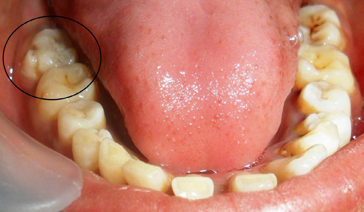
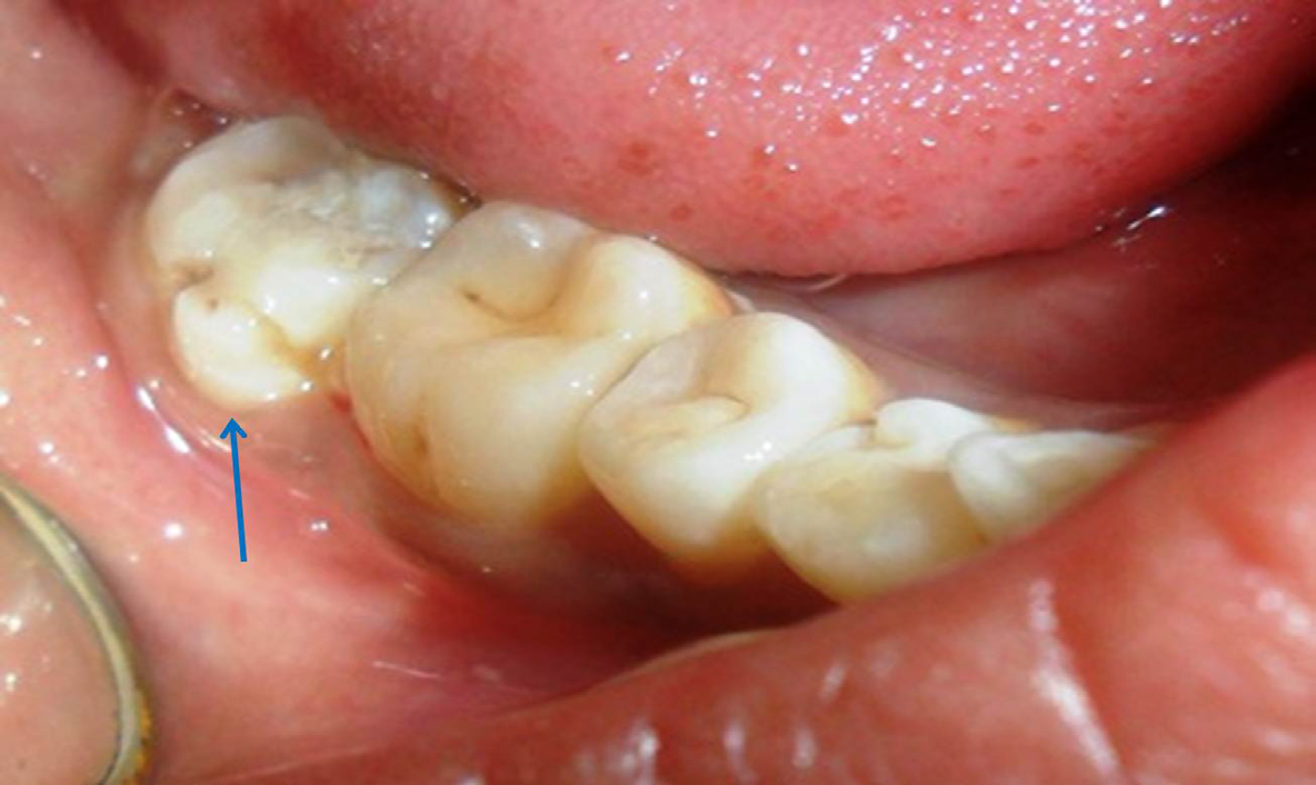
A 15-year-old female patient reported to a private dental clinic complaining of bad odour in the mouth. Patient was moderately built and nourished with well behavior. There were no signs and symptoms of any systemic, metabolic or syndromic disorders observed. On intraoral examination complete set of erupted permanent teeth excluding third molars were noticed. All second molars were erupted with deposits of plaque and calculus was also noticed in relation to molars and anterior teeth. On further examination of all teeth, it was found that in relation to mandibular right second molar, an additional cusp like structure was observed attached to its mesiobuccal cusp on buccal surface extending from cervical margin to the middle third and from middle to mesial aspect of buccal surface (Figure 1). A developmental groove was noticed running along with additional cusp and buccal surface of the molar which was discolored due to deposition of stains (Figure 2). The cusp of this additional structure was blunt having one small developmental pit at the upper aspect. No other peculiar findings were observed apart from this. On upper arch and contralateral side, no such structure was present. So finally, based on literature search, this accessory cusp like structure was diagnosed as a ‘Protostylid’ or ‘Protoconidal Cingulum.’ Complete oral prophylaxis was performed to remove plaque and calculus and proper oral hygiene instructions were given to the patient. As protostylid was not causing any functional problems, a pit and fissure sealant application was carried out to seal the deep pit and groove in order to prevent occurrence of caries.
Discussion
The occurrence of prototstylid was first shown by Dahlberg [1] in Eskimos skull and De Jonge Cohen [8] called them as “mesio-buccal edge prominence”. Numerous authors have studied the expression of protostylid in fossil hominins. Dart RA [9] in 1948 described this feature on a mandibular second molar belonging to Makapansgat, South Africa, and described it as “a laterally-disposed enamel ridge” separated from the protoconid (mesiobuccal cusp) by a cingular furrow. In 2004, Hlusko LJ [10] stated that the two terms “protostylid” and “protoconidal cingulum” can be used interchangeably when describing features on the enamel surface of the buccal face of hominin lower molars. Based on Hlusko LJ [10] investigation (2004), it was assumed that compared to cusp of Carabelli, protostylid never attains the status of a pronounced form of Carabelli’s cusp in modern humans, compared to prehistoric or early hominins in them protostylid presentation was more pronounced . However, contrary to this, a recent Indian case report illustrated occurrence of protostylid on the buccal surface of mandibular second molar (30-year-old female patient) which was almost similar to cusp of Carabelli and authors classified it as Type 6 (as protostylid was strongly developed, bulbous so that the tooth appeared to have an extra cusp on the buccal surface) based on Hanihara’s classification system [7].
On radiographic examination, it appeared as a radiopaque area extending from the occlusal surface up to the cementoenamel junction with absence of any pulpal extensions. This is the pathognomonic feature of protostylid which distinguishes it from the talon cusp, in that presence of enamel; dentine and pulp extension (pulpal horns) is well appreciated in talon cusps on the radiographs (Intra oral periapical radiographs) compared to protostylid. In the case described here too the protostylid appeared quite prominent but not comparable to the Appadurai R, et al. [7] case. The author of this paper (Nagaveni NB) recently published a case of protostylid in the mandibular left first molar (14-year- old male patient with Indian origin) which was classified as Grade 5 based on Hanihara’s classification [6]. Apart from Hanihara’s classification, other classification systems have been suggested for protostylid in the literature by different authors. Dahlberg AA [2] classified them into four types as grade 0, 1, 2 and 3 and it is described in detail in Table 1. Based on this classification, the protostylid of the present case was categorized as Grade 3. Later The Arizona State University Dental Anthropology System gave various plaques (grading) to classify varied expressions of protostylid which is illustrated in detail in Table 2 [5]. In this plaque about nine types of protostylid have been shown with 0 to 1 representing absence of this trait, and from 2 to 8 denotes presence of protostylid.
| Types | Description |
|---|---|
| Grade 0 | No pit or positive expression on buccal surface of lower molar |
| Grade 1 | Buccal pit (a pit of varying sizes, situated around the midpoint of the crown in the protoconid-hypoconid inter-lobal groove |
| Grade 2 | A slight mesial deviation of the groove separating the protoconid and hypoconid. |
| Grade 3 | Pronounced forms of the cingular trait. But never comparable to more pronounced forms of cusp of Carabelli. |
Table 1: Dahlberg classification on Protostylid.
| Plaques | Description |
| 0 | Protostylid is absent |
| 1 | Protostylid is absent, smooth buccal surface |
| 2 | Pit in a buccal fissure (point P or foramen cecum) |
| 3 | Buccal fissure curved towards distal side |
| 4 | Distal furrow from the vestibular furrow |
| 5 | Secondary groove more pronounced |
| 6 | Secondary groove stronger |
| 7 | Secondary groove extends across most of the buccal side of the mesiobuccal cusp (a weak or small cusp) |
| 8 | Cusp with a free apex. |
Table 2: The Arizona State University Dental Anthropology System’s Plaque (types) on different expressions of Protostylid.
Gaspersic D [11] in 1997, studied the 32 dental casts of extracted molars exhibiting a protostylid pit expression and compared them to original teeth clinically which was again confirmed histologically. He reported that protostylid occurred more frequently on the third molars (51.2%). 70.3% of protostylids related to the surface irregularities and are much readily observable by the protostylid pit. Finally, he concluded that identification of the protostylid is difficult because of its poor expressivity in third molars. Occurrence of accessory cusp on buccal surface of maxillary molars is termed as “Paramolar Tubercle” [12]. Therefore, protostylid dental trait should not be confused with paramolar tubercles. The exact etiology behind the occurrence of protostylid is not known, however it is speculated that it might develop as a result of enamel being laid down over a template, the membrana praeformativa during the formation of the tooth crown. It is expressed due to over activity of dental lamina, originating from the outer folding of inner enamel epithelium and focal hyperplasia of peripheral cells of mesenchymal dental papilla during morpho-differentiation stage of tooth development. Genetic studies have speculated that PAX and MSX genes are responsible for the development of protostylid [13].
Presence of protostylid has clinical significance because this contributes to the diagnoses of many early hominin taxa as it represents a distinctive expressions and incidences of discrete dental traits at the outer enamel surface (OES) [4]. They also play an important role in studies of the taxonomy and inferred phylogenetic relationships of Plio-Pleistocene African hominin taxa. Skinner MM, et al [4] in his investigation has stated that morphological development of dental traits and their interpretations of their variability within and among taxa can be obtained by examination of the enamel- dentine junction (EDJ). Therefore, he used micro-computed tomography to obtain non-destructive images of EDJ in teeth associated with protostylid. From this study, Skinner MM, et al [4] suggested that the deposition of enamel can alter protostylid morphology at the EDJ. Clinically protostylid do not cause any significant problems with respect to the function and occlusion. However, clinicians should be aware of the functional problems arising from protostylid such as temporomandibular joint problems [6, 7].
Protostylid is associated with abundant importance in forensic odontology. Dental tissues or structures remain unchanged even after a long period of their stay in extreme environments such as immersion under water, exposure to biological agents, buried under soil. Therefore, from forensic point of view, protostylids represents a useful structure for research in forensic odontology and this can be used to show whether two persons are genetically related if they found in two coeval individuals in a population. Protostylid trait is a constant feature of the human dentition, only the degree of expression is variable in individuals and populations as it is genetically controlled. They have a great significance anthropologically as well as in forensic sciences and every effort should be made to preserve this unique crown characteristic. Study on this trait ca sheds light on the evolutionary development and diversification of a population. Protostylids are an unique identifying feature for an individual and may play an important role in human identification in mass casualties, identification of a deceased individual in circumstances when all other human remain shave been destroyed, also in classification of bite marks on bodies and inanimate objects. Not only protostylids even other crown and root morphological traits are important from forensic and anthropological point of view [14, 15, 16, 17].
Conclusion
As protostylids represents an expression trait of ethnic marker among populations, knowledge about its occurrence among all clinicians is very important to conclude with proper diagnosis and also to provide appropriate treatment when required. For researchers it is a focus of interest to perform more evidence-based research in order to enrich the treasure of knowledge pertaining to protostylids in modern humans.
References
-
Dahlberg AA (1950) The evolutionary significance of the protostylid. Am J Phys Anthropol 8(1): 15-25.
-
Dahlberg AA (1945) The changing dentition of man. The Journal of the American Dental Association 32(11): 676- 690.
-
Scott GR (1978) The relationship between Carabelli’s trait and the protostylid. J Dent Res 57(4): 570.
-
Skinner MM, Wood BA, Hublin JJ (2009) Protostylid expression at the enamel-dentine junction and enamel surface of mandibular molars of Paranthropus robustus and Australopithecus africanus. J Hum Evol 56(1): 76-85.
-
Turner CG II, Nichol CR, Scott GR (1991) Scoring procedures for key morphological traits of the permanent dentition: the Arizona State University Dental Anthropology System. In: Kelley M, Larsen CS (Eds.), Advances in Dental Anthropology. Wiley Liss, New York, USA, pp: 13-31.
-
Nagaveni NB (2023) Protoconidal Cingulum/Protostylid in permanent mandibular first molar – Report of a rare dental morphological trait. Clin Pathol 7(1): 000168.
-
Appadurai R, Lingeshwar D, Shelloni MM, Valli MS (2018) Permanent mandibular protostylid: A rare developmental anomaly and its overview. Indian J Dent Res 29(2): 244-246.
-
Cohen DJ (1923) Some reflections following the researchers of Gottardi. Magzines Dent Img 8(35): 5-18.
-
Dart RA (1948) The adolescent mandible of Australopithecus promethecus. Am J Phys Anthropol 6(4): 391-411.
-
Hlusko LJ (2004) Protostylid variation in Australopithecus. J Hum Evol 46(5): 579-594.
-
Gaspersic D (1997) “Identification of Protostylid.” Anthropol Anz 55(1): 43-53.
-
Nagaveni NB, Umashankara KV, Radhika NB, Garewal RS (2009) Paramolar Tubercle: case reports and literature review. Inter J Dent Anthropol 14: 12-18.
-
Scott GR Dental morphology: A genetic study of American White families and variation in living Southwest Indians. PhD dissertation, Department of Anthropology, Arizona State University. Tempe.
-
Nagaveni NB (2008) Occurrence of cusp 7 (Metaconulid) in permanent lower first molars – report of 4 cases and review of literature. Inter J Dent Anthropol 13: 22-27.
-
Nagaveni NB, Umashankara KV, Radhika NB (2012) A retrospective analysis of accessory roots in mandibular molars of Indian pediatric patients. Inter J Dent Anthropol 20: 38-46.
-
Nagaveni NB, Umashankara KV (2013) Maxillary molar with dens evaginatus and multiple cusps: Report of a rare case and literature review. Int J Oral Health Sci 3(2): 92-97.
-
Moreno S, Reyes MP, Moreno F (2016) Cusp expression of protostylid in deciduous and permanent molars. J Forensic Dent Sci 8(3): 155-163.
- The Indispensable Role of Informal Caregivers in Supporting the Aging Population
- Socio-Religious Significance of Kamakhya Temple in Guwahati, Assam
- Is Anthropology Possible?
- A Contribution to the History and Paleobiology of Harput/Elazığ Türkiye and Its Surroundings
- A Study on the Cowrie Shells of the Dimasas in Assam
- The Significance of International Organizations Cooperation in the Efficient Resolution of Global Conflict