Comparison of Percutaneous Repair with Open Surgical Repair in Rupture of Achilles Tendon
Objectives: Although there is a lot of study about the treatment of Achilles tendon ruptures, it is controversial how to perform the treatment of Achilles tendon rupture and follow-up. It is aimed to compare the results of classical rehabilitation after open surgical repair and early mobilization after percutaneous repair of patients who applied to our hospital with acute tendon rupture. Materials and Methods: A total of 44 patients (12 females; 32 males) applied to emergency service with Achilles tendon ruptures were evaluated between January 2011 and June 2016. The patient was scheduled to undergo surgery for those who have a total Achilles rupture detected in magnetic resonance and who’s the Thompson test was positive, who is with a palpable ecchymosis. The mean age of the patients is 41,3 (25-58). While patients in the percutaneous repair group were treated early by local anesthesia, the open surgical repair group was operated on an average of 2 days (1-4 days). Patients were followed for a mean of 24 months. Results: Patients were evaluated with Achilles tendon total rupture score (ATRS) and the visual analogue scale (VAS). In the 12th month check-ups, the patients had a magnetic resonance imaging test and it was found that there were no problems with them. The mean ATRS (Achilles tendon total rupture score) was 71,1 in the 0th month, 76,9 in the 3rd month, 81,8 in the 12th month and 91,5 in the 24th month. The mean VAS score was 8,6 in the 0th month, 5,6 in the 3rd month, 3.1 in the 12th month and 1,5 in the 24th months. It was seen that the patients were limping due to pain while walking. It was observed that there were no problems with the wound sites and the pain decreased at 6th -week control. There were no gaps in the Achilles rupture zone, but there was thickening in the healing zone. Physical therapy was not needed because Achilles tension was not seen in the patients. They were observed as they walked slightly with a limp. The mean ATRS score was 85,7 in the 0th month, 87,1 in the 3rd month, 90,3 in the 12th month and 91,9 in the 24th month. The mean VAS score was 8,0 in the 0th month, 3,8 in the 3rd month, 2,0 in the 12th month and 1,3 in the 24th month. The surgery duration was 48,1 minutes in group A and 15,0 minutes in group B, the difference between them was statistically significant (p<0,05). While the discharge duration was 3,3 days in group A, it was 1 day in group B, and the difference between them was statistically significant (p<0,05). While the return-to-work was 81,7 days in group A, it was 22,3 days in group B, the difference between them was statistically significant (p<0,05). While VAS value was statistically significantly lower at month 0th month (p<0,05), 3rd month (p<0,05) and at 12th month (p<0,05) in group B, but no statistically significant difference was found between groups at 24th month (p=0,176). While ATSR value was statistically significantly lower at month 0th month (p<0,05), 3rd month (p<0,05) and at 12th month (p<0,05) in group b, but no statistically significant difference was found between groups at 24th month (p=0,942). Conclusion: The main purpose of the treatment of Achilles tendon rupture is to return to the pre-injury quality of life. Open surgical tendon repair requires long-term rehabilitation for preoperative preparation, general or spinal anesthesia, postoperative wound care, prolonged splint-splint use, and subsequent ankle stiffness. Re-rupture and calf atrophy can be seen with conservative treatment and again a long time splint is required. With percutaneous Achilles tendon repair, the duration of surgery and the length of stay were significantly reduced. Hospital costs were significantly reduced. The return of the patient to the work was significantly accelerated. The actual patient costs can be reduced by providing minimum rehabilitation period with repair of percutaneous rheumatoid rupture and early mobilization or by the help of some approaches such as ambulatory treatment, avoiding the systemic side effects of anesthesia, shortening of operating room time, more effective use of rehabilitation units, not using the device and early return to work.
Introduction
The Achilles tendon is the largest tendon in the human body and is formed by the combination of gastrocnemius and tedious parts of the soleus muscles. Fascicles come together and are enveloped by the epithelium, and the rough structure of the tendon emerges. This structure is surrounded by Parthenon; the Parthenon is separated from the epitenon (epithelium) by a thin layer of liquid which allows the tendon to move by reducing friction. Tendon rotation plays an important role in Achilles tendon pathologies. Turned collagen fibers lead to the formation of high-stress concentrations in the tendon. This occurs mainly within the Achilles tendon, 2-5cm proximal to the site of attachment to the tendon calcaneus, which is the region where the Achilles tendon ruptures are most common. Achilles tendon ruptures occur mostly during sports activities (44%-83%) and are more frequent in males than in females (1.7:1-12:1) [1, 2, 3]. The left Achilles tendon rupture is more frequent than the right one; this is most likely due to the high prevalence of right-dominant individuals and accordingly the “push-off” of the left lower extremity. Achilles tendon rupture typically occurs in men in the third and fourth decades that work in the office and seldom play sports [4]. Previous complaints about Achilles tendon are rarely available. Patients generally describe a sudden, sharp pain in the affected leg or a feeling of hitting behind the leg [5, 6]. Some may express a sense of explosion-like sound behind the legs. The most obvious clinical finding seen after the rupture of the Achilles tendon is the pain, which is evident when the weight put on the leg. This is followed by difficulty in walking or disorder in walking form. During the physical examination, swelling and edema are detected along the tendon tracts. If the swelling is not very advanced, the gap created in the torn area can be easily palpated, especially in full tears. The Thompson test is the absence of plantar flexion of the foot after compression of the gastro soleus; this finding is 100% reliable in full ruptures. Even in full ruptures, active plantar flexion may occur at the foot due to the effect of tibia is posterior and long finger flexors, which can sometimes be misleading. An increase in the dorsiflexion of the ankle on the side to which it is torn as compared to the undamaged side can be detected [7] (Figures 1a & 1b).
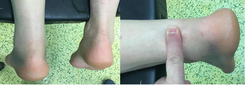
(a) (b) Figure 1a: Right Achilles tendon ruptures. Figure 1b: Tendon gap.
Purpose
Although there is a lot of study about the treatment of Achilles tendon ruptures, it is controversial how to perform the treatment of Achilles tendon rupture and follow-up. It is aimed to compare the results of classical rehabilitation after open surgical repair and early mobilization after percutaneous repair of patients who applied to our hospital with acute tendon rupture.
Method
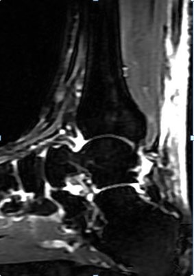
A total of 44 patients (12 females; 32 males) applied to emergency service with Achilles tendon rupture was evaluated between January 2011 and June 2016. Patients were examined and magnetic resonance imaging was performed. The patient was scheduled to undergo surgery for those who have a total Achilles rupture detected in magnetic resonance and who’s the Thompson test was positive, who is with a palpable ecchymosis. The mean age of the patients is 41,3 (25-58). Neither degenerative nor rheumatologic disease was seen in any of the patients nor was previous ache region pain detected. Patients were selected by three surgeons. When the patients applied to the emergency department, they were separated according to the doctor on duty and the operations were performed open surgical repair or percutaneous repair. While patients in the percutaneous repair group were treated early by local anesthesia, the open surgical repair group was operated on an average of 2 days (1-4 days). Patients were followed for a mean of 24 months and they were evaluated preoperatively, postoperatively at week 1st, 3rd, 6th and following in the 3rd, 12th and 24th month (Figure 2).
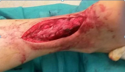
19 patients who underwent open surgery were operated on the first surgery day after admission to the hospital after general or spinal anesthesia preparation. Tourniquet was not used during surgery. Paratenon has been revealed with a dorsal incision made through the Achilles tendon rupture; while 1 prolene loop suture was used for the first six patients who underwent open surgical repair, the tendon repair was performed with 1 polydioxanone (PDS) loop suture for the others. Six patients needed to use plantaris tendon grafts to strengthen their tendon tendons. The skin was covered with prolene or stapler. Postoperatively, splints were applied on a 20-degree plantar flexion. After the first two days of medical dressing, patients were made a walk in 20-degree plantar flexion with an ankle stabilization device without stepping for three-week, with a half-load of three to six weeks and six weeks later made walked by putting whole weight on foot. Patients’ discharge times ranged from two to five days (Figures 3-5).
In 25 patients who underwent percutaneous Achilles tendon repair, eight split incisions were performed under local anesthesia to proximal and distal of the region where the tendon is ruptured. The suture was tied at the first entrance by using 1 polydioxanone firstly from distal to proximal then from proximal to distal while keeping the ankle in flexion with crossings. The dimpling’s formed in the skin were loosened with a fine-tipped clamp. The skin was covered with sterile strips. The Thompson test and active flexion were evaluated postoperatively. Patients were brought to their feet and made them walk when they were in the operating room. It was seen that the patients could easily be mobilized. The patients were discharged the same day by bandaging in soft dressing. After discharge, only two dressings were offered. In both groups, age, side, duration of operation, operation method, duration of discharge, achilles tendon total rupture score (ATRS), visual pain scale (VAS), duration of return to work, diameter measurement (calf measurement) of gastrocnemius of operated and not operated leg made 10 cm distal from the tibial tuberculum of the gastrocnemius without loading in the sitting position have been evaluated. We use SPSS 21 for statistical reports.
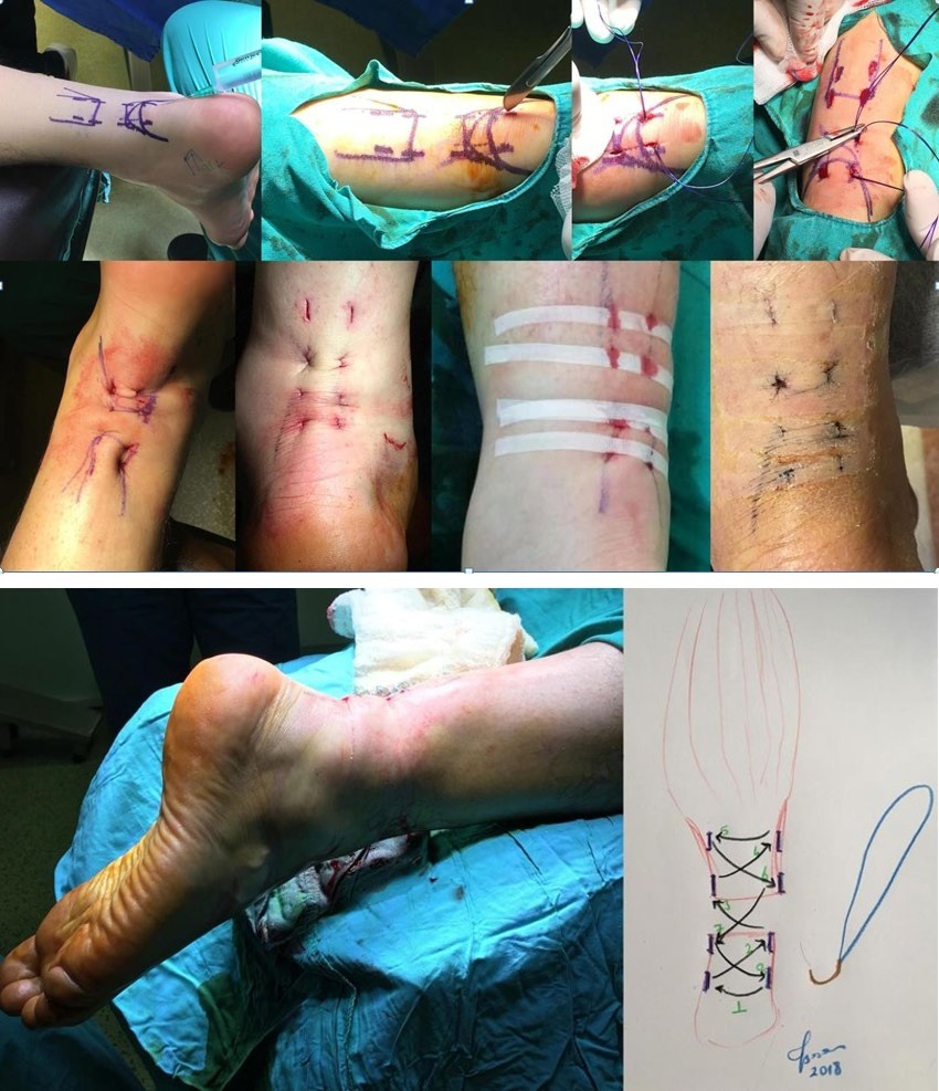
Figure 5a: Operative early motion of ankle. Figure 5b: Percutaneous Achilles tendon rupture repair suture techniques.
Findings
All of the patients were called to control the first week. Patients were evaluated with Achilles tendon total rupture score (ATRS) and the visual analogue scale (VAS) in the month of 0th, 3rd, 12nd, and 24th. In the 12th month check- ups, the patients had a magnetic resonance imaging test and it was found that there were no problems with them.
Patients who underwent open surgery were had their stitches removed between a mean of 25 days (21-35) and allowed to bath without plaster or equipment being wet. It was seen that the patients were limping when they walk after the stitches were removed. Physical therapy was initiated in 14 of the patients due to Achilles tension. Eight of the patients had reddened wound in the third week, but the infection could be treated with oral antibiotics. The chronic wound drainage has been seen in two patients. Debridement was applied to these patients and oral antibiotics were given to them. The mean ATRS (Achilles tendon total rupture score) was 71,1 in the 0th month, 76,9 in the 3rd month, 81,8 in the 12th month and 91,5 in the 24th month. The mean VAS score was 8,6 in the 0th month, 5,6 in the 3rd month, 3.1 in the 12th month and 1,5 in the 24th month. In four patients, skin irritation due to prolene suture node was observed. Sutures from these patients were taken in about 2 years.
Patients who had percutaneous Achilles tendon repair were evaluated at 1st week and wound site has been seen to be clean and they were allowed to bathe. During the third week of the patients, it was observed that the wound sites were clean. Patients were found to have active ankle flexion. It was seen that the patients were limping due to pain while walking. It was observed that there were no problems with the wound sites and the pain decreased at 6th-week control. There were no gaps in the achilles rupture zone, but there was thickening in the healing zone. Physical therapy was not needed because Achilles tension was not seen in the patients. They were observed as they walked slightly with a limp. The mean ATRS score was 85,7 in the 0th month, 87,1 in the 3rd month, 90,3 in the 12th month and 91,9 in the 24th month. The mean VAS score was 8,0 in the 0th month, 3,8 in the 3rd month, 2,0 in the 12th month and 1,3 in the 24th month. The first three patients were repaired with prolane. In one of these patients, prolane-bound skin tenderness was observed in the first year and the suture removed with local anesthesia. Any complications were not observed in any patient because PDS stitching was applied to the other patients.
There was no statistically significant difference between groups in terms of age, gender and surgical side between open surgery repair (group A) and percutaneous repair groups (group B). The surgery duration was 48,1 minutes in group A and 15,0 minutes in group B, the difference between them was statistically significant (p<0,05). While the discharge duration was 3,3 days in group A, it was 1 day in group B, and the difference between them was statistically significant (p<0,05). While the return-to-work was 81,7 days in group A, it was 22,3 days in group B, the difference between them was statistically significant (p<0,05). While VAS value was statistically significantly lower at month 0th month (p<0,05), 3rd month (p<0,05) and at 12th month (p<0,05) in group B, but no statistically significant difference was found between groups at 24th month (p=0,176). While ATSR value was statistically significantly lower at month 0th month (p<0,05), 3rd month (p<0,05) and at 12th month (p<0,05) in group b, but no statistically significant difference was found between groups at 24th month (p=0,942). Gastrocnemius (calf) atrophy was not detected in gastrocnemius circumferential measurements in the second group for the first year. In group A, general anesthesia was given to 16% of the patients and spinal anesthesia was given to 84% of patients. Local anesthesia has been applied to the entire B group (Table 1).
| Open Surgery | Percutaneous | p | |
|---|---|---|---|
| Surgery Time (minute) | 48,1 | 15,0 | p:0.000 |
| Discharge Time (day) | 3,3 | 1 | p:0.000 |
| VAS 0 | 8,6 | 8,0 | p:0.002 |
| VAS 3 month | 5,6 | 3,8 | p:0.000 |
| VAS 12 month | 3,1 | 2,0 | p:0.001 |
| VAS 24 month | 1,5 | 1,3 | p:0.176 |
| ATSR 0 | 71,1 | 85,7 | p:0.000 |
| ATSR 3 month | 76,9 | 87,1 | p:0.000 |
| ATSR 12 month | 81,8 | 90,3 | p:0.000 |
| ATSR 24 month | 91,5 | 91,9 | p:0.942 |
| Return to work (day) | 51,7 | 22,3 | p:0.000 |
Table 1: Open and Percutaneous Achilles tendon rupture repair statistical findings.
Discussion
Even though there is a lot of study about the treatment of Achilles tendon ruptures, the treatment of Achilles tendon rupture is still controversial. It can usually be grouped under three headings; conservative, open surgical repair and percutaneous Achilles tendon repair. With conservative treatment, besides patient costs reduced, there is no wound complication; however, muscle atrophy and rupture can be seen once again. Achilles tendon rupture requires the use of an ankle walking device for a period of time after an open surgical repair, and even if there is no wound complication, a long rehabilitation period is required and both delay the return of the patient to work and increase treatment costs. Ma and Griffith have developed a method of repairing Achilles tendon ruptures staying between open surgical repair and conservative treatment for percutaneous repair without opening the tendon rupture area. Percutaneous repair involves performing six micro dissections along the lateral and medial sides of the tendon and passing the suture material through these micro dissections. Ma and Griffith reported only two minor skin complications (non-infectious) in their series of 18 patients and reported that they did not experience re-rupture in any patient [8].
The percutaneous repair was later used by many authors and the modifications of the technique were developed and presented in various studies. Rowley and Scotland compared the patients who had conservative treatment-immobilization with plaster in the ankle joint (14 patients) with patients who underwent percutaneous repair (10 patients) after acute Achilles tendon rupture. It has been showed that returning to activities take shorter after the percutaneous procedure and they noted that normal plantar flexion power was regained to a large extent [9]. In this study, no other complication was encountered except for a sural nerve compression in a patient. Subsequent studies have reported lower success rates and higher complication rates for the percutaneous technique. Klein W, et al. [10] has reported that 13 percent of 38 patients who underwent through percutaneous technique caused sural nerve compression.
Hockenbury RT, et al. [11] have separated the samples of the open and the percutaneously repair into equal groups (n=5) in the in vitro study (Bunnell sewing technique) over fresh-frozen cadaveric Achilles tendons. They have shown that the open method provides twice as much repair power as compared to the percutaneous technique. The authors also reported that the sural nerve compression (60%) and tendon disorientation (80%) developed in tendons where percutaneous technique applied in tendons. Other studies comparing percutaneous and open repair results have generally shown similar results, with the power of percutaneous repair being reported to be lower and re- rupture rate higher than open repair [7].
In a study conducted by Doral et al 62 patients underwent endoscopic controlled percutaneous repair and physical therapy was started, but allowed to press down as much as possible with a walking device (moon brace), and the full press was allowed after 3 weeks. The average return to work was 11,7 weeks (10-13 weeks) [12]. In our study, return to work in open surgery repair patients 7 week (4- 11); percutaneous tendon repair was performed 3 weeks (2-4) in patients who underwent repair. In the study carried out by Rouvillain JL, et al. [13] it has been performed the percutaneous tendon repair described by Ma and Griffith under local anesthesia for 59 patients with Achilles tendon rupture. He was plastered for 3 weeks without letting step on. The other 3 weeks were given full load on the casted foot at 90 degrees. In the two patients, the re-ruptured has been seen. Sural nerve damage was not seen. They returned to work on average 85 days and returned to the sport in 5 months [13]. In our study, patients with percutaneous and open ascites repair were compared. However, early return to work was seen, since early rehabilitation started in percutaneous patients and the results were very good. Re- rupture and sural nerve damage were also not observed.
In a prospective randomized study with 39 patients, De la Fuente C, et al. [14] divided the Achilles tendon ruptured patients into two groups. Aggressive physical therapy started one day after surgery was compared with conventional physical therapy started 28 days after the operation and the aggressive physical therapy started early was found to have better clinical results and re-rupture was found to be 5% [14]. Our study was not prospectively designed. While the patients treated by percutaneous rehabilitation treated by aggressive treatment put weight on the feet immediately, patients who underwent open repair started giving partial load with the help of device after 3 weeks but started to give full load after 6 weeks. Better results were seen in patients who underwent percutaneous Achilles repair. There was no difference in the results of the 2nd year controls.
The results of open and percutaneous Achilles tendon repair were compared in Akpınar E, et al. [15] study with 90 patients. The results of both open repair and percutaneous repair were seen to be successful but they were not superior in terms of early recovery and return to work. However, when the study was examined, the patient was subjected to splinting for 2 days and afterward the splint was removed. It has not been put weight on the foot for 6 weeks under the control of angle adjustable device. After 6 weeks, it was put weight on the foot and physical therapy was started. In his two groups, one rupture for each was detected [15]. In our study, the results were better because of the early onset of percutaneous acupuncture repair, and the return to work occurred early.
In the study of Kadakia AR, et al. [16] although the number of re-rupture after percutaneous repair and limited open treatment is the same as open repair, much fewer complication rates are seen. There was no statistically significant difference in re-rupture rates in the reinforced Achilles repair with gastrocnemius fascia flap. There was no difference between the groups using PRP and those not used for biological strengthening. Promising developments are seen in animal studies with bone marrow, but more work needs to be done for the benefit [16]. If we consider the weaker side of our work, patients have not been operated on by the same surgeon; the patients haven’t been randomized. As the number of patients is low, the results can-not be generalized to the general assembly.
Results
The main purpose of the treatment of Achilles tendon rupture is to return to the pre-injury quality of life. Open surgical tendon repair requires long-term rehabilitation for preoperative preparation, general or spinal anesthesia, postoperative wound care, prolonged splint-splint use, and subsequent ankle stiffness. Re-rupture and calf atrophy can be seen with conservative treatment and again a long time splint is required. With percutaneous Achilles tendon repair, the duration of surgery and the length of stay were significantly reduced. Hospital costs were significantly reduced. The return of the patient to the work was significantly accelerated. The actual patient costs can be reduced by providing minimum rehabilitation period with repair of percutaneous rheumatoid rupture and early mobilization or by the help of some approaches such as ambulatory treatment, avoiding the systemic side effects of anesthesia, shortening of operating room time, more effective use of rehabilitation units, not using the device and early return to work. Studies with more patients work are needed.
Disclosure of Interest
No benefits or funds were received in support of this study. The authors report no conflict of interests.
References
-
Carden DG, Noble J, Chalmers J, Lunn P, Ellis J (1987) Rupture of the calcaneal tendon. The early and late management. J Bone Joint Surg Br 69(3): 416-420.
-
Cetti R, Christensen SE, Ejsted R, Jensen NM, Jorgensen U (1993) Operative versus nonoperative treatment of Achilles tendon rupture. A prospective randomized study and review of the literature. Am J Sports Med 21(6): 791-799.
-
Puddu G, Ippolito E, Postacchini F (1976) A classification of Achilles tendon disease. Am J Sports Med 4(4): 145- 150.
-
Hattrup SJ, Johnson KA (1985) A review of ruptures of the Achilles tendon. Foot Ankle 6(1): 34-38.
-
Bradley JP, Tibone JE (1990) Percutaneous and open surgical repairs of Achilles tendon ruptures. A comparative study. Am J Sports Med 18(2): 188-195.
-
DiStefano VJ, Nixon JE (1972) Achilles tendon rupture: Pathogenesis, diagnosis, and treatment by a modified pullout wire technique. J Trauma 12(8): 671-677.
-
Karahan M, Erol B (2004) Aşil tendon yırtıklarına yaklaşım. Totbid dergisi 3(1-2): 1-12.
-
Ma GW, Griffith TG (1977) Percutaneous repair of acute closed ruptured Achilles tendon: a new technique. Clin Orthop Relat Res 128: 247-255.
-
Rowley DI, Scotland TR (1982) Rupture of the Achilles tendon treated by a simple operative procedure. Injury 14(3): 252-254.
-
Klein W, Lang DM, Saleh M (1991) The use of the Ma- Griffith technique for percutaneous repair of fresh ruptured tendo Achillis. Chir Organi Mov 76(3): 223- 238.
-
Hockenbury RT, Johns JC (1990) A biomechanical in vitro comparison of open versus percutaneous repair of tendon Achilles. Foot Ankle 11(2): 67-72.
-
Doral MN, Bozkurt M, Turhan E, Ayvaz M, Atay OA, et al. (2009) Percutaneous suturing of the ruptured Achilles tendon with endoscopic control. Arch Orthop Trauma Surg 129(8): 1093-1101.
-
Rouvillain JL, Navarre T, Labrada-Blanco O, Garron E, Daoud W (2010) Percutaneous suture of acute Achilles tendon rupture: A study of 60 cases. Acta Orthop Belg 76(2): 237-242.
-
De la Fuente C, Lillo RPY, Carreño G, Marambio H (2015) Prospective randomized clinical trial of aggressive rehabilitation after acute Achilles tendon ruptures repaired with Dresden technique. Foot (Edinb) 26: 15- 22.
-
Akpınar E, Ceylan HH, Polat G, Ergin ON, Erdil M, et al. (2015) Comparison of Percutaneous and Open Techniques in Treatment of Acute Achilles Tendon Rupture. J Kartal TR 26(3): 243-247.
-
Kadakia AR, Dekker RG, Ho BS (2017) Acute Achilles Tendon Ruptures: An Update on Treatment. J Am Acad Orthop Surg 25(1): 23-31.
- Electrolyte Considerations for Athletes
- Comprehensive Rehabilitation in Adults with Diabetic Peripheral Neuropathy: A Literature Review on Frequency, Intensity, and Duration Parameters
- Exercise Duration and Its Association with ADHD Symptom Severity in Children and Adolescents: A Parent-Reported Survey Study
- Adaptation of the Adult Neurophysiology of Pain Questionnaire for Use in Pediatrics
- A Non-Pharmacological Multidisciplinary Pain Program within a Hospital Wellness Program: A Mixed Methods Study
- The Effect of Frenkel's Exercise with PNF on Functional Reach in Stroke Survivors: A Randomized Control Trial