Toxicity Interactions of Nanomaterials in Biological System: A Pressing Priority
Nanomaterials have made a rebellion in biomedical application especially treating several diseases due to its distinctive compositions. However, increased utilization of nanomaterials in biomedical applications has made an initiative to understand the possible interaction between the nanomaterials with the biological systems. These tiny particles enter into the body very easily and affect vulnerable systems which raise the interrogation of their potential effects on the susceptible organs. It is very crucial to comprehend the various exposure pathways, their movement, behavior and ultimate outcome. Specific and unique physicochemical properties, such as particle size and distribution, surface area, charge and coatings, particle shape/ structure, dissolution and aggregation, influence the nanomaterial interactions with cells. Toxicities in biological systems occurs as a result of a result of a variety of reasons including the production of ROS reactive oxygen species, degradation of the integrity of membrane and release of toxic metal ions thus preventing normal cell function. Various researchers have provided promising evidence that nanomaterial’s actively encompass and mediate chemical processes of cell, in addition to their passive interactions with cells. Certainly, it is very much essential to understand the possible toxic interactions of nanomaterial’s with the biological system as Nano toxicology. In this review, we emphasize the toxicological effects on different organs pertaining to nanomaterial-biological system interaction.
Introduction
An extensive research and production of nanomaterials (NMs) for various purposes has been increased. Human population either occupational or general exposing to various NMs cannot be ignored, and the risk associated with this exposure now has become a major public health concerns. These NM can enter into the bloodstream by efficiently crossing membrane barriers, distributes throughout the body, and exert its effects in different organs and tissues at the cellular and molecular levels [1]. Toxicological analysis of individual organs is important for mechanistic understanding, since, harmful effects coexist in distinct organ. Risk assessment should also be taken into account for the toxicological profile of NMs as a whole [2]. There is a need of Hazard information to reassure regulators, workers, and customers that these NM can be used safely as they are created and marketed. Intense scientific curiosity has been sparked by how nanoparticles enter and interact with the human body has been given much attention as well as increasing concern. Identified primary target organs for NPs toxicity are skin, lungs and reticuloendothelial system (RES), which includes the liver and spleen by Opsonisation. In vitro cytotoxicity studies are widely used to assess the toxicity and measure cellular uptake. Nanoparticles (NPs) can cross cell membranes due to their high surface area to volume ratio which may cause interactions in biological systems [3]. In the last few years, a substantial scientific work is focused to identify the potential toxicity of nanomaterials by studying the cellular pathways under in vitro and in vivo conditions.
Absorption of NP/NM into the biological system may alter the cellular system, including cell shape, cell differentiation, metabolism, cell mobility, and cellular immunity. If the NMs are accumulated in mitochondria can produce ROS and apoptosis and may induce hemolysis if it is absorbed into the blood circulation [4]. Consequently, the biological systems has some undesirable effects like dysfunction, deterioration of membrane integrity, and release of toxic metal ions which can bind with particular cell receptors and adopt specific conformations that prevent normal cell function, leading to produce different toxic effects like cytotoxicity, genotoxicity, and possibly cell necrosis [5]. Hence, it has become essential to identify the interface response of NPs/NMs with biological environment and its toxicological impact.
Interaction of Nanomaterials with Cells
Though there has been substantial progress in knowledge about the NP and cell interactions, the specific mechanisms by which nanomaterials pass through the lipid membrane of a particular cell type is crucial to understand [6]. Nanomaterials flow through the cellular membrane by penetration which is analogous to passive diffusion from extracellular to intracellular region in cell membrane [7]. Numerous studies have suggested that the primary mechanism for cellular uptake of NPs is endocytosis/phagocytosis.
Mechanism
Biological systems made with cells are called as compartment in which the lipid based membrane serves as diffusion barriers where the uptakes of particles take place. Most of the organic molecules enter into the cells by diffusion and the rate of cellular uptake depends upon the size and partition coefficient ($K_{ow}$) of a substance. The rapidly emerging field of Nanotoxicology attempts to translate concepts from chemical toxicology to Nano - objects.
The essential mechanisms followed for the removal of NPs from an organ are
- Uptake of Nanoparticles into Cells and Organs
- Soluble Nanoparticles: Trojan-Horse-Type Toxin Uptake
- Chemically Active Nanoparticles Inside Cells
- Solubility Determines Environmental Long-Term Fate [8]
Hazard Assessment
Physical and biological properties needs to be considered while determining the risk associated with the substances. NMs may enter into the body through different routes, such as inhalation, ingestion, or skin exposure. Once enters inside the body, NPs/NMs may interact with cells and tissues, including distant organs [9]. A substantial step while performing a hazard identification is to discover whether the compound of interest is classified as a genotoxic carcinogen, epigenetic carcinogen, or non-carcinogen. This could lead to adverse effects on multiple organs namely pulmonary system, cardiovascular system, skin, gastrointestinal system, immune system, and major organ systems including the central nervous system [10]. NPs, which entered into cells, can cause cytotoxic effect as well an immunotoxic effect might be caused by NPs when it gets accumulated inside the immune system cells like dendritic cells. Interaction of NPs with the nucleus and DNA can cause the production of reactive oxygen species (ROS), reactive nitrogen species (RNS) and inflammatory products.
Several in-vitro and in-vivo methods are performed to predict and evaluate the impact of NMs with the biological systems. The toxicological effects of the NMs interactions with several biological systems are classified as [11]
- Pulmonary Toxicity of Nanomaterials
- Cardiovascular Toxicity of Nanomaterials
- Dermal Toxicity of Nanomaterials
- Developmental Toxicity of Nanomaterials
- Immunotoxicity of Nanomaterials
- Neurotoxicity of Nanomaterials
- Gastro-Intestinal Effects of Nanomaterials
- Genotoxicity and Carcinogenicity of Nanomaterials
- Hepatotoxicity induced by nanomaterials
Among these toxicities, only few are discuss in the section below.
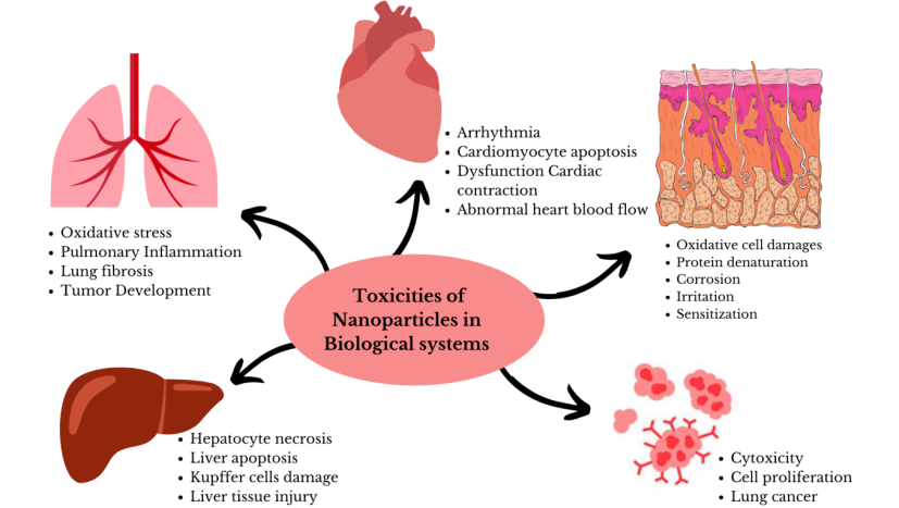
Pulmonary toxicity of nanomaterials: The respiratory system is the primary point of entry for nanoparticles into the human body. NMs may accumulate in the lungs and can interact with the pulmonary system produces several diseases which include cancer, genotoxicity, oxidative stress, fibrosis, and inflammation. If NMs are accumulated in the macrophages subsequently may produce minimal inflammatory consequences. Carbon nanoparticles/nanotubes (CNPs/CNTs) have received a lot of attention due to their light weight, very high surface area, durability, and wide range of applications [12]. The interactions of NPs with respiratory system have gained much interest by researchers as various natural and man-made fibrous particles are known to cause chronic and debilitating lung diseases. Oxidative stress is the most common toxic effect by CNTs in pulmonary system when compared to other NPs [13]. ROS have long been known to react with cellular macromolecules and disrupt the intracellular homeostasis. In vivo studies for MWCNTs (Multi walled carbon nanotubes) and SWCNTs (Single walled carbon nanotubes) were investigated by either pharyngeal aspiration or inhalation in 2-month-old C57Bl/6J male mice model. The result demonstrated that the level of glutathione was decreased whereas protein thiols and Malondialdehyde were increased. The observed result confirmed that administration of MWCNT and SWCNTs developed the local inflammation, oxidative stress and fibrosis induction [14]. Shvedova, et al. investigated the one single acute exposure of SWCNTs in Lewis lung carcinoma cells (LLC) and BEAS-2B cells (human bronchial epithelial cell line) [15]. The results indicated that SWCNTs up-regulated the lung tumor burden which leads to induce the myeloid-derived suppressor cells (MDSCs) and its derived TGFβ production. Luanpitpong, et al. investigated chronic exposure of Non-tumorigenic human lung epithelial cells with SWCNT at 0.02 μg/cm2 surface area dose [16]. The study results demonstrated that on chronic exposure to the human lung epithelial cells induced cancer stem cells lead to produce tumor development due to inactivation of p53 and caveolin-1 over-expression. Several studies demonstrated that administration of CNTs may act as tumor promoters.
Cardiovascular toxicity of nanomaterials: Heart appears to be less affected by short-term acute exposure of low levels of NPs, but on extended exposure at higher dose, damages the structure and functionality of the heart. NMs can accumulate in the heart tissue which mainly interferes with the steady state of cardiac electrophysiology, followed by damaging the mitochondria and myocardial cells. The study emphasized that the inhalation of NPs leads to produce cardiorespiratory diseases [17]. Presently, several NMs are widely used and frequently found in the environment. On exposure to these NMs in the workplace can cause harmful effects on the heart. Interaction between the cardiovascular system and NMs induce disrupted heart function which includes irregular heartbeat, arrhythmia, dysfunctional cardiac contraction, abnormal heart blood flow, and other cardiovascular issues. On the other side, maternal exposure can induce congenital cardiac contraction disorders, myocardial malformations, and developmental disorders of the heart’s structure in neonates. NMs affects the normal contraction and relaxation of the myocardium, altering cardiac hemodynamics, inducing cardiomyocytes to undergo oxidative stress, apoptosis, and inflammatory responses as well as disrupting the integrity of mitochondrial membranes, thereby inducing a variety of serious cardiac diseases. Small particle size of the NPs has greater penetration which can play a significant role in inducing stronger toxic effects and sever heart damage. The review results highlighted that on pulmonary exposure induces cardiovascular system damage in two pathophysiological phases. In the first phase, neuron regulated fast-onset and short lasting cardiovascular reactions are induced. Whereas in the second phase, chronic cardiovascular events like atherosclerosis are developed. In addition, NMs can alter the autonomic nervous activity [18]. Bostan, et al. reviewed the cardiac toxicity of iron oxide, titanium, carbon, silver and silica NPs and outlined that NMs can affect the heart tissue [19]. On exposure to CNT, it interfaces with cardiac electrophysiology, also generate a significant amount of free radicals, which result to oxidative damage and malfunction. Cardiomyocyte apoptosis and cardiac dysfunction are the other major effects of metal oxide NPs. It has found that particle exposure is closely related to cardiovascular disease [20]. Further, there are numerous study reports stressed that NMs can enter the heart tissue, accumulate and then cause damage.
Genotoxicity and carcinogenicity of nanomaterials: It has been regarded that when NPs or NPs containing fumes are inhaled it produces carcinogenicity to humans. Various investigational studies established the potential carcinogenicity of several engineered NMs to the human beings. However, genotoxicity is demonstrated by mutations and DNA damage such as breakage of DNA strand and oxidatively damaged DNA has been investigated in a number of models. NMs can produce genotoxicity by physical contact with the DNA nucleus and their subsequent direct interaction with the genome. This interaction may result in chromosomal damage, massive DNA malformations, DNA breaks, and other DNA lesions [21]. However, primary genotoxicity can be caused either directly or indirectly. The smallest NMs enter into the nucleus through nuclear pores by intracellular processes resembling endocytosis; subsequently break the nuclear membrane during mitosis which will allow the NMs to enter into the nucleus and then interact directly with DNA’s chromosomal structures. NMs may interact or bind to DNA molecules during interphase, preventing DNA replication and the DNA Transcription. NMs can disrupt the process of mitosis by interfering with the spindle apparatus, centrioles, and related proteins. This results in the development of micronuclei that may exhibit clastogenicity or aneugenicity. Secondary genotoxicity is considered to be the main mechanism of genotoxicity of NMs. It is mediated out by ROS inflammatory cells. NMs have the ability to activate phagocytes and produce an oxidative burst. Several genotoxic and cytotoxic effects on NMs were investigated and found to be linked to the exposure of NPs. In vitro studies evaluating potential nanotoxicity in human primary cells showed that titanium dioxide (TiO2), zinc oxide (ZnO), and cerium dioxide (CeO2) NPs induced both primary and oxidative DNA damage in salivary leukocytes, which were proved to be a suitable biomatrix for Nano genotoxicity studies using the comet assay [22]. Lactate dehydrogenase (LDH) assays were used to determine the cytotoxic effects of coated and uncoated graphene oxide (GO)
and reduced Graphene Oxide (rGO) on human chondrocytes. Graphene and graphene-based NMs have received the much attention in the nanotechnology industry due to their unique physical, chemical, and mechanical properties [23]. Different amorphous silica nanomaterial (aSiO2 NM) variants were found to cause oxidative DNA damage [24].
Hepatotoxicity induced by nanomaterials: More than 90% of NMs are translocated from other tissues and then accumulate in the liver [25]. The structure and operation of hepatic cells may be harmed by bio concentration of NPs. Reviewing the information on NP-induced hepatotoxicity is therefore crucial. The size of the NPs plays a crucial role in the hepatotoxicity. Methods of NMs administration can induce different toxicological consequences, but the oral route is the most pertinent. When NPs get accumulate in the liver exhibit the toxic effects, hepatic damage, also impairment of other cell including Kupffer cells [26]. Investigations involving IV injection may occasionally overestimate the true frequency of hepatotoxic effects due to higher bioavailability of NMs. In human liver (HepG2) cells, silica nanoparticles were observed to cause apoptosis, which was mediated by ROS through the p53, bax/bcl-2, and caspase pathways. These findings were validated by Jinhee choe, et al_. based on the metabolomic investigation done on the liver of ICR mice and human hepatoma cells (HepG2) treated with SiNPs. This study found that SiNP induce hepatotoxicity by glutathione metabolism along with oxidative stress and affect the CYP450 [27]. Hepatotoxic effects have been observed for NMs when they administered through oral, IV injection, as well as the inhalation route which was mimicked by intra tracheal instillation. This information is especially significant when human beings are occupational exposure situations to this NMs. Studies reveal that the colloidal gold nanoparticles are used to treat rheumatoid arthritis and to diagnosis cancer. During the treatment, the gold NPs are accumulated in the kupffer liver cells and caused liver tissue injury. In addition, toxicity of metal and metal oxide NPs were assessed in rat liver cell line BRL 3A model based on mitochondrial dysfunction and oxidative stress. The study results outlined that metal NPs produced higher toxic effect in the liver cells when compare to metal oxide NPs [28]. Hepatotoxicity investigational study was performed for TiO2 through _in vivo techniques. The histological examination results demonstrated that TiO2 are accumulated in the liver cells leads to increase the liver further producing increased hepatocyte necrosis [29].
Dermal toxicity of nanomaterials: Dermal toxicity to nanoparticles refers to the penetration of nanoparticles into skin produces irritation on the skin surface as well as the local effects of sensitization and any other potential systemic effects that may follow on such exposure. Skin absorption or dermal penetration of chemicals is a process of diffusion from the outer surface of the skin through the skin and finally into systemic circulation [30]. Metal based nanoparticles such as TiO2, ZnO, Ag and Au NPs are used in cosmetics, e.g., as UV-filters and to achieve antibacterial/ anti- inflammatory properties, accelerated wound healing, increase collagen production etc. At the same time, studies on cultured skin cells often indicated the toxic properties for these NPs. For TiO2 nanoparticles, one concern is their photocatalytic activity leading to ROS production. MeO-NPs and MeO-coated NMs could pose a severe hazards including oxidative damages in cell membrane lesions and protein denaturation when in direct exposure with skin. The toxicity of textiles coated with antimicrobial copper and zinc oxide (CuO, ZnO) NPs can be evaluated using an in vitro recreated 3D model of epidermis [31].
Various toxicities of the nanomaterial in the biological systems are shown in Figure 1.
Conclusion
Numerous genes and proteins are essential for the biological process, however it is still unclear how many of these biological components and processes come together. Biological investigations by utilizing NPs/NMs approaches can perspectives on how complex signal pathways affect the cells behavior and what happens at the molecular level. Further, NPs may also be utilized for better understanding of how these processes can be improved to achieve the task with the cell cooperation. Understanding the underlying metabolic pathways involved in disease and injury may be facilitated by NP monitoring and detection approaches. The development of effective and secure Nano medicine will be facilitated by a critical and logical evaluation of clinical needs and their impact on the biological system. Without doubt, a critical understanding of biological responses to engineered NMs is required along with organ toxicity investigational study needed to identify and comprehend their toxicity in order to avoid the negative consequences. In addition, to increasing current knowledge on the toxicological profile of these specific NPs, lay the groundwork for future research in this area also to address the nontoxicity evaluation challenges. The development and implementation of individualized Nano medicines should be aided by the toxicological assessment, despite the fact that there are many significant challenges, such as scientific and regulatory difficulties related to the production of nonmedical products. Therefore, it is realistic to consider the bedside delivery of tailored Nano medicine in the future.
References
-
Boverhof DR, David RM (2010) Nanomaterial characterization: considerations and needs for hazard assessment and safety evaluation. Analytical and bioanalytical chemistry 396(3): 953-961.
-
GubalaV, Johnston LJ, Krug HF, Moore CJ, Ober CK, et al. (2018) Engineered nanomaterials and human health: Part 2 Applications and nanotoxicology (IUPAC Technical Report). Pure and Applied Chemistry 90(8): 1325-1356.
-
Soro CA, Nesma T, Velasco PJ, Viñuela AL, Gomez HF, et al. (2019) Interactions of nanoparticles and biosystems: Microenvironment of Nnanoparticles and biomolecules in nanomedicine. Nanomaterials (Basel) 9(10): 1365.
-
Armstead L, Li B (2016) Nanotoxicity: emerging concerns regarding nanomaterial safety and occupational hard metal (WC-Co) nanoparticle exposure. International Journal of Nanomedicine 11: 6421-6433.
-
Jackson TC, Patani BO, Israel MB (2017) Nanomaterials and cell interactions: A Review. Journal of Biomaterials and Nano biotechnology 8(4): 220-228.
-
Behzadi S, Serpooshan V, Tao W, Hamaly MA, Alkawareek MY, et al. (2017) Cellular uptake of nanoparticles: journey inside the cell. Chemical Society reviews 46(14): 4218-4244.
-
Tripathi AC, Saraf SA, Saraf SK (2015) Carbon nanotropes: A contemporary paradigm in drug delivery. Materials 8(6): 3068-3100.
-
Stark WJ (2011) Nanoparticles in Biological Systems. Angew Chem Int 50(6): 1242-1258.
-
Gehr P (2018) Interaction of nanoparticles with biological systems. Colloids and Surfaces B: Biointerfaces. 172: 395-399.
-
Liu XQ, Rui-Zhi T (2017) Biological responses to nanomaterials: understanding nano-bio effects on cell behaviours. Drug Delivery 24(1): 1-15.
-
Ceccatelli S, Giuseppe B (2017) Neurological System. In: Fadeel B, et al. (Eds.), Adverse Effects of engineered Nanomaterials: Exposure toxicology, and impact on human health. 2nd (Edn.), Elsevier, pp: 468.
-
Dahm MM, Evans DE, Schubauer-Berigan MK, Birch ME, Fernback JE (2012) Occupational exposure assessment in carbon nanotube and nanofiber primary and secondary manufacturers. The Annals of occupational hygiene 56(5): 542-556.
-
Pacurari M, Lowe K, Tchounwou PB, Kafoury R (2016) A review on the respiratory system toxicity of carbon nanoparticles. International Journal of Environmental Research and Public Health 13(3): 325.
-
Khaliullin TO, Shvedova AA, Kisin ER, Zalyalov ER, Fatkhutdinova LM (2015) Evaluation of fibrogenic potenatial of industrial multi-walled carbon nanotubes in acute aspiration experiment. Bulletin of experimental biology and medicine 15(8): 684-687.
-
Shvedova AA, Kisin ER, Yanamala N, Tkach AV, Gutkin DW, et al. (2015) MDSC and TGFbeta are required for facilitation of tumor growth in the lungs of mice exposed to carbon nanotubes. Cancer research 75(8): 1615-1623.
-
Luanpitpong S, Wang L, Castranova V, Rojanasakul Y (2014) Induction of stem-like cells with malignant properties by chronic exposure of human lung epithelial cells to single-walled carbon nanotubes. Particle and Fibre Toxicology 11(22): 11-22.
-
Nagarajan M, Maadurshni GB, Tharani GK, Udhayakumar I, Kumar G, et al. (2022) Exposure to zinc oxide nanoparticles (ZnO-NPs) induces cardiovascular toxicity and exacerbates pathogenesis – Role of oxidative stress and MAPK signaling. Chemico-Biological Interactions 351: 109719.
-
Kan H, Pan D, Castranova V (2018) Engineered nanoparticle exposure and cardiovascular effects: the role of a neuronal-regulated pathway. Inhalation toxicology 30(9-10): 335-342.
-
Bostan HB, Rezaee R, Valokala MG, Tsarouhas K, Golokhvast K, et al. (2016) Cardiotoxicity of nano- particles. Life sciences 165: 91-99.
-
Cheng Y, Chen Z, Yang S, Liu T, Yin L, et al. (2021) Nanomaterials-induced toxicity on cardiac myocytes and tissues and emerging toxicity assessment techniques. Science of the Total Environment 800: 149584.
-
Valdiglesias V (2022) Cytotoxicity and genotoxicity of nanomaterials. Nanomaterials 12(4): 634.
-
Moratin H, Ickrath P, Scherzad A, Meyer TJ, Naczenski S, et al. (2021) Investigation of the immune modulatory potential of zinc oxide nanoparticles in human lymphocytes. Nanomaterials 11(3): 629.
-
Daniyal M, Liu B, Wang W (2020) Comprehensive review on graphene oxide for use in drug delivery system. Current medicinal chemistry 27(22): 3665-3685.
-
Brandão F, Costa C, Bessa MJ, Dumortier E, Chainiaux FD, et al. (2021) Genotoxicity and gene expression in the rat lung tissue following instillation and inhalation of different variants of amorphous silica nanomaterials (aSiO2 NM). Nanomaterials (Basel) 11(6): 1502.
-
Rana SVS (2020) A comprehensive assessment of hepatotoxicity induced by engineered nanoparticles- A Review. Journal of Toxicology and Risk Assessment 6: 035.
-
Boas VV, Vinken M (2021) Hepatotoxicity induced by nanomaterials: Mechanisms and _in vitro_ models. Archives of Toxicology 95(1): 27-52.
-
Chatterjee N, Jeong J, Yoon D, Kim S, Choi J (2018) Global metabolomics approach in _in vitro_ and _in vivo_ models reveals hepatic, glutathione depletion induced by amorphous silica nanoparticles. Chemico-biological interactions 293: 100-106.
-
Hussain S, Hess K, Gearhart J, Geiss KT, Schlager J (2005) In vitro toxicity of nanoparticles in BRL 3A rat liver cells. Toxicology in vitro : An International Journal Published in Association with BIBRA 19(7): 975-983.
-
Gupta SM, Tripathi M (2011) A review of TiO2 nanoparticles. Chinese Science Bulletin 56: 1639-1657.
-
Niska K, Zielinska E, Radomski MW, Inkielewicz Stepniak I (2018) Metal nanoparticles in dermatology and cosmetology: Interactions with human skin cells. Chemico-biological interactions 295: 38-51.
-
Bengalli R, Colantuoni A, Perelshtein I, Gedanken A, Collini M, et al. (2021) _In vitro_ skin toxicity of CuO and ZnO nanoparticles: Application in the safety assessment of antimicrobial coated textiles. Nano Impact 21: 100282.
- Effects of 5-HTP and Melatonin on the Sleep Cycle of Medical Students
- Adsorption of Bisphenol A on NH4OH- Modified Rice Husk and Sugar Cane Bagasse Biochar
- Comparative Assessment of the Reinforcement Efficiency of Palm Fruit Fibre and Coconut Fibre in High Density Polyethylene (HDPE) Matrix Composite
- Importance of Bio Compounds Naturally Present in Food with Functionality in Animal Metabolism
- Sub-Acute Study on the Cardiotoxic Effects of Monosodium Glutamate Ingestion in Albino Rat
- Weight Management and Its Natural Solutions: A Review